From Failure to Function: Staged Surgical Salvage of an Infected Twice-Failed Repair of Grade IV Obstetric Rectovaginal Tear - A case report
- Vatsal Tanwar , Junior Resident, Department of General Surgery, Jawaharlal Nehru Medical College and KLES Dr. Prabhakar Kore Hospital & Medical Research Center, Belagavi.
- Sudarshan Chaugale , Associate professor in Department of General Surgery (Surgical Gastroenterology), Jawaharlal Nehru Medical College and KLES Dr. Prabhakar Kore Hospital & Medical Research Center, Belagavi.
- Farzana Begaum Dharwad , Assistant professor in Department of Obstetrics & Gynecology, Jawaharlal Nehru Medical College and KLES Dr. Prabhakar Kore Hospital & Medical Research Center, Belagavi
Article Information:
Abstract:
Background: Obstetric Grade IV rectovaginal tears are rare but devastating complications of vaginal delivery, often resulting in rectovaginal fistula, fecal incontinence, recurrent perineal infections, and severe impairment of quality of life. Failed primary repair, particularly in resource-limited settings, further complicates management and increases the risk of poor functional outcomes. Infected wounds may lead to irreversible loss of anal sphincter tissue if not managed promptly with expert, staged surgical intervention. Case Presentation: A 27-year-old primiparous underwent full-term normal vaginal delivery however due to baby weighing 3.6kg she had to undergo extensive episiotomy for obstructed labour and landed up with grade 4 Obstetric perineal tear. Initial perineal repair was performed immediately after delivery at the same primary health centre, however in view of persistent perineal soiling with feces she again underwent perineal repair on POD 8 however the stitches failed to hold and the patient presented with persistent perineal pain and rectovaginal fistula with infected wound due to feces as shown in Figure (1). In view of failed twice rectovaginal tear repair she was referred to Surgical Gastroenterology for further management. Magnetic resonance imaging of the pelvis revealed extensive perineal edema, a rectovaginal fistula, and a full-thickness tear of the anal canal involving both internal and external anal sphincters, consistent with a Grade IV obstetric perineal injury. Considering the severity of tissue damage and ongoing contamination, a staged surgical approach was adopted. The patient underwent diversion sigmoid colostomy with tissue debridement and definitive repair of the rectovaginal septum and anal sphincter followed by delayed stoma reversal after adequate healing (after 2months). At follow-up, the patient demonstrated restored anal sphincter tone, normal bowel continence, and no recurrence of rectovaginal fistula, with significant improvement in quality of life. This case highlights the effectiveness of staged surgical management in complex Grade IV failed repair of an infected obstetric rectovaginal tears and emphasizes the importance of staged surgical repair
Keywords:
Article :
INTRODUCTION:
Obstetric perineal injuries continue to represent a significant source of maternal morbidity worldwide, particularly in low- and middle-income countries where access to skilled obstetric care and timely recognition of complications may be limited. Within this spectrum, obstetric anal sphincter injuries (OASIS) constitute the most severe forms, encompassing third- and fourth-degree perineal tears (1). A Grade IV perineal tear is defined by complete disruption of the vaginal epithelium, perineal body, anal sphincter complex, and rectal mucosa, frequently resulting in the development of a rectovaginal fistula. Although the overall reported incidence of OASIS ranges from 0.5% to 5% of vaginal deliveries, Grade IV tears are relatively uncommon but are associated with profound physical, psychological, and social morbidity (2).
Rectovaginal fistula following childbirth is a devastating complication that leads to fecal incontinence, recurrent perineal infections, sexual dysfunction, and marked impairment of quality of life. The condition is associated with considerable emotional distress and social stigma, particularly among young women of reproductive age (3). Obstetric trauma remains one of the leading etiologies of rectovaginal fistula in developing regions and is commonly associated with prolonged or obstructed labor, instrumental delivery, fetal macrosomia, shoulder dystocia, or inadequate perineal support during childbirth. Delayed diagnosis, improper primary repair, and secondary infection further increase the risk of fistula formation and long-term sphincter dysfunction (4).
The management of Grade IV obstetric perineal tears poses a substantial clinical challenge. While primary repair at the time of delivery is considered ideal, secondary surgical intervention becomes necessary when injuries are missed, inadequately repaired, or complicated by tissue edema and fecal contamination (5). Failed initial repairs are associated with higher rates of wound dehiscence, persistent fistula formation, and long-term fecal incontinence. Consequently, careful patient selection, accurate delineation of injury extent, and appropriate timing of surgical intervention are critical determinants of successful outcomes (6).
Advances in imaging modalities, particularly magnetic resonance imaging of the pelvis, have significantly enhanced the evaluation of complex perineal injuries. MRI provides detailed visualization of the rectovaginal septum, anal sphincter complex, and surrounding soft tissue involvement, thereby facilitating precise surgical planning (7). In cases characterized by extensive tissue damage and active contamination, a staged surgical approach incorporating fecal diversion with wound debridement followed by definitive repair is increasingly advocated. Temporary diverting colostomy reduces fecal contamination, minimizes infection risk, and promotes optimal tissue healing prior to sphincteroplasty and fistula repair (8).
Despite the severity of these injuries, the literature detailing outcomes of staged surgical management for obstetric Grade IV rectovaginal tears remains limited, particularly in resource-constrained settings. Available evidence largely consists of small case series and retrospective studies, with a lack of standardized treatment protocols (9). This highlights the need for detailed case reports that address diagnostic challenges, surgical decision-making, and long-term functional outcomes.
The present case report describes the successful staged management of a Grade IV obstetric rectovaginal tear with complete anal sphincter disruption in a young woman following normal vaginal delivery. By outlining the clinical presentation, imaging findings, surgical strategy, and postoperative outcome, this report aims to contribute to the existing evidence base and underscore the importance of individualized, multidisciplinary care in achieving favorable anatomical and functional results.
CASE PRESENTATION:
A 27-year-old woman, para 1, presented to the Department of Surgical Gastroenterology with complaints of severe perineal pain and involuntary passage of fecal matter through the vagina. She had a history of a twice-repaired Grade IV obstetric rectovaginal tear following an extensive episiotomy performed for obstructed labor during a vaginal delivery of a 3.6-kg neonate at an outside healthcare facility. The patient reported persistent perineal discomfort, foul-smelling vaginal discharge, and significant social distress secondary to fecal incontinence. She had undergone primary perineal suturing on two occasions, one week apart, at the referring center; however, both repairs failed, and the wound subsequently became infected, necessitating referral for further specialized management.
Patient History and Clinical Background
The patient had no significant past medical history and no known comorbid conditions such as diabetes mellitus, hypertension, or connective tissue disorders. She had no history of prior surgical interventions before the index obstetric event. There was no history suggestive of inflammatory bowel disease, chronic constipation, or pre-existing anorectal pathology. Her antenatal period was uneventful, with regular antenatal care and no reported complications. The delivery was full-term and spontaneous. Approximately two weeks postpartum, following two failed repairs of a Grade IV obstetric perineal tear, she again noted fecal staining per vagina, accompanied by worsening perineal pain and difficulty maintaining local hygiene, resulting in a significant negative impact on her quality of life.
Physical Examination
On admission, the patient was conscious, oriented, and hemodynamically stable. General physical examination revealed a moderately built and adequately nourished young woman with no evidence of pallor, icterus, or lymphadenopathy. Her vital parameters were within normal limits, with a blood pressure of 110/70 mmHg and a pulse rate of 92 beats per minute. Abdominal examination demonstrated a soft, non-distended abdomen without tenderness or palpable masses. Bowel sounds were normal.
The sutures from the previous repairs failed to hold, and the patient subsequently presented with persistent perineal pain and an infected rectovaginal fistula with fecal contamination, as demonstrated in Figure 1. Local perineal examination revealed an infected perineal wound with fecal soiling and complete disruption of the anal sphincter complex and rectovaginal septum. Digital rectal examination demonstrated loss of the anal sphincter complex between the 11 o’clock and 1 o’clock positions, with markedly reduced sphincter tone. A full-thickness rectovaginal wall defect extending proximally for approximately 4 cm was noted. The perineum was edematous, with inflamed and infected tissues, along with residual sutures from previous failed repairs.
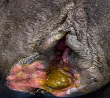
Figure 1: Preoperative clinical photograph showing a failed repair of a Grade IV obstetric rectovaginal tear with complete disruption of the perineal body, rectovaginal septum, and anal sphincter complex, resulting in an infected rectovaginal fistula with fecal contamination.
Diagnostic Evaluation
Baseline laboratory investigations, including complete blood count, renal function tests, liver function tests, and serum electrolytes, were within normal limits, with no evidence of systemic infection or metabolic derangement. Imaging evaluation was performed using magnetic resonance imaging of the pelvis to accurately delineate the extent of the injury. MRI revealed extensive edema of the perineal region, predominantly involving the tissues surrounding the vagina and rectum. An irregular defect was identified in the posterior aspect of the lower vaginal wall, communicating with the anterior rectal wall, thereby confirming the presence of a rectovaginal fistula. In addition, a vertical full-thickness tear measuring approximately 4 cm was noted at the 12 o’clock position of the anal canal, involving both the internal and external anal sphincter muscles. These imaging findings were consistent with a Grade IV obstetric perineal tear.
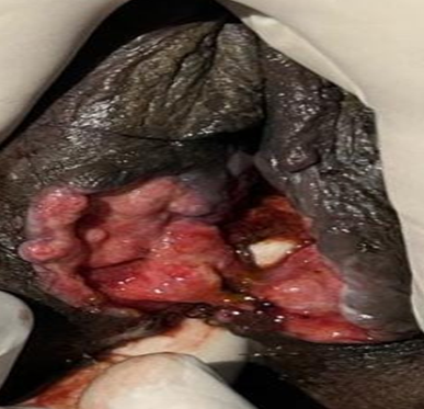
Figure 2: Preoperative clinical photograph demonstrating a failed repair of Grade IV obstetric rectovaginal tear with complete disruption of the perineal body and rectovaginal septum, resulting in a rectovaginal fistula and anal sphincter involvement. A suture holding the anal sphincter.
Surgical Planning and Initial Management Considering the high risk of repair failure in the setting of ongoing fecal contamination and inflamed, sloughed tissues, a staged surgical approach was planned. Following pre-anesthetic evaluation and informed consent, the patient underwent a diverting sigmoid colostomy with thorough debridement of devitalized perineal tissues, followed by definitive repair of the rectovaginal septum and anal sphincter complex using interrupted 3-0 and 4-0 polydioxanone (PDS) sutures.
Intraoperatively, the rectal and vaginal walls were carefully dissected and repaired separately using interrupted 3-0 PDS sutures. Sphincteroplasty was then performed with meticulous identification and approximation of the internal and external anal sphincter muscles to restore anatomical continuity and sphincter function.
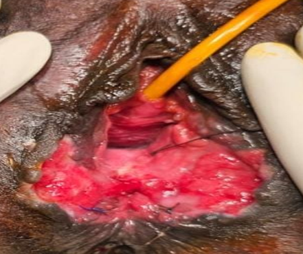
Figure 3: On-table preoperative view showing a Grade IV obstetric rectovaginal tear with complete disruption of the perineal body and rectovaginal septum, prior to surgical dissection and repair.
Surgical Procedure
After multidisciplinary evaluation and obtaining informed consent, the patient was taken up for surgery under general anesthesia and positioned in the lithotomy position. Following meticulous aseptic preparation and bladder decompression with an indwelling Foley catheter, a thorough examination of the perineal region confirmed a Grade IV rectovaginal tear with complete disruption of the rectovaginal septum and anal sphincter complex. A staged surgical approach was therefore undertaken.
The rectovaginal plane was carefully dissected to separate the posterior vaginal wall from the anterior rectal wall. The rectal mucosa was repaired first using interrupted absorbable sutures, followed by separate closure of the vaginal mucosa using interrupted 3-0 polydioxanone (PDS) sutures. The internal and external anal sphincter muscles were then clearly identified, mobilized, and reconstructed with meticulous approximation using interrupted 3-0 PDS sutures. Subsequently, the perineal body was reconstructed in layers to restore normal anatomical integrity. Adequate hemostasis was achieved, and the perineal wound was closed with well-approximated skin sutures, completing the procedure uneventfully.
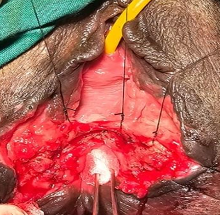
Figure 4: Intraoperative view demonstrating Grade IV obstetric rectovaginal tear with complete disruption of the rectovaginal septum and anal sphincter complex. The posterior vaginal wall and anterior rectal wall are clearly delineated following dissection, with identification of internal and external anal sphincter components prior to definitive layered repair and sphincteroplasty.
Postoperative Course and Follow-Up
The postoperative period was uneventful. The patient was gradually initiated on oral liquids and advanced to a soft diet as tolerated. Stoma function was satisfactory, and meticulous perineal wound care was maintained. She was mobilized early and discharged in stable condition with detailed instructions regarding stoma care, perineal hygiene, and scheduled follow-up. Regular outpatient follow-up demonstrated satisfactory wound healing, absence of infection, and progressive improvement in local symptoms.
After an interval of approximately two months, during which clinical evaluation confirmed restoration of perineal integrity and improvement in anal sphincter tone, the patient was electively readmitted for stoma reversal. The procedure was performed under general anesthesia without complications. Post–stoma reversal recovery was smooth, and the patient resumed normal bowel habits. At subsequent follow-up visits, she remained continent, with no evidence of recurrent rectovaginal fistula, and reported a marked improvement in overall quality of life. A silicone Foley urinary catheter was placed post-operatively and retained for two months postoperatively to maintain continuous urinary diversion. This measure was undertaken to minimize urinary contamination of the reconstructed perineal wound and rectovaginal repair site, thereby promoting optimal tissue healing and reducing the risk of wound breakdown or infection.
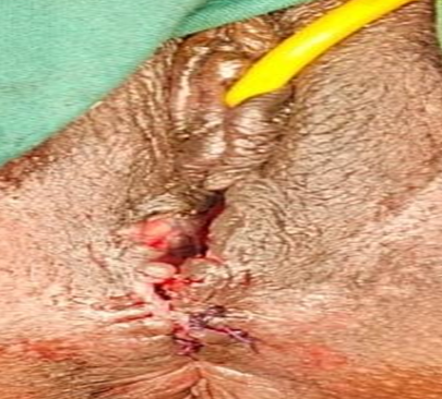
Figure 5: Immediate postoperative view following staged repair of Grade IV obstetric rectovaginal tear showing reconstructed perineal body, restored anal sphincter complex, and well-approximated perineal wound with intact vaginal and anal separation.
Eight-week postoperative follow-up demonstrated well-healed perineum with restored perineal body, intact rectovaginal septum, healthy vaginal mucosa, and anatomically aligned anal verge. There was no evidence of wound dehiscence, infection, or recurrent rectovaginal fistula. Figure (7) Postoperative clinical photograph showing a laparoscopy-assisted diversion sigmoid colostomy. The end colostomy was well-matured with healthy mucosa and intact peristomal skin. Laparoscopic port-site scars are visible, with no evidence of local infection or stoma-related complications.
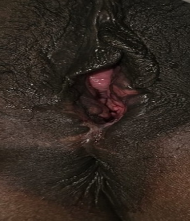
Figure 6: Eight-week postoperative follow-up
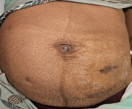
Figure 7: Postoperative - A laparoscopy-assisted diversion sigmoid colostomy.
DISCUSSION:
Obstetric anal sphincter injuries represent a severe form of childbirth-related trauma and are a major contributor to long-term maternal morbidity. Grade IV perineal tears, characterized by disruption of the rectovaginal septum and complete injury to both the internal and external anal sphincters, are uncommon but carry significant functional and psychosocial consequences (10). The present case highlights the complexity of managing such injuries, particularly when initial repair fails and the patient presents late with an infected wound, fecal incontinence, and a rectovaginal fistula.
Delayed recognition remains one of the principal challenges in the management of severe obstetric perineal injuries. Missed or inadequately repaired tears at the time of delivery can result in tissue breakdown, secondary infection, and fistula formation (11). In the current case, primary perineal repair was attempted twice at an outside facility but failed, most likely due to ongoing fecal contamination, tissue edema, and suboptimal surgical exposure. This underscores the importance of meticulous perineal examination immediately following vaginal delivery and the need for early referral to specialized centers when severe injuries are suspected.
Accurate delineation of the extent of injury is critical for successful management. Magnetic resonance imaging plays a pivotal role in evaluating complex perineal injuries, particularly in delayed presentations. MRI provides detailed visualization of sphincter integrity, fistulous tracts, and surrounding soft-tissue inflammation, thereby facilitating appropriate surgical planning (12). In this patient, MRI confirmed complete sphincter disruption with rectovaginal communication, supporting the decision to proceed with a staged surgical approach.
The choice between immediate primary repair and staged surgical management remains a subject of debate. While primary repair is recommended in acute settings with minimal contamination, delayed presentations with inflamed and infected tissues are associated with a high risk of repair failure (13). In such situations, fecal diversion has been shown to reduce local sepsis, protect the repair, and promote optimal tissue healing (14). In the present case, a diverting sigmoid colostomy effectively controlled contamination and facilitated successful repair of both the rectovaginal septum and anal sphincter complex.
Sphincteroplasty remains the cornerstone of treatment in patients with complete anal sphincter disruption. Precise identification and separate reconstruction of the internal and external anal sphincters are essential for restoring continence (15). Delayed stoma reversal following adequate healing and functional recovery further contributes to favorable outcomes. The excellent postoperative continence and absence of fistula recurrence in this patient emphasize the effectiveness of this staged surgical strategy (16).
Beyond anatomical restoration, the psychosocial impact of obstetric rectovaginal injuries warrants careful consideration. Young women affected by fecal incontinence often experience social isolation, anxiety, and depression (17). Early definitive management, coupled with appropriate counseling and psychosocial support, is therefore an integral component of comprehensive care. Multidisciplinary collaboration involving surgical gastroenterologists, gynecologists, radiologists, and stoma care nurses is crucial in achieving optimal outcomes (18).
This case contributes to the limited literature on staged surgical management of failed and infected Grade IV obstetric rectovaginal tears, particularly in resource-limited settings. It reinforces the principle that individualized, staged treatment planning guided by imaging findings and clinical severity can result in excellent anatomical and functional recovery, even in complex and delayed presentations.
CONCLUSION:
Grade IV obstetric rectovaginal tears are rare but severe childbirth-related injuries associated with substantial physical and psychological morbidity. Early recognition, accurate assessment of the extent of injury, and appropriate surgical planning by experienced specialists are essential for achieving favorable outcomes. In delayed presentations complicated by failed primary repair and active contamination, a staged surgical approach comprising fecal diversion, definitive repair of the rectovaginal septum and anal sphincter complex, and delayed stoma reversal provides optimal conditions for tissue healing. The present case demonstrates that meticulous surgical technique, supported by multidisciplinary care, can result in excellent anatomical restoration and functional recovery, with resolution of fecal incontinence and a marked improvement in quality of life. This case underscores the importance of specialized management in tertiary care centers for complex obstetric perineal injuries.
REFERENCES:
1. Darmody E, Bradshaw C, Atkinson S. Women’s experience of obstetric anal sphincter injury following childbirth: an integrated review. Midwifery. 2020,91:102820.
2. Chia CC, Huang SC. Third- and fourth-degree perineal laceration in vaginal delivery. Taiwan J Obstet Gynecol. 2012,51(1):148–152.
3. Touhidi Nezhad F, Jalali R, Karimi F. Women’s experiences of rectovaginal fistula: an ethno-religious experience. BMC Womens Health. 2020,20(1):130.
4. Das B, Snyder M. Rectovaginal fistulae. Clin Colon Rectal Surg. 2016,29(1):50–57.
5. Goh JTW, Natukunda H, Singasi I, Kabugho E, Browning A, Krause HG. Surgical repair and follow-up of chronic fourth-degree obstetric perineal tear (total perineal defect) in two centres in eastern Africa. Int Urogynecol J. 2021,32(9):2437–2445.
6. Satora M, Żak K, Frankowska K, Misiek M, Tarkowski R, Bobiński M. Perioperative factors affecting the healing of rectovaginal fistula. J Clin Med. 2023,12(19):6421.
7. Huebner M, Rall K, Brucker SY, Reisenauer C, Siegmann-Luz KC, Delancey JOL. The rectovaginal septum: visible on magnetic resonance images of women with Mayer-Rokitansky-Küster-Hauser syndrome (Müllerian agenesis). Int Urogynecol J. 2014,25(3):323–330.
8. Lightner AL, Pemberton JH. The role of temporary fecal diversion. Clin Colon Rectal Surg. 2017,30(3):178–183.
9. Tang J, Fu D, Wang X, Wang X. Obstetric rectal buttonhole tear: case series, literature review, and management recommendations. Int J Womens Health. 2025,17:4837–4848.
10. Spinelli A, Laurenti V, Carrano FM, Gonzalez-Díaz E, Borycka-Kiciak K. Diagnosis and treatment of obstetric anal sphincter injuries: new evidence and perspectives. J Clin Med. 2021,10(15):3261.
11. Ramar CN, Vadakekut ES, Grimes WR. Perineal lacerations. StatPearls. Treasure Island (FL): StatPearls Publishing, 2024.
12. Gallego JC, Echarri A. Role of magnetic resonance imaging in the management of perianal Crohn’s disease. Insights Imaging. 2018,9(1):47–58.
13. Frykberg RG, Banks J. Challenges in the treatment of chronic wounds. Adv Wound Care (New Rochelle). 2015,4(9):560–582.
14. Falih Noori I. Rectovaginal fistulas, outcomes of various surgical repair and predictive factors of healing success: a retrospective cohort study of 40 cases. Int J Surg Open. 2021,32:100335.
15. Berg MR, Gregussen H, Sahlin Y. Long-term outcome of sphincteroplasty with separate suturing of the internal and the external anal sphincter. Tech Coloproctol. 2019,23(12):1163–1170.
16. Barugola G, Bertocchi E, Leonardi A, Almoudaris AM, Ruffo G. Post surgical rectovaginal fistula: who really benefits from stoma diversion? Updates Surg. 2021,73(1):165–171.
17. Wilson SM, Sikkema KJ, Watt MH, Masenga GG, Mosha MV. Psychological symptoms and social functioning following repair of obstetric fistula in a low-income setting. Matern Child Health J. 2016,20(5):941–950.
18. Jin Y, Tian X, Li Y, Jiménez-Herrera M, Wang H. Effects of continuous care on health outcomes in patients with stoma: a systematic review and meta-analysis. Asia Pac J Oncol Nurs. 2022,9(1):21–33.