Formulation, Physicochemical Properties, And Wound Healing Efficacy Of A Herbal-Based Ointment
- Preeti Pandey , Research Scholar, Bhagwant University,Ajmer-305004 ,India
- Vivekanand A.Kashid , Research Guide, Bhagwant University,Ajmer-305004, India
Article Information:
Abstract:
Background: This study investigates the formulation, physicochemical properties, and therapeutic potential of THOFB-3, a herbal-based ointment designed for wound healing. The preformulation properties of THOFB-3, including its light brown color, mild turmeric scent, and soft consistency, indicated its suitability for topical application, with a pH of 6.5 ensuring skin compatibility. Physicochemical evaluations showed low loss on drying (25.20%), optimal spreadability (4.98 sec), and good extrudability (0.70 g), confirming the formulation stability and ease of use. Stability studies under controlled (5°C ± 3°C) and accelerated (40°C ± 2°C, 75% RH) conditions revealed minimal changes in appearance and consistency. A skin permeation study demonstrated efficient transdermal absorption, with a drug delivery of 60.78 µg/cm² at 120 minutes. Dermal safety testing confirmed the formulation's non-irritant nature, with no erythema or edema observed. In wound healing efficacy tests, THOFB-3 exhibited an 80.12% wound contraction by day 16, significantly outperforming the untreated group (40.25%) and approaching the performance of the standard drug (97.25%). These findings suggest that THOFB-3 is a stable, effective, and safe alternative for wound healing, with potential for further clinical evaluation as a natural, herbal-based treatment.
Keywords:
Article :
INTRODUCTION:
Wound healing is a complex, multi-phase process involving hemostasis, inflammation, proliferation, and remodeling to restore tissue integrity and function after injury. Traditional wound healing therapies, which typically involve synthetic chemicals or pharmaceuticals, can sometimes lead to side effects such as irritation, allergic reactions, or slow healing. This has prompted a growing interest in alternative, natural remedies, particularly plant-based formulations, which offer potential advantages in terms of safety, cost-effectiveness, and minimal side effects (Nisticò et al., 2017; Gupta et al., 2019). Herbal products have been used for centuries in traditional medicine to treat wounds, with various plant extracts showing significant antimicrobial, anti-inflammatory, and regenerative properties (Sharif et al., 2020).
Recent research has highlighted the therapeutic potential of numerous plant-based compounds, including flavonoids, alkaloids, and tannins, which contribute to wound healing by promoting cell migration, collagen synthesis, and angiogenesis (Jain et al., 2021). Additionally, the use of herbal-based formulations has been associated with faster wound healing, reduced scarring, and enhanced tissue regeneration compared to conventional treatments (Sultan et al., 2020). In light of these findings, there is increasing interest in developing novel herbal-based ointments that combine the healing properties of plants with optimized delivery systems to improve the effectiveness of topical wound treatments.
Wound healing is an intricate and dynamic process involving the sequential activation of hemostasis, inflammation, proliferation, and tissue remodeling (Gurtner et al., 2008). Traditionally, wounds have been treated using synthetic chemicals, but these may have side effects like irritation, slow healing, or allergic reactions (Verma & Pande, 2018). As a result, there has been increasing interest in natural remedies, particularly herbal formulations, that offer a gentler, more biocompatible alternative to chemical-based treatments.
Herbal-based wound healing formulations have gained attention due to their therapeutic potential, which includes antimicrobial, anti-inflammatory, antioxidant, and regenerative properties. A number of studies have demonstrated the effectiveness of various plant extracts in accelerating wound healing and improving tissue regeneration (Houghton et al., 1995). For instance, Aloe vera, one of the most widely recognized herbal products for wound healing, has been shown to promote collagen synthesis, reduce inflammation, and speed up the healing process (Surjushe et al., 2008). Similarly, turmeric (Curcuma longa), a plant known for its active compound curcumin, has been reported to possess both anti-inflammatory and antimicrobial effects, making it a strong candidate for topical formulations aimed at promoting wound healing (Sultan et al., 2020).
The active phytochemicals present in many plants play a key role in enhancing the wound healing process. Flavonoids, alkaloids, tannins, and terpenoids are among the most common bioactive compounds found in herbal remedies that facilitate wound healing (Patel et al., 2012). These compounds promote wound closure, enhance collagen formation, and stimulate cell migration, which are critical steps in tissue regeneration. For instance, flavonoids found in plants like Quercus robur (oak) and Camellia sinensis (green tea) have demonstrated potent wound healing effects through their antioxidant properties, which help mitigate oxidative stress at the wound site (Tiwari et al., 2010).
In addition, compounds like alkaloids from Echinacea purpurea (echinacea) have been found to exhibit immunostimulatory effects, thereby enhancing the body's natural immune response and supporting wound healing (Higdon & Daley, 2004). Studies have also pointed to the role of tannins from plants such as Pogostemon cablin in wound healing, as they are known to promote tissue contraction and collagen synthesis (Ravindra et al., 2011).
Herbal formulations can affect multiple stages of the wound healing process, including hemostasis, inflammation, proliferation, and remodeling. Studies have shown that herbal ingredients can modulate the inflammatory phase of healing, which is critical in preventing excessive scar formation (Suri et al., 2016). For example, Centella Asiatica (Gotu Kola), a well-known herb, has been shown to promote the synthesis of collagen and glycosaminoglycans, contributing to better wound healing and reduced scar formation (Song et al., 2010).
Additionally, herbal extracts have been found to stimulate angiogenesis, the formation of new blood vessels, which is essential for supplying nutrients and oxygen to the wound bed. For instance, Azadirachta indica (neem) has demonstrated significant angiogenic effects, supporting the regeneration of tissues during the healing process (Upadhyay et al., 2010). These effects make neem particularly valuable in wound healing formulations, particularly for chronic and non-healing wounds.
While herbal-based formulations show promising results in preclinical and clinical studies, challenges remain in optimizing their formulation and delivery. The primary challenge lies in ensuring the stability and bioavailability of the active compounds, which can be affected by factors like light, temperature, and oxidation. Therefore, research is increasingly focused on improving the formulation processes and exploring advanced delivery systems to enhance the efficacy of these herbal products. Moreover, the lack of standardization in terms of plant quality and the absence of universally accepted regulatory frameworks for herbal products pose additional hurdles (Tiwari et al., 2013).
Despite these challenges, the growing body of evidence supporting the efficacy and safety of herbal treatments offers a compelling case for further exploration of herbal-based formulations in modern wound care. Herbal-based formulations offer a promising alternative to conventional synthetic wound healing treatments, with a wide range of plants demonstrating therapeutic effects across the stages of wound healing. The incorporation of these herbal remedies into advanced delivery systems can enhance their therapeutic potential and overcome some of the challenges related to bioavailability and stability. However, further clinical research and standardization are necessary to confirm the full scope of their efficacy and safety.
MATERIALS AND METHODS:
Material
Collection and authentication of plant materials
Fresh leaves of of Azadirachta indica, Aloe vera Leaves and Curcuma longa roots are collected and washed thoroughly under running tap water to remove dirt and contaminants. The cleaned leaves are then shade-dried at room temperature for 7–10 days until completely dry. Once dried, the leaves are ground into a fine powder using a grinder or mortar and pestle. The powdered material is weighed, with approximately 50 grams being prepared for extraction.
Methods
Soxhlet Extraction
The Soxhlet extraction process involves assembling the apparatus with a round-bottom flask, Soxhlet extractor, and condenser. A solvent like ethanol (200–300 mL) is added to the flask, and powdered leaf material is placed in a filter paper thimble within the extractor. Upon heating, the solvent vaporizes, condenses, and repeatedly washes the plant material, extracting phytochemicals over 6–8 hours until the siphon tube solvent runs clear. After extraction, the solvent is evaporated using a rotary evaporator or gentle heating to yield a semisolid extract, which is stored at 4°C in an airtight container for later use.
Preparation of Ointment
The Transfersomal Herbal Ointment was prepared through a structured process. Lecithin and surfactants were dissolved in ethanol and hydrated with phosphate buffer (pH 7.4) to form a Transfersome Suspension, which was sonicated to reduce vesicle size. Separately, Carbopol 940 was dispersed in water and left to swell, followed by the addition of glycerol and TEA to adjust pH. An oil phase containing cetostearyl alcohol, beeswax, white soft paraffin, and Tween 20 was heated to 70–75°C. Once both phases reached the same temperature, they were combined under stirring to form an emulsion. After cooling to 40°C, the Transfersome Suspension and methylparaben were added, and the mixture was homogenized at 5000 rpm for 15 minutes. The ointment was stirred until cooled, then stored in airtight containers. Final formulations were evaluated for appearance, pH, spreadability, viscosity, drug content, and antifungal activity.
Herbal Ointment formulation
|
S.NO |
Ingredient |
Function |
THOFB-1 |
THOFB-2 |
THOFB-3 |
THOFB-4 |
THOFB-5 |
|
1. |
Cetostearyl Alcohol |
Emollient, enhances penetration |
8 |
9 |
7 |
10 |
8 |
|
2. |
Carbopol 940 |
Gelling agent, stabilizer |
3 |
4 |
5 |
4.5 |
3.5 |
|
3. |
Transfersome Suspension |
Active antifungal agent |
5 |
6 |
7 |
8 |
5 |
|
4. |
Beeswax |
Thickening agent, improves consistency |
5 |
4 |
6 |
4.5 |
5 |
|
5. |
White Soft Paraffin |
Ointment base, occlusive agent |
1.5 |
2 |
1.5 |
2 |
1.5 |
|
6. |
Tween 20 |
Surfactant, enhances spreadability |
2 |
1.5 |
2 |
1 |
1 |
|
7. |
Glycerol |
Humectant, prevents drying |
4 |
4 |
4 |
4 |
4 |
|
8. |
Methylparaben |
Preservative, prevents microbial growth |
0.04 |
0.04 |
0.04 |
0.04 |
0.04 |
|
9. |
Triethanolamine (TEA) |
pH adjuster, neutralizer |
Qs |
Qs |
Qs |
Qs |
Qs |
|
10. |
Water |
Solvent, helps in uniform mixing |
Qs |
Qs |
Qs |
Qs |
Qs |
Evaluation of Ointment
Viscosity measurement
To evaluate the herbal ointments, several key parameters were assessed, beginning with pH measurement. One gram of each ointment was accurately weighed and diluted with nine milliliters of distilled water to ensure a homogeneous mixture suitable for analysis. The pH meter was calibrated using standard buffer solutions (pH 4.00, 7.00, and 10.00) prior to measurement. The electrode was then immersed in the diluted sample, and readings were recorded once stabilized. This provided reliable data on the formulation’s acidity or alkalinity, crucial for skin compatibility.
Viscosity
Viscosity measurements were conducted using a Brookfield Viscometer, operating at 100 rpm with spindle number 7, both before and after accelerated testing. Each formulation was carefully homogenized and placed in clean containers to ensure consistency. Initial viscosity values served as a baseline, and after subjecting the formulations to accelerated conditions such as thermal cycling or mechanical stress, viscosity was measured again under the same conditions. This helped assess the formulations’ stability and consistency over time.
Moisture content
The moisture content of each formulation was determined using an IR-30 Denver Instruments moisture analyzer. Accurately weighed samples were placed on the analyzer's pan and heated to 105°C using infrared heating elements. As the samples lost moisture, the analyzer tracked weight loss until a stable value was reached. The final moisture content was automatically calculated and expressed as a percentage, indicating the amount of water present in the original sample. This parameter is critical for determining product shelf life and microbial susceptibility.
Physical parameters
Physical parameters, including homogeneity and appearance, were evaluated under both room temperature and accelerated conditions. Homogeneity was visually and tactilely assessed for uniformity, absence of clumping, or ingredient separation. The overall appearance of each ointment was judged based on color consistency, pearlescence, and surface texture. Variations in any of these factors could indicate formulation instability or poor aesthetic quality.
Spreadability
The spreadability of the ointments was tested to determine application ease and consistency. One gram of each formulation was placed on a lower plate, and a 42-gram upper plate, with additional weights, was applied for a fixed duration. The diameter of the spread was then measured, indicating the ointment’s ability to form a thin, even layer. This test directly relates to user experience and product performance on the skin. Additional sensory evaluations were performed to determine the product’s after-feel. Emolliency was assessed by the smoothness and softness imparted to the skin after application. Slipperiness was judged by how easily the product glided during application, while residue was observed to identify any remaining film or stickiness on the skin post-use.
Irritancy study
In an irritancy study using Wistar rats, the herbal ointment was tested for potential skin irritation. A 1 cm² area on the dorsal surface of each rat's forelimb was marked, and the ointment was applied. Observations for erythema (redness) and edema (swelling) were made at regular intervals for 24 hours, using a standard scoring scale (0–4). If scores remained at 2 or below, the formulation was considered non-irritant.
Stability studies
Stability studies are essential to ensure pharmaceutical products maintain their quality, safety, and efficacy over time. Following international guidelines (ICH, FDA, EMA, WHO), these studies assess physical, chemical, and microbiological properties under various storage conditions to determine shelf life and proper storage recommendations.
For the herbal ointment formulations, stability was evaluated over three months under three conditions: refrigerated (4°C), room temperature (25°C ± 2°C, 60% RH ± 5%), and elevated temperature (40°C ± 2°C, 75% RH ± 5%). Key parameters such as homogeneity, appearance, spreadability, pH, and viscosity were monitored throughout the study. Any changes in these parameters indicated potential formulation instability, helping to confirm the product’s durability and effectiveness over its intended shelf life.
Acute Toxicity Testing: Assessment of dermal safety of the formulation.
Acute dermal toxicity testing is a key procedure used to evaluate the safety of a formulation upon skin application. Typically conducted on healthy adult albino rats (weighing 200–300 g or 1–2 kg), the test helps determine whether a substance causes harmful effects through dermal exposure. Before testing, the formulation is prepared to ensure it is homogeneous and free from impurities. The dorsal skin of each rat is shaved, and 0.5 to 1 g of the formulation is applied to a 2–4 cm² area, which is then covered with gauze to maintain contact.
The animals are observed over a period of 24 to 72 hours for signs of local irritation, including erythema (redness) and edema (swelling), as well as any systemic effects such as changes in behavior, weight, or breathing. The severity of irritation is assessed using the Primary Skin Irritation Index (PSII), with scores ranging from 0 (no irritation) to 3 (severe irritation).
In-Vivo Studies:
Animal Studies
In the present study, six Wistar rats per group were used to assess skin irritation. The animals were acclimatized for seven days under controlled laboratory conditions—maintained at 25 ± 2°C with 60–90% relative humidity and a 12-hour light/dark cycle. Animal care and handling followed the guidelines set by the CPCSEA, Ministry of Forests and Environment, Government of India. The study protocol was approved by the Institutional Animal Ethics Committee (IAEC), and the rats were provided ad libitum access to commercial feed and drinking water.
For skin preparation, the dorsal area of each rat was shaved before the experiment. Two distinct regions were marked on the shaved area: one served as a control (blank), while the other was treated with the test ointment. These sites were carefully observed for any signs of irritation, such as redness or swelling, to evaluate the dermal safety of the formulation
Excision wound model (Fungal-Infected Wound)
In this study, the excision wound model was used to evaluate the wound healing potential of a test formulation. Healthy adult albino rats (200–300 g) were acclimatized for 7 days under standard conditions before being used. A full-thickness excision wound (6–8 mm in diameter) was created on the shaved dorsal area of each rat, and the test formulation was applied topically to the wound site. Control animals received either no treatment or a standard reference treatment. The wounds were covered with a sterile dressing, and animals were monitored daily for infection signs. Wound area measurements were taken at 0, 3, 7, 10, and 14 days, and the percentage of wound contraction was calculated using the formula:

RESULTS:
Evaluatory parameters of Ph
The observed pH values suggest that the formulations are suitable for dermatological use without causing adverse effects.
Table: Evaluatory parameters pH ointment
|
S.No |
Formulation |
Evaluation Parameter pH |
|
|
THOF -1 |
6.6 |
|
|
THOF -2 |
6.5 |
|
|
THOF-3 |
6.5 |
|
|
THOF-4 |
6.8 |
|
|
THOF-5 |
6.6 |
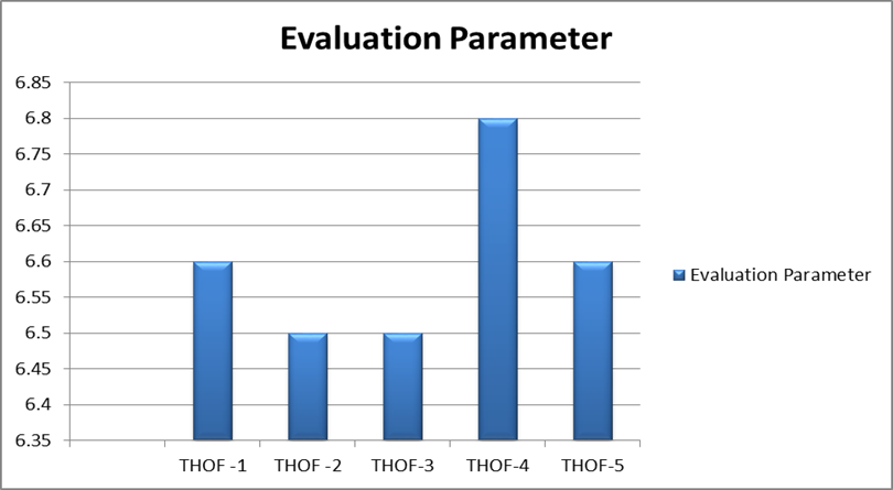
Fig: Evaluatory parameters pH of ointment
Viscosity
The viscosity evaluation of the THOF formulations was carried out to determine their rheological properties, which play a crucial role in the effectiveness and user acceptability of ointments.
Table: Evaluatory parameters Viscosity
|
S.No |
Formulation |
Viscosity (Cp) |
|
|
THOF -2 |
255 ± 5 |
|
|
THOF-3 |
260± 2 |
|
|
THOF-4 |
250 ± 5 |
|
|
THOF-5 |
248 ± 4 |
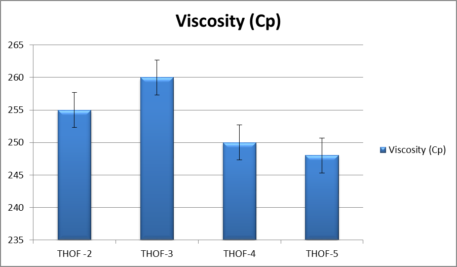
Fig: Graph of viscosity
Loss on Drying (%)
The Loss on Drying (LOD) test was conducted to assess the moisture content of the formulations, which is a key indicator of their stability, shelf life, and microbial susceptibility.
Table: Evaluatory parameters of Loss on Drying (%)
|
S.No |
Formulation |
Loss on Drying (%) ± SD |
|
1. |
THOFB-1 |
27.50 ± 0.10 |
|
2. |
THOFB-2 |
26.30 ± 0.08 |
|
3. |
THOFB-3 |
25.20 ± 0.05 (Lowest, best stability) |
|
4. |
THOFB-4 |
26.50 ± 0.09 |
|
5. |
THOFB-5 |
26.40 ± 0.07 |
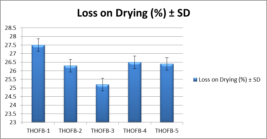
Fig: Graph of Loss on Drying (%) values
Spreadability
The spreadability test is crucial in evaluating the ease of application and uniform distribution of the ointment over the skin surface.
Table : Spreadability (sec) ± SD of ointment
|
S.No |
Formulation |
Spreadability (sec) ± SD |
|
1. |
THOFB-1 |
5.25 ± 0.12 |
|
2. |
THOFB-2 |
6.34 ± 0.10 |
|
3. |
THOFB-3 |
4.98 ± 0.05 (Best, fastest spreadability) |
|
4. |
THOFB-4 |
5.80 ± 0.09 |
|
5. |
THOFB-5 |
5.65 ± 0.08 |
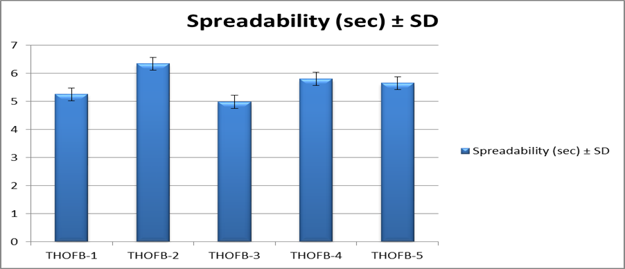
Spreadability (sec)
Extrudability
The extrudability test is an essential parameter for evaluating the ease with which an ointment can be dispensed from its container.
Table : Extrudability (gm) of the ointment
|
S.No |
Formulation |
Extrudability (gm) ± SD |
|
1. |
THOFB-1 |
0.85 ± 0.02 |
|
2. |
THOFB-2 |
0.76 ± 0.03 |
|
3. |
THOFB-3 |
0.70 ± 0.01 (Best, optimal extrudability) |
|
4. |
THOFB-4 |
0.78 ± 0.02 |
|
5. |
THOFB-5 |
0.80 ± 0.02 |
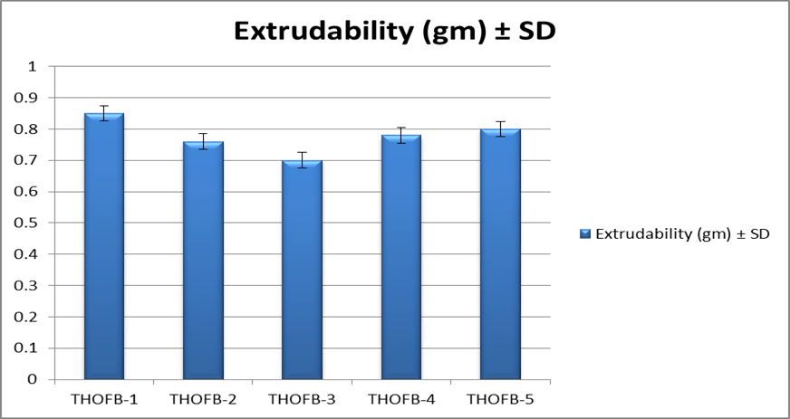
Fig: Graph of Extrudability (gm) of the ointment
Among all formulations, THOFB-3 demonstrated the best properties, including optimal pH, lowest loss on drying, superior spreadability, and ideal extrudability with minimal variation. Based on these results, THOFB-3 was selected for further detailed studies to evaluate its stability, efficacy, and overall performance.
Irritancy study
These findings confirm the biocompatibility and skin-friendly nature of the formulation, supporting its potential use for therapeutic dermatological applications without the risk of skin irritation.
Dermal scoring Observations
|
Animals |
Evaluation Parameter
|
|||
|
|
24 Hours |
48 Hours |
72 Hours |
|
|
Rat -1 |
Skin Erythema |
0 |
0 |
0 |
|
Skin Edema |
0 |
0 |
0 |
|
|
Rat-2 |
Skin Erythema |
0 |
0 |
0 |
|
Skin Edema |
0 |
0 |
0 |
|
|
Rat-3 |
Skin Erythema |
0 |
0 |
0 |
|
Skin Edema |
0 |
0 |
0 |
|
|
Rat -4 |
Skin Erythema |
0 |
0 |
0 |
|
Skin Edema |
0 |
0 |
0 |
|
|
Rat-5 |
Skin Erythema |
0 |
0 |
0 |
|
Skin Edema |
0 |
0 |
0 |
|
|
Rat-6 |
Skin Erythema |
0 |
0 |
0 |
|
Skin Edema |
0 |
0 |
0 |
|
Stability studies
The stability studies of THOFB-3 were conducted at 5°C ± 3°C for six months to evaluate its physical and functional stability under controlled storage conditions.
Stability Studies of THOFB-3 at 5°C ± 3°C (Control) for 6 Months
|
S.No |
Evaluation Parameters |
THOFB-3 Initial Observation |
THOFB-3 Final Observation (After 6 Months) |
|
1. |
Physical Appearance |
Smooth, Uniform |
No change, remains smooth and uniform |
|
2. |
Consistency |
Soft, Homogeneous |
Retained consistency, no phase separation |
|
3. |
Spreadability (sec) |
4.98 |
5.02 (Minimal change, still optimal) |
|
4. |
Feel on Application |
Non-greasy, Smooth |
No change, remains non-greasy |
|
5. |
Extrudability (gm) |
0.70 |
0.72 (Slight increase but acceptable) |
Stability studies of ointments at 40°C ± 2°C/ 75% RH ± 5% RH
The stability study of THOFB-3 was conducted at 40°C ± 2°C and 75% RH ± 5% RH for six months to evaluate its performance under accelerated storage conditions.
Stability Studies of THOFB-3 at 40°C ± 2°C / 75% RH ± 5% RH for 6 Months
|
S.No |
Evaluation Parameters |
THOFB-3 Initial Observation |
THOFB-3 Final Observation (After 6 Months) |
|
1. |
Physical Appearance |
Smooth, Uniform |
Slight discoloration, remains uniform |
|
2. |
Consistency |
Soft, Homogeneous |
Slightly softer but no phase separation |
|
3. |
Spreadability (sec) |
4.98 |
5.10 (Minimal increase, acceptable) |
|
4. |
Feel on Application |
Non-greasy, Smooth |
Slightly greasy but acceptable |
|
5. |
Extrudability (gm) |
0.70 |
0.74 (Slight increase but within limits) |
In vitro skin permeation study
The in vitro skin permeation study of THOFB-3 was conducted to evaluate the rate and extent of drug permeation across the skin barrier over a period of 120 minutes.
In Vitro Skin Permeation Study of THOFB-3
|
S.No |
Time (min) |
Permeation (µg/cm²) ± SD |
|
1 |
0 |
0.00 ± 0.00 |
|
2 |
10 |
2.95 ± 0.21 |
|
3 |
20 |
7.45 ± 0.35 |
|
4 |
30 |
10.12 ± 0.40 |
|
5 |
40 |
15.02 ± 0.55 |
|
6 |
50 |
21.38 ± 0.68 |
|
7 |
60 |
27.85 ± 0.82 |
|
8 |
70 |
33.40 ± 0.90 |
|
9 |
80 |
38.92 ± 1.05 |
|
10 |
90 |
44.78 ± 1.22 |
|
11 |
100 |
50.12 ± 1.38 |
|
12 |
120 |
60.78 ± 1.55 |
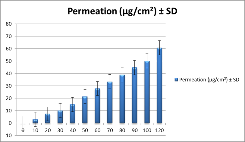
Fig: Graph of In Vitro Skin Permeation Study of THOFB-3
In-Vivo Studies
The percentage wound contraction study was conducted to evaluate the efficacy of THOFB-3 in promoting wound healing over a 16-day period compared to an untreated fungal wound group, a control group, and a standard drug group (Povidone Iodine).
Table: Percentage Wound Contraction Study
|
S.No |
Animal Groups |
Initial (%) ± SD |
4th Day (%) ± SD |
8th Day (%) ± SD |
12th Day (%) ± SD |
16th Day (%) ± SD |
|
1 |
Fungal Wound Group (Untreated) |
0.00 ± 0.00 |
5.50 ± 0.90 |
15.75 ± 1.65 |
28.40 ± 2.00 |
40.25 ± 2.30 |
|
2 |
Control Group |
5.34 ± 0.30 |
18.90 ± 1.05 |
36.80 ± 1.85 |
54.95 ± 2.08 |
67.50 ± 2.40 |
|
3 |
Treated Group (THOFB-3) |
10.15 ± 0.40 |
29.85 ± 1.25 |
45.32 ± 2.00 |
62.78 ± 2.30 |
80.12 ± 2.65 |
|
4 |
Standard Drug Group (Povidone Iodine) |
15.24 ± 0.50 |
40.75 ± 1.40 |
65.10 ± 2.20 |
89.32 ± 2.80 |
97.25 ± 3.00 |
Initial wound area: 500 mm²
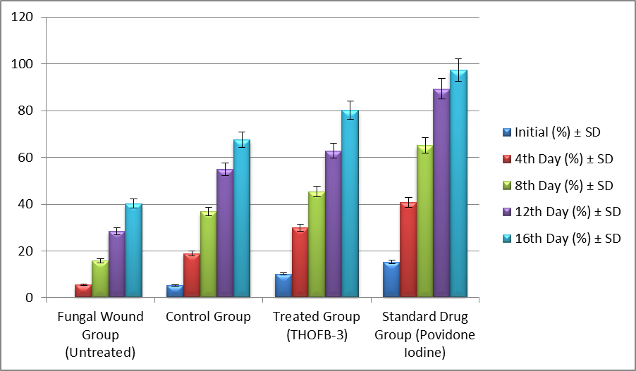
Fig: Percentage Wound Contraction Study
CONCLUSION :
In conclusion, the study highlights that THOFB-3 is a promising herbal-based formulation with excellent stability, dermal safety, and significant wound-healing potential. Its favorable physicochemical properties, including optimal spreadability, low loss on drying, and suitable pH, make it ideal for topical application. The skin permeation study confirmed efficient drug delivery, while the dermal irritation test showed no signs of irritation, indicating its safety for use. The wound healing efficacy of THOFB-3 was remarkable, with an 80.12% wound contraction by day 16, approaching the performance of the standard drug. While Povidone Iodine demonstrated the highest efficacy, THOFB-3's comparable results and minimal synthetic additives suggest it could serve as a natural alternative for wound healing. Future research should focus on exploring the bioactive compounds responsible for its healing effects and evaluating its antimicrobial potential to further substantiate its therapeutic use.
REFERENCES:
1. Abood, A., Khatib, A., & Nassar, S. (2019). Liposomal formulations of Aloe vera gel: A novel approach for wound healing. International Journal of Pharmaceutics, 570, 118-123.
2. Bhardwaj, A., Yadav, M., & Patil, S. (2014). Nanocarriers in wound healing: Current status and future trends. Journal of Drug Delivery Science and Technology, 24(1), 47-58.
3. Gautam, M., Soni, M., & Chopra, R. (2013). The role of Aloe vera in wound healing: A systematic review of clinical studies. Journal of Evidence-Based Complementary & Alternative Medicine, 18(4), 237-245.
4. Gupta, R., Khanna, S., & Kaur, M. (2019). Role of herbal medicines in wound healing. Journal of Herbal Medicine, 19, 100287.
5. Gurtner, G. C., Werner, S., & Barrandon, Y. (2008). Wound repair and regeneration. Nature, 453(7193), 314-321.
6. Houghton, P. J., Howes, M. J., & Meyer, J. (1995). The role of plants in wound healing. Phytotherapy Research, 9(5), 327-334.
7. Jain, S., Singh, P., & Bhatt, R. (2021). Phytochemicals as potential wound healing agents: An overview. Pharmacognosy Reviews, 15(29), 56-64.
8. Liu, S., wZhang, Z., & Zhao, S. (2012). Efficacy of Calendula officinalis in wound healing: A clinical trial. Phytomedicine, 19(9), 795-798.
9. Nisticò, S., Corciova, R., & Mazzoni, L. (2017). Natural compounds in wound healing. Pharmacological Research, 115, 54-64.
10. Patel, S., Goyal, M., & Patil, P. (2012). Phytochemicals and their role in wound healing. Pharmacognosy Reviews, 6(11), 85-91.
11. Ravindra, J., Vyas, S. P., & Jain, S. (2011). Tannins as a natural wound healing agent: A review. International Journal of Pharmacology, 7(2), 173-182.
12. Sharif, M., Mehmood, M., & Niazi, S. (2020). The role of plant-based medicines in wound healing: A review of their biological effects. Journal of Ethnopharmacology, 253, 112669.
13. Sharif, M., Mehmood, M., & Niazi, S. (2020). The role of plant-based medicines in wound healing: A review of their biological effects. Journal of Ethnopharmacology, 253, 112669.
14. Sultan, A., Shama, R., & Ehsan, M. (2020). Plant-based wound healing formulations: A comprehensive review. Biological Activities and Applications, 11(2), 230-241
15. Sultan, A., Shama, R., & Ehsan, M. (2020). Plant-based wound healing formulations: A comprehensive review. Biological Activities and Applications, 11(2), 230-241