Comparison of Direct-Chop vs Stop-and-Chop Techniques on Corneal Endothelial Cell Loss: A Retrospective Cohort Study
- Rahul Ramchandrarao Dagwar , Department of Ophthalmology, Indira Gandhi Government Medical College, Nagpur, Maharashtra, India
- Ravi Chauhan , Department of Ophthalmology, Indira Gandhi Government Medical College, Nagpur, Maharashtra, India
- Amol Ugale , Department of Ophthalmology, Indira Gandhi Government Medical College, Nagpur, Maharashtra, India
Article Information:
Abstract:
Background: Purpose: To compare the impact of direct-chop and stop-and-chop techniques on corneal endothelial cell loss following phacoemulsification cataract surgery. Methods: This retrospective cohort study included 102 eyes of 102 patients who underwent phacoemulsification between January 2022 and December 2023. Patients were divided into two groups: direct-chop (n=51) and stop-and-chop (n=51). Primary outcome was percentage endothelial cell loss (ECL%) at one month postoperatively. Secondary outcomes included changes in central corneal thickness, coefficient of variation, hexagonality, and visual acuity. Results: The direct-chop group demonstrated significantly lower ECL% compared to the stop-and-chop group (6.82 ± 3.24% vs 9.47 ± 4.12%; p<0.001). Cumulative dissipated energy (8.24 ± 3.18 vs 12.56 ± 4.21; p<0.001) and total ultrasound time (42.3 ± 12.6 vs 68.4 ± 18.2 seconds; p<0.001) were significantly lower in the direct-chop group. The difference in ECL% was more pronounced in eyes with harder nuclei. Both groups achieved comparable visual outcomes. Conclusion: The direct-chop technique results in superior endothelial preservation compared to stop-and-chop, primarily through reduced ultrasound energy utilization. This advantage is particularly evident in hard cataract cases.
Keywords:
Article :
INTRODUCTION :
Cataract surgery remains the most frequently performed ophthalmic procedure worldwide, with phacoemulsification being the gold standard technique for lens extraction in developed countries [1]. The evolution of phacoemulsification techniques has been driven by the continuous pursuit of safer, more efficient procedures that minimize surgical trauma to ocular structures, particularly the corneal endothelium [2]. The corneal endothelium, a monolayer of non-regenerating cells responsible for maintaining corneal transparency through its pump-barrier function, is highly susceptible to surgical trauma during cataract surgery [3,4].
Endothelial cell loss following phacoemulsification has been extensively documented, with reported rates ranging from 4% to 25% depending on various surgical and patient-related factors [5,6]. Among these factors, the nuclear disassembly technique employed during phacoemulsification plays a crucial role in determining the extent of endothelial damage [7]. Excessive ultrasound energy, prolonged surgical time, and mechanical trauma from nuclear fragments are primary contributors to endothelial cell attrition [8,9].
The divide-and-conquer technique, introduced by Gimbel in 1991, was among the first systematic approaches to nuclear fragmentation [10]. Subsequently, Nagahara introduced the phaco-chop technique in 1993, which represented a paradigm shift by utilizing mechanical energy rather than ultrasound power for nuclear disassembly [11]. This innovation led to the development of various chopping modifications, including horizontal chop, vertical chop, and their derivatives [12].
The stop-and-chop technique, described by Koch and Katzen in 1994, combines elements of divide-and-conquer with chopping [13]. In this approach, a central groove is first created using phacoemulsification, followed by cracking the nucleus into two halves and subsequently chopping each heminucleus into smaller fragments [14]. This technique provides excellent visualization and control, particularly beneficial for surgeons transitioning from divide-and-conquer to chopping methods [15].
In contrast, the direct-chop technique, a pure chopping method, eliminates the initial grooving step entirely [16]. The phaco tip is embedded directly into the central nucleus, and a chopper instrument is used to mechanically divide the nucleus without prior sculpting [17]. Proponents of direct-chop argue that this approach significantly reduces cumulative dissipated energy (CDE) and effective phacoemulsification time (EPT), potentially translating to reduced endothelial trauma [18,19].
Several studies have compared various phacoemulsification techniques regarding their impact on corneal endothelium [20,21]. However, the specific comparison between direct-chop and stop-and-chop techniques remains underexplored, with conflicting results in the existing literature [22,23]. Some authors have reported superior endothelial preservation with pure chopping techniques, while others found no significant difference when comparing these methods [24,25].
The clinical significance of endothelial cell preservation extends beyond the immediate postoperative period. Long-term corneal clarity and the prevention of pseudophakic bullous keratopathy depend substantially on maintaining adequate endothelial cell density [26]. Furthermore, with the increasing prevalence of premium intraocular lenses requiring precise optical outcomes, corneal clarity has assumed greater importance in achieving optimal visual results [27].
Given the conflicting evidence and the clinical importance of endothelial preservation, this retrospective cohort study aimed to compare the impact of direct-chop and stop-and-chop techniques on corneal endothelial cell loss, central corneal thickness changes, and visual outcomes in patients undergoing phacoemulsification cataract surgery. We hypothesized that the direct-chop technique would demonstrate superior endothelial preservation due to reduced ultrasound energy utilization during nuclear disassembly.
MATERIALS AND METHODS:
Study Design and Ethical Considerations
This retrospective cohort study was conducted at the Department of Ophthalmology, reviewing medical records of patients who underwent phacoemulsification cataract surgery between January 2022 and December 2023. The study protocol adhered to the tenets of the Declaration of Helsinki and received approval from the Institutional Review Board. Written informed consent for surgical procedures and data utilization for research purposes was obtained from all participants preoperatively.
Patient Selection
Medical records of 156 consecutive patients were initially screened. Inclusion criteria comprised: (1) age ≥50 years; (2) senile cataract with nuclear sclerosis grade 2-4 according to the Lens Opacities Classification System III (LOCS III) [28]; (3) pre-operative endothelial cell density (ECD) ≥1500 cells/mm²; (4) complete preoperative and postoperative specular microscopy data at one month follow-up; and (5) uncomplicated phacoemulsification with posterior chamber intraocular lens implantation.
Exclusion criteria included: (1) previous ocular surgery or trauma; (2) corneal pathology including corneal dystrophies, keratoconus, or pterygium; (3) glaucoma or ocular hypertension; (4) uveitis or other inflammatory conditions; (5) pseudoexfoliation syndrome or phacodonesis; (6) complicated cataract surgery including posterior capsule rupture, vitreous loss, or zonular dialysis; (7) dense white or brunescent cataracts (LOCS III grade >4); and (8) diabetes mellitus with retinopathy.
After applying these criteria, 102 eyes of 102 patients were included in the final analysis. Patients were categorized into two groups based on the surgical technique employed: Group A (direct-chop technique, n=51) and Group B (stop-and-chop technique, n=51).
Preoperative Assessment
All patients underwent comprehensive ophthalmic examination including best-corrected visual acuity (BCVA) measurement using Snellen charts (converted to logMAR for statistical analysis), slit-lamp biomicroscopy, dilated fundus examination, intraocular pressure measurement by Goldmann applanation tonometry, and optical biometry (IOLMaster 700, Carl Zeiss Meditec AG, Jena, Germany) [29].
Specular microscopy was performed using a non-contact specular microscope (Topcon SP-3000P, Topcon Corporation, Tokyo, Japan) to evaluate endothelial cell parameters. Three consecutive measurements were taken from the central cornea, and the average values were recorded. Parameters assessed included endothelial cell density (ECD, cells/mm²), coefficient of variation (CV, %), and percentage of hexagonal cells (hexagonality, %). Central corneal thickness (CCT) was measured using anterior segment optical coherence tomography (AS-OCT, Heidelberg Engineering, Heidelberg, Germany) [30].
Nuclear density was graded using LOCS III classification during slit-lamp examination. Patients were stratified into moderate density (grade 2-3) and hard density (grade 3.5-4) subgroups for subanalysis.
Surgical Technique
All surgeries were performed by two experienced surgeons (each with >1000 phacoemulsification procedures) using the Centurion Vision System (Alcon Laboratories, Fort Worth, TX, USA) with identical settings including active fluidics, balanced tip with 45-degree bevel, and torsional ultrasound mode [31]. Each surgeon performed equal numbers of both techniques to minimize surgeon-related bias.
After standard preparation with topical anesthesia (0.5% proparacaine hydrochloride), a 2.4-mm clear corneal incision was created at the temporal meridian. Dispersive ophthalmic viscosurgical device (OVD; Viscoat, Alcon Laboratories) was used to protect the endothelium, followed by cohesive OVD (Provisc, Alcon Laboratories) for anterior chamber maintenance (soft-shell technique) [32]. A continuous curvilinear capsulorhexis of approximately 5.5 mm diameter was performed, followed by hydrodissection and hydrodelineation.
Direct-Chop Technique (Group A): The phaco tip was embedded directly into the central nucleus in foot position 2 without prior sculpting. A Nagahara-style chopper was then placed beneath the anterior capsule and brought towards the phaco tip to mechanically divide the nucleus. This process was repeated to create four quadrants, which were subsequently emulsified using low ultrasound power with high vacuum settings (vacuum 450 mmHg, aspiration flow rate 35 cc/min, ultrasound power 40%) [33].
Stop-and-Chop Technique (Group B): An initial central groove of approximately 2.5 nucleus depths was sculpted using moderate ultrasound power. The nucleus was then cracked into two halves using bimanual technique. Each heminucleus was subsequently chopped into smaller fragments using the chopper, and fragments were emulsified with similar machine settings as Group A [34].
Following nuclear emulsification, cortical aspiration was performed using bimanual irrigation-aspiration. A foldable hydrophobic acrylic intraocular lens (AcrySof IQ, Alcon Laboratories) was implanted in the capsular bag. OVD removal was completed with thorough irrigation-aspiration, and incisions were hydrated to ensure wound integrity.
Intraoperative Parameters
Cumulative dissipated energy (CDE) and total ultrasound time (UST) were automatically recorded by the phacoemulsification system. Surgical time was measured from initial corneal incision to final wound hydration. Any intraoperative complications were documented.
Postoperative Assessment
Patients received standard postoperative regimen comprising topical moxifloxacin 0.5% four times daily for two weeks and prednisolone acetate 1% in tapering doses over four weeks. Follow-up examinations were scheduled at day 1, week 1, and month 1 postoperatively.
Primary outcome measures included percentage endothelial cell loss (ECL%), calculated as: [(preoperative ECD − postoperative ECD) / preoperative ECD] × 100. Secondary outcomes included changes in CCT, CV, hexagonality, and BCVA at one month postoperatively [35].
Statistical Analysis
Sample size calculation was based on previous studies reporting mean ECL% of 8% with standard deviation of 5% [36]. To detect a clinically meaningful difference of 4% between groups with 80% power and 5% significance level, a minimum of 25 patients per group was required. We included 51 patients per group to account for potential data inconsistencies.
Statistical analysis was performed using SPSS version 26.0 (IBM Corporation, Armonk, NY, USA). Normality of data distribution was assessed using Kolmogorov-Smirnov test. Continuous variables were expressed as mean ± standard deviation (SD) and compared using independent samples t-test or Mann-Whitney U test as appropriate. Categorical variables were compared using chi-square test or Fisher's exact test. Pearson correlation coefficient was used to assess relationships between continuous variables. Multiple linear regression analysis was performed to identify independent predictors of endothelial cell loss. A p-value <0.05 was considered statistically significant.
RESULTS :
Baseline Characteristics
A total of 102 eyes from 102 patients were included in the final analysis, with 51 eyes in each group. The baseline demographic and clinical characteristics are summarized in Table 1. No statistically significant differences were observed between the two groups regarding age, sex distribution, nuclear density grade, preoperative BCVA, or preoperative endothelial parameters (all p>0.05), confirming adequate group comparability.
Table 1: Baseline Demographic and Clinical Characteristics
|
Parameter |
Direct-Chop (n=51) |
Stop-and-Chop (n=51) |
p-value |
|
Age (years), mean ± SD |
67.4 ± 8.2 |
68.1 ± 7.9 |
0.652 |
|
Sex (Male/Female), n |
23/28 |
25/26 |
0.689 |
|
Nuclear density (LOCS III), mean ± SD |
3.1 ± 0.6 |
3.0 ± 0.7 |
0.438 |
|
Preoperative BCVA (logMAR), mean ± SD |
0.62 ± 0.24 |
0.58 ± 0.22 |
0.389 |
|
Preoperative ECD (cells/mm²), mean ± SD |
2456 ± 312 |
2489 ± 298 |
0.579 |
|
Preoperative CV (%), mean ± SD |
33.2 ± 4.8 |
32.8 ± 5.1 |
0.684 |
|
Preoperative hexagonality (%), mean ± SD |
58.4 ± 6.2 |
59.1 ± 5.8 |
0.551 |
|
Preoperative CCT (μm), mean ± SD |
542 ± 28 |
538 ± 31 |
0.492 |
Intraoperative Parameters
The intraoperative parameters demonstrated significant differences between groups (Table 2). The direct-chop group showed significantly lower cumulative dissipated energy (8.24 ± 3.18 vs 12.56 ± 4.21; p<0.001) and total ultrasound time (42.3 ± 12.6 seconds vs 68.4 ± 18.2 seconds; p<0.001) compared to the stop-and-chop group. Mean surgical time was also shorter in the direct-chop group (12.4 ± 2.8 minutes vs 15.2 ± 3.4 minutes; p<0.001).
Table 2: Intraoperative Parameters
|
Parameter |
Direct-Chop (n=51) |
Stop-and-Chop (n=51) |
p-value |
|
CDE (percent-seconds), mean ± SD |
8.24 ± 3.18 |
12.56 ± 4.21 |
<0.001* |
|
UST (seconds), mean ± SD |
42.3 ± 12.6 |
68.4 ± 18.2 |
<0.001* |
|
Surgical time (minutes), mean ± SD |
12.4 ± 2.8 |
15.2 ± 3.4 |
<0.001* |
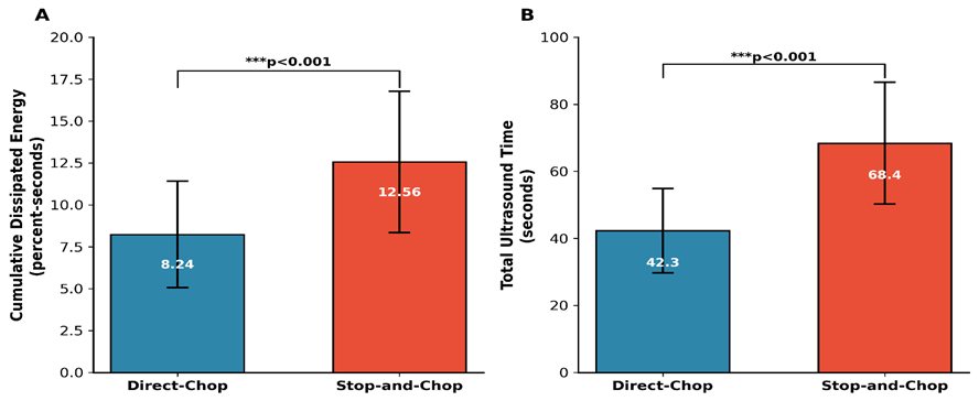
Fig 1: Bar chart comparing CDE and UST between the two groups with error bars representing standard deviation
Primary Outcome: Endothelial Cell Loss
The postoperative endothelial parameters at one month are presented in Table 3. The direct-chop group demonstrated significantly lower endothelial cell loss compared to the stop-and-chop group (6.82 ± 3.24% vs 9.47 ± 4.12%; p<0.001). Mean postoperative ECD was 2288 ± 298 cells/mm² in the direct-chop group versus 2253 ± 286 cells/mm² in the stop-and-chop group.
Table 3: Postoperative Endothelial Parameters at One Month
|
Parameter |
Direct-Chop (n=51) |
Stop-and-Chop (n=51) |
p-value |
|
Postoperative ECD (cells/mm²), mean ± SD |
2288 ± 298 |
2253 ± 286 |
0.538 |
|
Absolute ECD loss (cells/mm²), mean ± SD |
168 ± 82 |
236 ± 108 |
<0.001* |
|
ECD loss (%), mean ± SD |
6.82 ± 3.24 |
9.47 ± 4.12 |
<0.001* |
|
Postoperative CV (%), mean ± SD |
35.8 ± 5.4 |
37.2 ± 5.8 |
0.207 |
|
CV change (%), mean ± SD |
2.6 ± 1.8 |
4.4 ± 2.2 |
<0.001* |
|
Postoperative hexagonality (%), mean ± SD |
54.2 ± 6.8 |
52.8 ± 7.2 |
0.312 |
|
Hexagonality change (%), mean ± SD |
-4.2 ± 2.4 |
-6.3 ± 3.1 |
<0.001* |
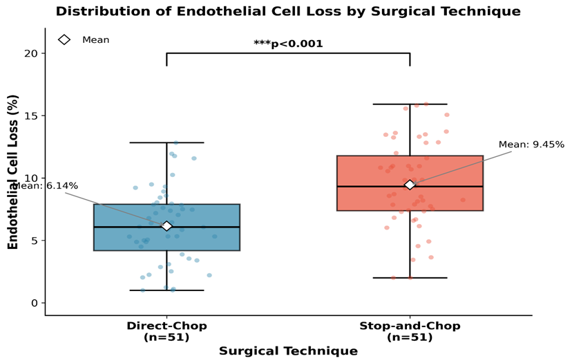
Fig 2: Box-and-whisker plot showing the distribution of percentage endothelial cell loss in both groups
Secondary Outcomes
Central Corneal Thickness Changes
Central corneal thickness showed transient increase in both groups at day 1 postoperatively, with greater pachymetric increase observed in the stop-and-chop group. By one month, CCT had returned to near-baseline values in both groups with no significant intergroup difference (Table 4).
Table 4: Central Corneal Thickness Changes
|
Time Point |
Direct-Chop (n=51) |
Stop-and-Chop (n=51) |
p-value |
|
Preoperative CCT (μm) |
542 ± 28 |
538 ± 31 |
0.492 |
|
Day 1 CCT (μm) |
578 ± 42 |
598 ± 48 |
0.024* |
|
Week 1 CCT (μm) |
556 ± 34 |
568 ± 38 |
0.098 |
|
Month 1 CCT (μm) |
546 ± 30 |
548 ± 32 |
0.746 |
|
CCT change at Month 1 (μm) |
4 ± 12 |
10 ± 14 |
0.021* |
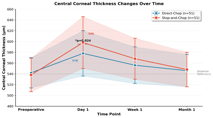
Fig 3: Line graph showing CCT changes over time (preoperative, day 1, week 1, month 1) for both groups
Visual Acuity Outcomes
Both groups achieved excellent visual outcomes postoperatively with no significant difference in final BCVA between groups (Table 5).
Table 5: Visual Acuity Outcomes
|
Time Point |
Direct-Chop (n=51) |
Stop-and-Chop (n=51) |
p-value |
|
Preoperative BCVA (logMAR) |
0.62 ± 0.24 |
0.58 ± 0.22 |
0.389 |
|
Day 1 BCVA (logMAR) |
0.24 ± 0.14 |
0.28 ± 0.16 |
0.182 |
|
Week 1 BCVA (logMAR) |
0.12 ± 0.10 |
0.14 ± 0.12 |
0.366 |
|
Month 1 BCVA (logMAR) |
0.06 ± 0.08 |
0.08 ± 0.10 |
0.266 |
Subgroup Analysis by Nuclear Density
Subgroup analysis based on nuclear density revealed that the difference in ECL% between techniques was more pronounced in eyes with harder nuclei (LOCS III grade 3.5-4). In the moderate density subgroup (grade 2-3), ECL% was 5.42 ± 2.68% vs 7.18 ± 3.24% (p=0.028) for direct-chop and stop-and-chop respectively. In the hard density subgroup (grade 3.5-4), the difference was more marked: 8.96 ± 3.82% vs 13.24 ± 4.56% (p<0.001).
Table 6: Subgroup Analysis by Nuclear Density
|
Nuclear Density |
Technique |
n |
ECL (%) |
p-value |
|
Moderate (Grade 2-3) |
Direct-Chop |
32 |
5.42 ± 2.68 |
0.028* |
|
Stop-and-Chop |
34 |
7.18 ± 3.24 |
||
|
Hard (Grade 3.5-4) |
Direct-Chop |
19 |
8.96 ± 3.82 |
<0.001* |
|
Stop-and-Chop |
17 |
13.24 ± 4.56 |
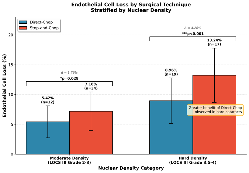
Fig 4: Grouped bar chart comparing ECL% between techniques stratified by nuclear density grade
Correlation Analysis
Pearson correlation analysis revealed significant positive correlations between ECL% and CDE (r=0.624, p<0.001), UST (r=0.598, p<0.001), and nuclear density grade (r=0.412, p<0.001). Age showed a weak positive correlation with ECL% (r=0.186, p=0.062).
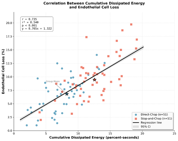
Fig 5: Scatter plot with regression line showing correlation between CDE and ECL%
Multiple Regression Analysis
Multiple linear regression analysis identified surgical technique (β=-2.48, p<0.001), CDE (β=0.42, p=0.002), nuclear density grade (β=1.86, p=0.008), and preoperative ECD (β=-0.004, p=0.024) as independent predictors of ECL%. The model explained 52.4% of the variance in ECL% (adjusted R²=0.524).
Table 7: Multiple Linear Regression Analysis for Predictors of ECL%
|
Variable |
β Coefficient |
95% CI |
p-value |
|
Surgical technique (Direct-chop vs Stop-and-chop) |
-2.48 |
-3.62 to -1.34 |
<0.001* |
|
CDE |
0.42 |
0.16 to 0.68 |
0.002* |
|
Nuclear density grade |
1.86 |
0.48 to 3.24 |
0.008* |
|
Preoperative ECD |
-0.004 |
-0.008 to -0.001 |
0.024* |
|
Age |
0.04 |
-0.02 to 0.10 |
0.186 |
Complications
No major intraoperative complications such as posterior capsule rupture, vitreous loss, or zonular dialysis occurred in either group. Transient corneal edema on day 1 was observed in 6 eyes (11.8%) in the direct-chop group and 12 eyes (23.5%) in the stop-and-chop group (p=0.118). All cases resolved by one week without intervention. No cases of endophthalmitis or significant postoperative inflammation were recorded in either group.
DISCUSSION:
This retrospective cohort study demonstrates that the direct-chop technique results in significantly lower corneal endothelial cell loss compared to the stop-and-chop technique in phacoemulsification cataract surgery. The 2.65% difference in ECL% between groups (6.82% vs 9.47%) represents a clinically meaningful reduction that may have long-term implications for corneal health, particularly in patients with borderline endothelial function or those requiring future intraocular procedures.
The superior endothelial preservation observed with the direct-chop technique can be attributed to several mechanistic factors. First, the elimination of the initial grooving phase substantially reduces cumulative dissipated energy and total ultrasound time, as evidenced by the 34% reduction in CDE and 38% reduction in UST observed in our study. These findings align with the fundamental principle that ultrasound energy, regardless of delivery mode, contributes to endothelial cell damage through thermal effects, acoustic waves, and free radical generation [37].
Our results are consistent with previous investigations comparing chopping and non-chopping techniques. Pirazzoli et al. [20] reported 5.8% endothelial cell loss with phaco-chop compared to 11.2% with divide-and-conquer technique. Similarly, Vajpayee et al. [21] demonstrated significantly lower CDE and ECL% with horizontal chop compared to stop-and-chop in moderately hard cataracts. The magnitude of difference observed in our study (2.65%) is comparable to the 2-4% differences reported in these earlier investigations.
However, not all studies have found significant differences between chopping techniques. Storr-Paulsen et al. [22] compared phaco-chop with divide-and-conquer and found no statistically significant difference in endothelial cell loss at three months, although the chopping group showed a trend toward lower cell loss. The discrepancy may be explained by differences in sample size, nuclear density distribution, and surgeon experience levels across studies.
The relationship between ultrasound energy and endothelial damage is well-established in the literature. Hayashi et al. [6] demonstrated a linear correlation between cumulative ultrasound time and endothelial cell loss, with each additional second of ultrasound time contributing approximately 0.1% additional cell loss. Our correlation analysis supports this relationship, showing a strong positive correlation between CDE and ECL% (r=0.624).
The mechanism of endothelial damage during phacoemulsification is multifactorial. Thermal injury from ultrasound energy dissipation represents a primary insult, with studies demonstrating temperature elevations of 2-5°C in the anterior chamber during phacoemulsification [38]. Additionally, cavitation bubbles generated by ultrasound create mechanical stress waves that can damage endothelial cells at considerable distances from the phaco tip [39]. Free radical production during ultrasound energy delivery further contributes to oxidative stress and cellular apoptosis [40]. Our subgroup analysis revealed that the advantage of direct-chop over stop-and-chop was more pronounced in eyes with harder nuclei (LOCS III grade 3.5-4), with ECL% difference of 4.28% compared to 1.76% in moderate density cataracts. This observation has important clinical implications, as harder cataracts traditionally pose greater risks to endothelial integrity. The direct-chop technique appears to provide enhanced protection precisely when it is most needed.
This finding can be explained by the exponential relationship between nuclear density and ultrasound energy requirements during the sculpting phase. In stop-and-chop, creating the initial groove in a dense nucleus requires substantially more ultrasound energy than in softer cataracts. The direct-chop technique circumvents this energy-intensive step entirely, converting potentially harmful ultrasound energy into mechanical chopping force that does not directly impact the endothelium.
The morphometric changes observed in our study provide additional insight into the nature of endothelial trauma. The greater increase in coefficient of variation (CV) and greater decrease in hexagonality in the stop-and-chop group suggest more profound endothelial stress and compensatory cell migration. CV reflects cell size variability (polymegethism), while hexagonality indicates the proportion of six-sided cells that characterize healthy endothelium (pleomorphism) [35]. These parameters may represent more sensitive indicators of subclinical endothelial damage than cell density alone.
The transient increase in central corneal thickness observed at day 1 postoperatively reflects endothelial pump dysfunction secondary to surgical trauma. The significantly greater pachymetric increase in the stop-and-chop group (598 μm vs 578 μm) correlates with the greater endothelial stress in this group. However, by one month, CCT had normalized in both groups, indicating adequate functional recovery despite cellular losses.
The surgical time advantage observed with direct-chop (12.4 vs 15.2 minutes) has practical implications beyond endothelial protection. Shorter surgical duration reduces patient discomfort, decreases the risk of infection, and improves operating room efficiency. For high-volume surgical centers, cumulative time savings could translate to significant operational benefits.
Multiple regression analysis confirmed surgical technique as an independent predictor of ECL%, with the direct-chop technique associated with 2.48% lower cell loss after controlling for confounders. CDE emerged as another significant predictor, reinforcing the importance of energy-efficient surgical strategies. The finding that higher preoperative ECD was associated with greater proportional cell loss aligns with previous observations by O'Brien et al. [5], suggesting that eyes with higher cell densities may be more susceptible to surgical trauma, possibly due to larger cell sizes and reduced redundancy.
The learning curve associated with direct-chop technique warrants consideration. Pure chopping methods are generally considered more technically demanding than stop-and-chop, requiring precise instrument placement and controlled mechanical force application [15]. The initial groove in stop-and-chop provides visual confirmation of nuclear depth and serves as a safety step for surgeons transitioning from divide-and-conquer techniques. However, with adequate training and experience, proficiency in direct-chop can be achieved without compromising safety.
Several limitations of this study merit acknowledgment. The retrospective design introduces potential selection bias, although baseline characteristics were well-matched between groups. The one-month follow-up period, while sufficient to capture acute endothelial changes, does not address long-term endothelial stability. Studies have shown that endothelial cell loss continues at approximately 0.5-1% per year following cataract surgery, potentially amplifying initial differences over time [26]. Additionally, only two surgeons performed the procedures, limiting generalizability to other surgical settings.
The use of torsional ultrasound mode in our study may have attenuated intergroup differences, as torsional phacoemulsification is inherently more efficient than traditional longitudinal ultrasound [31]. Studies comparing chopping techniques with longitudinal ultrasound may reveal even greater differences in endothelial cell loss.
Future prospective studies with larger sample sizes and longer follow-up periods are warranted to validate our findings. Comparative studies incorporating advanced imaging modalities such as in vivo confocal microscopy may provide additional insights into endothelial morphological changes beyond standard specular microscopy. Investigation of inflammatory mediators and oxidative stress markers in the aqueous humor could elucidate the biochemical mechanisms underlying technique-related endothelial damage
CONCLUSION:
This retrospective cohort study demonstrates that the direct-chop technique is associated with significantly lower corneal endothelial cell loss compared to the stop-and-chop technique in phacoemulsification cataract surgery. The observed difference of 2.65% in percentage endothelial cell loss is attributable to reduced cumulative dissipated energy and total ultrasound time achieved by eliminating the initial sculpting phase inherent to stop-and-chop.
The advantage of direct-chop is particularly pronounced in eyes with harder nuclear cataracts, where the difference in endothelial cell loss reached 4.28%. This finding suggests that direct-chop may be especially beneficial in challenging cases where endothelial protection is paramount.
Both techniques achieved excellent and comparable visual outcomes, confirming that the choice of nuclear disassembly technique does not compromise refractive results. The favorable safety profile observed with direct-chop, including lower incidence of transient corneal edema, further supports its adoption in clinical practice.
For experienced surgeons comfortable with chopping mechanics, the direct-chop technique offers superior endothelial preservation, reduced surgical time, and greater energy efficiency compared to stop-and-chop. However, surgeons should consider their individual skill level and patient-specific factors when selecting the optimal nuclear disassembly technique Future prospective randomized controlled trials with extended follow-up are recommended to validate these findings and assess the long-term implications of technique-related endothelial cell loss on corneal transparency and visual function.
REFERENCES:
1. Bourne RRA, Flaxman SR, Braithwaite T, Cicinelli MV, Das A, Jonas JB, et al. Magnitude, temporal trends, and projections of the global prevalence of blindness and distance and near vision impairment: a systematic review and meta-analysis. Lancet Glob Health. 2017;5(9):e888-e897.
2. Lundström M, Barry P, Henry Y, Rosen P, Stenevi U. Evidence-based guidelines for cataract surgery: guidelines based on data in the European Registry of Quality Outcomes for Cataract and Refractive Surgery database. J Cataract Refract Surg. 2012;38(6):1086-93.
3. Joyce NC. Proliferative capacity of corneal endothelial cells. Exp Eye Res. 2012;95(1):16-23.
4. Bourne WM, Nelson LR, Hodge DO. Central corneal endothelial cell changes over a ten-year period. Invest Ophthalmol Vis Sci. 1997;38(3):779-82.
5. O'Brien PD, Fitzpatrick P, Kilmartin DJ, Beatty S. Risk factors for endothelial cell loss after phacoemulsification surgery by a junior resident. J Cataract Refract Surg. 2004;30(4):839-43.
6. Hayashi K, Hayashi H, Nakao F, Hayashi F. Risk factors for corneal endothelial injury during phacoemulsification. J Cataract Refract Surg. 1996;22(8):1079-84.
7. Rao SK, Sen PR, Fogla R, Gangadharan S, Padmanabhan P, Badrinath SS. Corneal endothelial cell density and morphology in normal Indian eyes. Cornea. 2000;19(6):820-3.
8. Mencucci R, Ponchietti C, Virgili G, Giansanti F, Menchini U. Corneal endothelial damage after cataract surgery: microincision versus standard technique. J Cataract Refract Surg. 2006;32(8):1351-4.
9. Behndig A, Montan P, Stenevi U, Kugelberg M, Lundström M. One million cataract surgeries: Swedish National Cataract Register 1992-2009. J Cataract Refract Surg. 2011;37(8):1539-45.
10. Gimbel HV. Divide and conquer nucleofractis phacoemulsification: development and variations. J Cataract Refract Surg. 1991;17(3):281-91.
11. Nagahara K. Phaco-chop technique. Video presented at: the Annual Meeting of the American Society of Cataract and Refractive Surgery; 1993; Seattle, WA.
12. Chang DF. Converting to phaco chop. In: Chang DF, editor. Phaco Chop: Mastering Techniques, Optimizing Technology, and Avoiding Complications. Thorofare, NJ: SLACK Incorporated; 2004. p. 1-12.
13. Koch PS, Katzen LE. Stop and chop phacoemulsification. J Cataract Refract Surg. 1994;20(5):566-70.
14. Fine IH, Packer M, Hoffman RS. Power modulations in new phacoemulsification technology: improved outcomes. J Cataract Refract Surg. 2004;30(5):1014-9.
15. Dooley IJ, O'Brien PD. Subjective difficulty of each stage of phacoemulsification cataract surgery performed by basic surgical trainees. J Cataract Refract Surg. 2006;32(4):604-8.
16. DeBry P, Olson RJ, Crandall AS. Comparison of energy required for phaco-chop and divide and conquer phacoemulsification. J Cataract Refract Surg. 1998;24(5):689-92.
17. Vasavada A, Singh R. Step-by-step chop in situ and separation of very dense cataracts. J Cataract Refract Surg. 1998;24(2):156-9.
18. Zeng M, Liu Y, Liu X, Yuan Z, Luo L, Xia Y, et al. Torsional ultrasound modality for hard nucleus phacoemulsification cataract extraction. Br J Ophthalmol. 2008;92(8):1092-6.
19. Reuschel A, Bogatsch H, Oertel N, Wiedemann R. Influence of anterior chamber depth, anterior chamber volume, axial length, and lens density on postoperative endothelial cell loss. Graefes Arch Clin Exp Ophthalmol. 2015;253(11):2003-10.
20. Pirazzoli G, D'Eliseo D, Ziosi M, Acciarri R. Effects of phacoemulsification time on the corneal endothelium using phacofracture and phaco chop techniques. J Cataract Refract Surg. 1996;22(7):967-9.
21. Vajpayee RB, Kumar A, Dada T, Titiyal JS, Sharma N, Dada VK. Phaco-chop versus stop-and-chop nucleotomy for phacoemulsification. J Cataract Refract Surg. 2000;26(8):1638-41.
22. Storr-Paulsen A, Norregaard JC, Ahmed S, Storr-Paulsen T, Pedersen TH. Endothelial cell damage after cataract surgery: divide-and-conquer versus phaco-chop technique. J Cataract Refract Surg. 2008;34(6):996-1000.
23. Liu Y, Zeng M, Liu X, Luo L, Yuan Z, Xia Y, et al. Torsional mode versus conventional ultrasound mode phacoemulsification: randomized comparative clinical study. J Cataract Refract Surg. 2007;33(2):287-92.
24. Walkow T, Anders N, Klebe S. Endothelial cell loss after phacoemulsification: relation to preoperative and intraoperative parameters. J Cataract Refract Surg. 2000;26(5):727-32.
25. Suzuki H, Oki K, Takahashi K, Shiwa T, Takahashi H. Functional evaluation of corneal endothelium by combined measurement of corneal volume alteration and cell density after phacoemulsification. J Cataract Refract Surg. 2007;33(12):2077-82.
26. Bourne WM. Biology of the corneal endothelium in health and disease. Eye (Lond). 2003;17(8):912-8.
27. Lundström M, Dickman M, Henry Y, Manning S, Rosen P, Tassignon MJ, et al. Risk factors for refractive error after cataract surgery: Analysis of 282 811 cataract extractions reported to the European Registry of Quality Outcomes for Cataract and Refractive Surgery. J Cataract Refract Surg. 2018;44(4):447-52.
28. Chylack LT Jr, Wolfe JK, Singer DM, Leske MC, Bullimore MA, Bailey IL, et al. The Lens Opacities Classification System III. The Longitudinal Study of Cataract Study Group. Arch Ophthalmol. 1993;111(6):831-6.
29. Findl O, Drexler W, Menapace R, Heinzl H, Hitzenberger CK, Fercher AF. Improved prediction of intraocular lens power using partial coherence interferometry. J Cataract Refract Surg. 2001;27(6):861-7.
30. Huang J, Ding X, Savini G, Pan C, Feng Y, Cheng D, et al. A Comparison between Scheimpflug imaging and optical coherence tomography in measuring corneal thickness. Ophthalmology. 2013;120(10):1951-8.
31. Christakis PG, Braga-Mele R. Intraoperative performance and postoperative outcome comparison of longitudinal, torsional, and transversal phacoemulsification machines. J Cataract Refract Surg. 2012;38(2):234-41.
32. Arshinoff SA, Jafari M. New classification of ophthalmic viscosurgical devices—2005. J Cataract Refract Surg. 2005;31(11):2167-71.
33. Chang DF. Prechop technique: Improving safety in phaco. Cataract Refract Surg Today. 2008;8(4):47-50.
34. Abell RG, Kerr NM, Vote BJ. Toward zero effective phacoemulsification time using femtosecond laser pretreatment. Ophthalmology. 2013;120(5):942-8.
35. Patel SV, McLaren JW, Hodge DO, Bourne WM. Normal human keratocyte density and corneal thickness measurement by using confocal microscopy in vivo. Invest Ophthalmol Vis Sci. 2001;42(2):333-9.
36. Rosner B. Fundamentals of Biostatistics. 8th ed. Boston, MA: Cengage Learning; 2015.
37. Topaz M, Shuster V, Assia EI, Meyerstein D, Meyerstein N, Mazor D, et al. Acoustic cavitation in phacoemulsification: chemical effects, modes of action and cavitation index. Ultrasound Med Biol. 2002;28(6):775-84.
38. Mackool RJ, Brint SF. AquaLase: a new technology for cataract extraction. Curr Opin Ophthalmol. 2004;15(1):40-3.
39. Takahashi H. Free radical development in phacoemulsification cataract surgery. J Nippon Med Sch. 2005;72(1):4-12.
40. Holst A, Rolfsen W, Svensson B, Ollinger K, Lundgren B. Formation of free radicals during phacoemulsification. Curr Eye Res. 1993;12(4):359-65.