"The Hidden twin": Fetus in feto
- Mamidala Vyshnavi Netha , JR- III Dept of Paediatrics, J. N. Medical College, KLE Academy of Higher Education and Research, Deemed to be University, Belagavi, Karnataka, India-590010.
- Swapnil Pattanshetti , Associate Professor, Dept of Paediatric Surgery, J. N. Medical College, KLE Academy of Higher Education and Research, Deemed to be University, Belagavi, Karnataka, India-590010
- Manisha Bhandarkar , Professor Dept of Neonatology, J. N. Medical College, KLE Academy of Higher Education and Research, Deemed to be University, Belagavi, Karnataka, India-590010
- Ramchandra Bhatt , Associate Professor Dept of Neonatology, J. N. Medical College, KLE Academy of Higher Education and Research, Deemed to be University, Belagavi, Karnataka, India-590010
- Shraddha Gupta , JR- III Dept of Paediatrics, J. N. Medical College, KLE Academy of Higher Education and Research, Deemed to be Univsersity, Belagavi, Karnataka, India-590010
Article Information:
Abstract:
Background: Fetus in fetu (FIF) is an extremely rare congenital anomaly, characterized by the presence of a malformed parasitic twin enclosed within the body of its host. It most commonly presents in infancy as an abdominal mass, and must be distinguished from teratomas due to differences in management and prognosis. Case Presentation: A full-term female neonate (2.95 kg) developed abdominal distension, non-bilious vomiting, and absent meconium within 48 hours of birth. Examination revealed a firm, midline mass. Imaging showed a retroperitoneal, encapsulated mass with calcified fetal parts, suggestive of fetus in fetu. Exploratory laparotomy revealed a pedunculated mass displacing bowel, supplied by a branch of the aorta. Complete excision was performed. Gross and histopathological examination confirmed a parasitic fetus with rudimentary limbs and vertebral axis. Postoperative recovery was uneventful.
Keywords:
Article :
INTRODUCTION:
Fetus in fetu (FIF) is an extremely rare developmental anomaly defined as the presence of a malformed parasitic twin enclosed within the body of its host (1). The condition is thought to arise from aberrant embryogenesis in diamniotic, monochorionic, monozygotic twin pregnancies, where unequal division of the totipotent inner cell mass results in the inclusion of one embryo within the other (2). The most common site for the parasitic twin is the retroperitoneal space, although other locations such as the cranial cavity, scrotum, and mediastinum have also been documented (3,4).
The classical clinical presentation involves a newborn or infant with a progressively enlarging abdominal mass. (5). Ultrasound and CT scan can reveal characteristic findings such as a well-encapsulated mass with developed axial skeleton and limb buds, differentiating it from teratomas, which lack an organized vertebral axis. This distinction is critical, as FIF is considered benign and curable by complete
surgical excision, whereas teratomas carry a risk of malignant transformation (3,6).
Histopathologically, the presence of multiple differentiated tissues of ectodermal, mesodermal, and endodermal origin further substantiates the diagnosis. While most cases are diagnosed postnatally, antenatal detection is increasing with advances in fetal imaging (4,7,8).
This report presents a rare case of FIF in a full-term neonate who developed features of intestinal obstruction in the immediate postnatal period. Surgical exploration led to the successful removal of the encapsulated parasitic twin. The case is discussed in the context of relevant literature, with emphasis on diagnostic challenges, embryological basis, and the importance of early intervention.
CASE PRESENTATION AND MANAGEMENT:
Clinical Features
A full-term female neonate, delivered by elective cesarean section and weighing 2.95 kg, initially had a normal postnatal transition. By the second day of life, she developed progressive abdominal distension, poor feeding tolerance, and repeated non-bilious vomiting, with no passage of meconium since birth. Examination revealed a hemodynamically stable, alert infant with a tense, distended abdomen and a firm, non-tender mass in the epigastric and umbilical regions. Bowel sounds were sluggish, the rectum was empty on digital examination, and no external anomalies were noted.
Imaging and Preoperative Diagnosis
Ultrasonography revealed a retroperitoneal, encapsulated mass with mixed echogenicity and hyperechoic foci suggestive of vertebral structures and long bones. Antenatal scans showed a cystic lesion in the 2nd trimester and spine in the 3rd, raising suspicion of fetus in fetu. CECT confirmed a 9×7×5 cm well-defined mass with a vertebral axis, rudimentary limbs, and fluid-filled sac, displacing adjacent bowel without infiltration. A feeding vessel arose from the aorta. No malignant features or elevated AFP were noted. Findings were diagnostic of fetus in fetu.
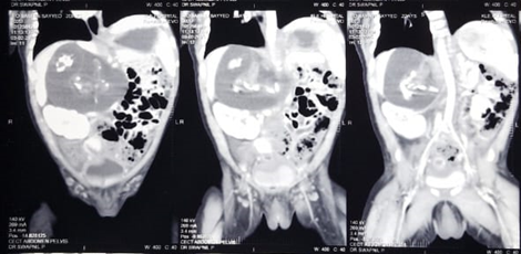
Fig 1: CECT abdomen (coronal view) showing a well-encapsulated retroperitoneal mass with
vertebral axis, limb buds and rudimentary fetal parts likely to be fetus in fetu.
Surgical Management
An exploratory laparotomy was performed under general anesthesia on day four of life. A right upper transverse abdominal incision provided access. Intra-operatively, a retroperitoneal, well-encapsulated, pedunculated mass was visualized. The lesion was compressing adjacent bowel loops and displacing the duodenum superiorly. A feeding vessel, arising from a branch of the aorta, supplied the mass and was carefully isolated, ligated, and divided. The mass was excised in toto without rupture of the capsule. On gross examination, it exhibited a well-formed vertebral axis, identifiable limb buds with digits, a rudimentary cranial vault, and fluid resembling amniotic contents.
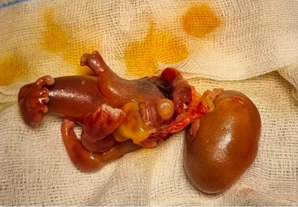
Fig 2: Fetus in Feto
Histopathological Examination
Microscopic examination confirmed fetus in fetu, showing well-differentiated vertebral and long bones, neural tissue, primitive gastrointestinal structures with mucosal lining, skin, cartilage, muscle, and respiratory-type epithelium. No features of malignancy, necrosis, or immature elements were observed, supporting a benign, fully differentiated parasitic fetal mass.
Postoperative Course
The neonate had an uneventful recovery. Nasogastric decompression and IV fluids were maintained for 48 hours. Bowel sounds returned by postoperative day two, with passage of meconium. Feeds were started on day three and gradually increased. The baby remained stable, with no signs of infection or electrolyte imbalance, and was discharged on day six with outpatient follow-up.
DISCUSSION:
Fetus in fetu (FIF) is a striking congenital anomaly with a unique embryological origin (9). It is believed to result from aberrant twinning during the second
week of gestation, where one twin envelops the other
due to persistent vitelline circulation anastomoses. The host fetus develops normally while the parasitic twin, enclosed within the host’s body, halts development due to lack of independent blood supply. The retroperitoneum is the most commonly involved site owing to the derivation of the superior mesenteric artery from the vitelline circulation (9,10).
The hallmark of FIF is the presence of an encapsulated mass containing well-differentiated fetal components, particularly a vertebral column, limb buds, and sometimes organogenesis. These features distinguish it from teratomas, which lack an axial skeleton and may carry a malignant potential. Willis’s criteria suggest that a vertebral axis and limb arrangement is essential to confirm FIF over a teratoma (11).
In this case, the neonate presented with intestinal obstruction from a retroperitoneal mass compressing adjacent bowel loops, leading to distension and vomiting typical of FIF. Imaging was diagnostic: ultrasonography showed a mixed echogenic encapsulated lesion with calcified skeletal elements, while CECT confirmed a vertebral axis, limb buds, and a fluid-filled sac. These findings were pathognomonic and guided surgical planning.
Complete surgical excision, including the capsule, is the treatment of choice to prevent recurrence. Although benign, incomplete removal or immature elements may pose a risk of malignant transformation. Postoperative monitoring includes clinical follow-up and serum AFP assessment. In this case, AFP was normal, and histopathology revealed organized fetal tissues (vertebrae, long bones, neural, gastrointestinal, and respiratory elements) without malignancy.
While most cases present in infancy, FIF can occasionally remain undiagnosed until later childhood or adulthood, presenting incidentally or with compressive symptoms such as jaundice or urinary obstruction (12,13). This case reinforces the classical imaging, surgical, and histopathologic features of FIF and emphasizes the importance of early diagnosis in neonates with unexplained abdominal distension or obstruction.
CONCLUSION:
Fetus in fetu is a benign yet rare anomaly that can mimic neonatal obstruction. Early recognition and imaging help differentiate it from teratomas by identifying vertebral axis. Complete surgical excision confirms diagnosis and ensures cure. This case underscores the importance of considering FIF in neonatal abdominal mass evaluation.
REFERENCES:
1. Gude D, Rayudu BR, Bansal D, Sashidhar C. Revisiting fetus-in-fetu. Ann Saudi Med. 2012 Jul;32(4):427–9. doi:10.5144/0256-4947.2012.427
2. Boyce MJ, Lockyer JW, Wood CB. Foetus in foetu: serological assessment of monozygotic origin by automated analysis. J Clin Pathol. 1972;25(9):793–8. doi:10.1136/jcp.25.9.793
3. Hong SS, Goo HW, Jung MR, Kim HJ, Kim EAR, Kim KS, et al. Fetus in fetu: Three-dimensional imaging using multidetector CT. AJR Am J Roentgenol. 2002 Dec;179(6):1481–3. doi:10.2214/ajr.179.6.1791481
4. Arlikar JD, Mane SB, Dhende NP, Sanghavi Y, Valand AG, Butale PR. Fetus in fetu: Two case reports and review of literature. Pediatr Surg Int. 2009 Mar;25(3):289–92. doi:10.1007/s00383-008-2322-1
5. Sunita S, Kamal R, Navtej, Meenu G, S M, Rajeev S. Fetus-in-fetu presenting as acute intestinal obstruction. Indian J Pathol Microbiol. 2010 Jan;53(1):128–9. doi:10.4103/0377-4929.59197
6. Patankar T, Fatterpekar GM, Prasad S, Maniyar A, Mukherji SK. Fetus in fetu: CT appearance - Report of two cases. Radiology. 2000 Mar;214(3):735–7. doi:10.1148/radiology.214.3.r00mr12735
7. Gangopadhyay AN, Srivastava A, Srivastava P, Gupta DK, Sharma SP, Kumar V. Twin fetus in fetu in a child: A case report and review of the literature. J Med Case Rep. 2010;4:96. doi:10.1186/1752-1947-4-96
8. Sharma N, Verma P, Pathak P. 27 cm fetus in fetu presenting in a 36-year-old man: report of a rare case and brief review of literature. Indian J Surg. 2007 Aug;69(4):155–7. doi:10.1007/s12262-007-0032-6
9. Gupta SK, Singhal P, Arya N. Fetus-in-fetu: A Rare Congenital Anomaly. J Surg Tech Case Rep. 2010;2(2):77–9. doi:10.4103/2006-8808.72612
De Lagausie P, De Napoli Cocci S, Stempfle N, Truong QD, Vuillard E, Ferkadji L, et al. Highly differentiated teratoma and fetus-in-fetu: A single pathology? J Pediatr Surg. 1997 Jan;32(1):115–6. doi:10.1016/S0022-3468(97)90216-7
Lewis RH. Foetus in foetu and the retroperitoneal teratoma. Arch Dis Child. 1961 Apr;36(186):220–6. doi:10.1136/adc.36.186.220
Nicolini U, Dell’Agnola CA, Ferrazzi E, Motta G. Ultrasonic prenatal diagnosis of fetus in fetu. J Clin Ultrasound. 1983 Jul-Aug;11(6):321–2. doi:10.1002/jcu.1870110609
Prescher LM, Butler WJ, Vachon TA, Henry MC, Latendresse T, Ignacio RC. Fetus in fetu: Review of the literature over the past 15 years. J Pediatr Surg Case Rep. 2015 Dec;3(12):554–62. doi:10.1016/j.epsc.2015.10.002