Melasma in Kashmir: An Epidemiological study
- Tabasum Fatima , MD Scholar, Regional Research Institute of Unani Medicine, Naseem Bagh Campus, University of Kashmir, Hazratbal, Srinagar-190006, J&K, India.
- shameem.A.Rather , Professor, Regional Research Institute of Unani Medicine, Naseem Bagh Campus, University of Kashmir, Hazratbal, Srinagar-190006, J&K, India
Article Information:
Abstract:
Abstract: Background: Kalaf is a common, acquired, chronic, symmetrically reticulated hyper pigmentary disorder characterized by brown to black macules on the exposed areas of the skin. It is prevalent in females between the age groups of 30-55 years. The treatment of melasma is often very challenging associated with high relapse rates. As such the disease has no serious implications on the health of the patients but it has a negative impact on the quality of life and it is also associated with low self-esteem. The disease also has social stigma attached to it causing tremendous stress for the patients. A demographic study was conducted at RRIUM Srinagar to see the prevalence of Melasma on the basis of skin tone, skin type, distribution of lesions, colour of lesions, erythema and seasonal variation.
Keywords:
Article :
INTRODUCTION :
Melasma is derived from Greek word “melas” meaning black and is also called Cholasma which is derived from the word “cholezin” meaning green. 1Melasma is called as Kalaf in Unani System of Medicine and is described by various ancient physicians. Kalaf is a disease which is caused by disturbance in the quality and quantity of Sawdāʾ(black bile). Various eminent Unani scholars have described the pathogenesis of the disease. In the famous book “The Cannon of Medicine” Ibn Sῑnā has described that Kalaf is caused by the accumulation of dead blood cells under the skin. 2Various other eminent Unani physicians have written that factors like morbid matters, cold melancholic blood and burnt bile also contribute in the development of the lesions. 3,4,5According to modern concept, melasma is a common, chronic, acquired, hyper pigmentary disorder characterized by brown spots or macules on the sun exposed areas of the body most commonly face6 .
The prevalence of Melasma is between 1.5-33.3% with the prevalence in pregnancy being about 50-70%. 7It is most commonly seen in darker people with Fitzpatrick’s skin type IV-V. It is more commonly seen in females as compared to males. The most commonly affected age group is between 30-55 years. 8The most common patients seen in the dermatological clinic are acquired hyper pigmentary disorders of the skin, which are associated with psychological stress and among these Melasma (Kalaf) is the most common. 9Although it is not associated with any health hazard, it has a negative influence on the quality of life and low self-esteem. 10,11,12Treatment of melasma is often very challenging associated with high relapse rates. The disease has no significant effect on the body but it has tremendous effect on the quality of life of the individual and is associated with social stigma. 13Melasma has high prevalence in East Asians, Indians, Pakistani, Middle East, Africans, Hispanics and Brazilian due to greater exposure to sunlight14,15,16
Risk Factors
The risk factors of Melasma described in all classical literature are as follows:-
1. Exposure to sunlight.
2. Pregnancy and amenorrhea.
3. Bad diet intake (saqeel and raddi ghiza).
4. Diseases of the liver and spleen.
5. Prolonged high fever.
6. Unhygienic conditions.
7. Consumption of alcohol 2,17,18,19,20,21,9,22
Dark skin coloured people are at the maximum risk of increased melanogenesis. 1,8,23,24,10,25,26It is most prevalent in the summer months and its prevalence decreases in the autumn and winter seasons25.
According to clinical features four types of melasma are seen
a) Centrofacial pattern
It is the most common pattern, seen in about 65% cases of melasma. It involves the cheeks, forehead, nose and upper lips.
b) Malar pattern
This pattern is seen in 20% of melasma patients. It is seen mostly in the malar area commonly on the cheeks and nose.
c) Mandibular pattern
It is seen in 15% cases of melasma with common involvement on the ramus of mandibular region.
d) Extra facial pattern
This pattern is commonly seen on extensor surfaces of the arm, neck and upper one third of the trunk10 .
MATERIALS AND METHODS:
A clinical study was conducted at the Regional Research Institute of Unani Medicine, Naseem Bagh, Srinagar, J&K. Recruitment of patients was done during the study duration of 12 months, starting in March 2021, from outpatient and inpatient departments (OPD/IPD) of the RRIUM hospital after obtaining proper ethical clearance from Institutional ethical committee Regional Research Institute of Unani Medicine, Habbak, Naseem Bagh. Clinically diagnosed patients of Melasma between the age group of 15 to 50 years of all genders were included in the study. Patients with a history of cardiovascular, endocrine, renal, liver diseases, skin sensitivity, pregnancy, lactation, those on oral contraceptives and those on anti-coagulants were excluded from the trial. Sample size was calculated using GPOWER software (Version 3.0.10), it was estimated that the number of patients required in each group with 80% power, 80% effect size and 5% significance level is 36. Thirty-six (36) patients were taken for the study after obtaining written consent.
RESULTS AND DISCUSSION:
Demographic Characteristics
All the patients registered for the study had skin tone either fair or medium; none of the patient was having light or dark skin tone (Fig. 1.). Out of 36 patients, 20 patients (55.56%) were having medium skin tone while, 16 patients (44.44%) were having fair skin tone. These findings are in agreement with the findings reported by Hamdi et al. (2008)27 that most the patients having melasma were having fair and medium skin tone. 28It is well documented that people having fair and medium skin tone have higher chances of development of melasma than those having lighter or darker skin tone reference. Also the prevalence of fair coloured in Kashmir region could also be a cause of more fair skinned people in our study.
In Fig.2. it is seen that Fitzpatrick scale was followed to determine the skin type of patients registered for the study reference. 29In the present study all the patients registered for the study were having skin types between III and IV as per Fitzpatrick scale. 19 patients (52.78%) were having type IV skin while 17 patients (47.22%) were having type III skin type, out of 36 patients (Fig.2.). Various authors have reported that melasma is not common in people where skin type falls in the far ends of the Fitzpatrick scale; however people whose skin type is in the middle range of the scale i.e. III and IV are more likely to develop melasma25,26.
In the present study majority of the patients that is 33 (91.67%) were having symmetrical lesions, while only 3 patients out of 36 (8.33%) were having asymmetric lesions (Fig.3.). The highest prevalence of symmetrical lesions indicated that there was symmetrical pattern of melasma in our study. Melasma is symmetrical lesions as mentioned in Fitzpatrick’s textbook of dermatology6.
The colour of melasma lesions in patients was found either to be light brown or dark brown and none of the patients had bluish grey or black colour of the lesions. Out of 36 patients 19 (52.78%) where having dark brown colour of the lesion, while 17 (47.22%) were having light brown colour of lesion (Fig.4.). Light brown and dark brown are the most common lesion colours reported by Yalamanchili et al. (2015)30.
In the present study the predominant pattern of lesions observed among the patients where centro facial melasma and malar. Out of 36 patients centrofacial melasma was observed in 23 patients (63.89%), malar melasma in 12 patients (33.33%) while mandibular melasma was observed only in 1 patient (2.78%) (Fig 5). KrupaShankar et al. (2014)31; Qazi et al. (2017)32 also reported the centrofacial and malar patterns comprised the majority of the lesions of melasma patients in their study33,32.
In the present study out of 36 patients 25 (69.44%) reported aggravation of lesions in summer while 11 patients (30.56%) reported no impact of seasonal variation on melasma improvement (Fig.6.). During summer more sun exposure and heat intolerance can be the majority of the contributing factors for aggravation of melasma patients Bagherani et al. (2015) .KrupaShankar et al. (2014)10,31 also reported aggravation of lesions in melasma patient during summer months
In the present study erythema was absent in 28 patients (77.28%) and present in only 8 patients (22.22%) out of 36 patients (Fig.7.). Although existence of erythema within the melasma lesion is reported in many studies, but the relationship between degree of erythema and pigmentation within the melasma lesion is not clarified in such studies Kim et al. (2007)34 and Lee et al. (2010)13. Park et al. (2013)35 observed positive correlation between erythema and pigmentation in melasma lesions but at the same time reported that further studies are needed to understand the biological mechanism behind the clinical changes observed in such studies. Since the sample size in our study was comparatively smaller, therefore as such no concrete inference could be drawn whether erythema has a positive or negative relationship in melasma therefore in depth studies on much larger sample size must be conducted in future to correlate the presence of erythema in melasma patients.
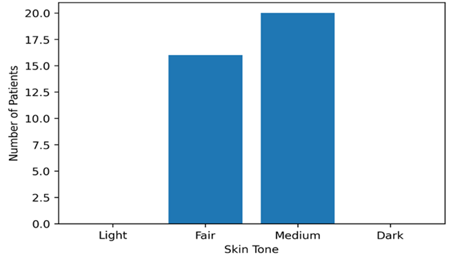
Fig.1. Distribution of patients as per skin tone
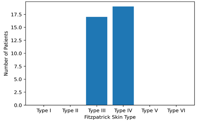
Fig.2. Distribution of patients as per Fitzpatricks skin type
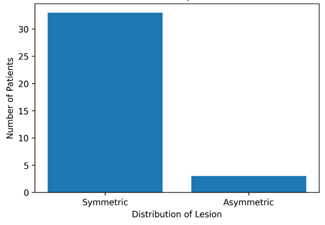
Fig.3. Distribution of patients as per distribution of lesion
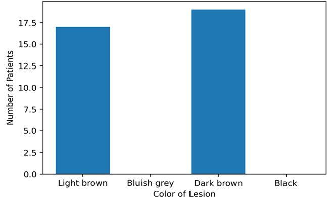
Fig.4. Distribution of patients as per color of lesion
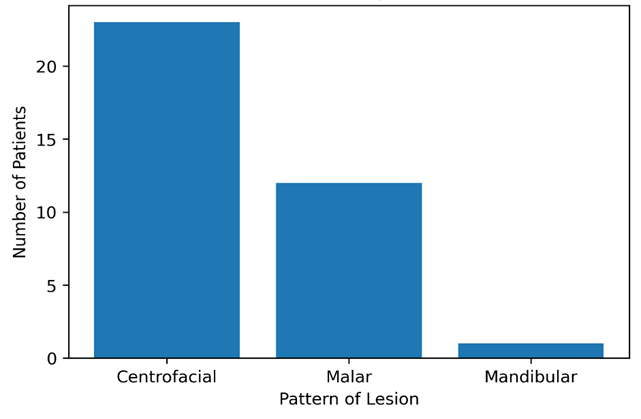
Fig.5. Distribution of patients as per pattern of lesion
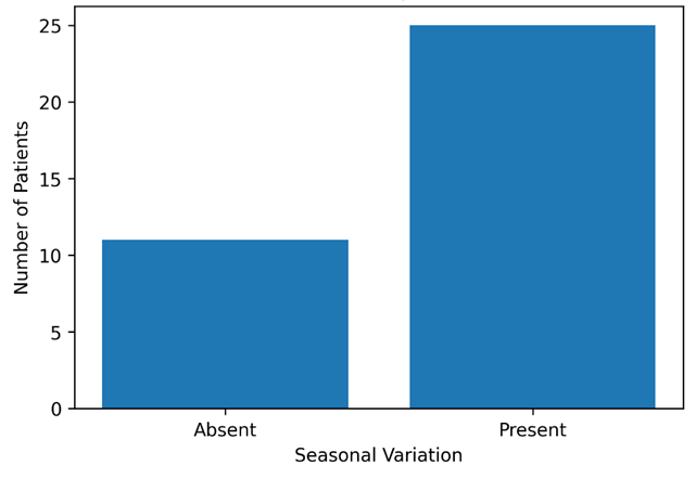
Fig.6. Distribution of patients as per seasonal variation
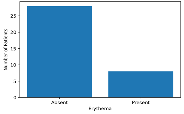
Fig.7. Distribution of patients as per Erythema
CONCLUSION:
The present study showed that out of 36 patients enrolled maximum patients had medium to fair skin with Fitzpatricks skin type of III & IV. The lesions were mostly centrofacial, symmetrical, with light and dark brown lesions with no erythema. It was further observed that the disease is prevalent in people who were exposed to sun and UV rays.
LIMITATIONS OF THE STUDY:
The sample size of the study is small, further studies are needed with larger sample sizes and also multi location studies are to be conducted to confirm the demography.
REFERENCES:
1. Handel A. C, Miot L. D and Miot H. A. Melasma: a clinical and epidemiological review. An. Bras. Dermatol., 2014; 89: 771-782.
2. Ibn Sina. Al-Qanun-Fil-Tibb (Urdu translation by Kantoori G. H). Vol-2. New Delhi: Aijaz Publishing House; 2010: 1422-1424.
3. Bhagdadi I. H. Kitabul Mukhtarat Fit Tib (Urdu translation by C. C. R. U. M). Vol-4. New Delhi: Ministry of Health & Family Welfare, Govt. of India; 2007: 118-119.
4. Tabri A. M. Moalijat al Buqratiya (Urdu translation by C. C. R. U. M). Vol-2. New Delhi: Ministry of Health & Family Welfare, Govt. of India; 1995: 241-243.
5. Anonymous. Tarjuma Sharah Asbab (Urdu translation by Hakeem K. R. Ahmed). Vol-4. New Delhi: C. C. R. U. M, Ministry of Health & Family Welfare, Govt. of India; 2010: 262-271.
6. Wolff K, Goldsmith L. A, Katz S. I and et al. Fitzpatrick’s dermatology in general medicine. 7th ed. Vol-1. USA: McGraw Hill; 2008: 635.
7. Sarkar R, Arora P, Garg V. K and et al. Melasma update. Indian. Dermatol. Online. J., 2014; 5: 426.
8. James W. D, Berg T. G and Elston D. M. Andrews’ diseases of the skin and clinical dermatology. 10th ed. Philadelphia: Saunders Elsevier; 2006: 854-855.
9. Mojtabaee M, Mokaberinejad R, Adhami S and et al. Nutritional advice for patients with melasma in Iranian traditional medicine. J. Skin. Stem. Cell., 2016; 3: 1.
10. Bagherani N, Gianfaldoni S and Smoller B. An overview on melasma. Pigmentary. Disord., 2015; 2: 218.
11. Ball Arefiev K. L and Hantash B. M. Advances in the treatment of melasma: a review of the recent literature. Dermatol. Surg., 2012; 38: 1-14.
12. Desai S. R. Hyperpigmentation therapy: a review. J. Clin. Aesthet. Dermatol., 2014; 7: 13-17.
13. Lee H. I, Lim Y. Y, Kim B. J and et al. Clinicopathologic efficacy of copper bromide plus/yellow laser for treatment of melasma in Asian patients. Dermatol. Surg., 2010; 36: 885-893.
14. Grimes P. E, Ijaz S, Nashawati R and et al. New oral and topical approaches for the treatment of melasma. Int. J. Womens. Dermatol., 2019; 5: 30-36.
15. Rodrigues M and Pandya A. G. Melasma: clinical diagnosis and management options. Australas. J. Dermatol., 2015; 56: 151-163.
16. Ishiy P. S, Silva L. R, Penha M. A and et al. Skin diseases reported by workers from UNESP campus at Rubião Jr, Brazil. An. Bras. Dermatol., 2014; 89: 529-531.
17. Qaumri A. H. M. Ghina Munna (Urdu translation by C. C. R. I. U. M). New Delhi: Ministry of Health & Family Welfare; 2008: 435-454.
18. Ahmed H. H. J. Tazkirah-i-Jaleel (Urdu translation by C. C. R. U. M). New Delhi: Ministry of Health & Family Welfare; 2008: 434-436.
19. Qurrah S. I. Tarjumae Zakheera Sabit Ibne Qurrah (Urdu translation by Hakim S. A. Ali). Aligarh: Litho Colour Printers; 1987: 34-35.
20. Razi A. M. B. Z. Al Hawi Fil Tib (Urdu translation by Hakeem M. Y. Siddiqui). Vol-23, Part-2. Aligarh: Saba Publishers; 1994: 14-17.
21. Khan A. Al-Haziq. Karachi: Madina Publishing Company; 1983: 550-552.
22. Aquil Z, Khalid M, Qamaruddin and et al. Potential role of Unani medicinal plants in management of Kalaf. J. Skin., 2019; 3: 1-6.
23. Sidharth S. Etiopathogenesis of melasma. In: Sarkar S, editor. Melasma a monograph. 1st ed. New Delhi: Jaypee Publishers; 2015: 6-14.
24. Ogbechie-Godec O. A and Elbuluk N. Melasma: an up-to-date comprehensive review. Dermatol. Ther., 2017; 7: 305-318.
25. Guinot C, Cheffai S, Latreille J and et al. Aggravating factors for melasma: a prospective study. J. Eur. Acad. Dermatol. Venereol., 2010; 24: 1060-1069.
26. Hexsel D, Lacerda D. A, Cavalcante A. S and et al. Epidemiology of melasma in Brazilian patients. Int. J. Dermatol., 2014; 53: 440-444.
27. Al-Hamdi K. I, Hasony H. J and Jareh H. L. Melasma in Basrah: a clinical and epidemiological study. Med. J. Basrah. Univ., 2008; 26: 1-5.
28. Kang H. Y and Ortonne J. P. What should be considered in treatment of melasma. Ann. Dermatol., 2010; 22: 373-378.
29. Fors M, González P, Viada C and et al. Validity of the Fitzpatrick skin phototype classification in Ecuador. Adv. Skin. Wound. Care., 2020; 33: 1-5.
30. Yalamanchili R, Shastry V and Betkerur J. Clinico-epidemiological study and quality of life assessment in melasma. Indian. J. Dermatol., 2015; 60: 519.
31. KrupaShankar D. S, Somani V. K, Kohli M and et al. A cross-sectional multicentric study of melasma in India. Dermatol. Ther., 2014; 4: 71-81.
32. Qazi I, Dogra N. K and Dogra D. Melasma: a clinical and epidemiological study. Int. J. Contemp. Med. Res., 2017; 4: 2087-2090.
33. Esteve E, Saudeau L, Pierre F and et al. Physiological cutaneous signs in normal pregnancy. Ann. Dermatol. Venereol., 1994; 121: 227-231.
34. Kim E. H, Kim Y. C, Lee E. S and et al. The vascular characteristics of melasma. J. Dermatol. Sci., 2007; 46: 111-116.
35. Park G. H, Lee J. H, Choi J. R and et al. Degree of erythema in melasma lesions and response to Nd:YAG laser. J. Dermatolog. Treat., 2013; 24: 297-299.