Porous Tantalum As A Next-Generation Biomaterial For Dental Implants - A Case Report
- Godavari Deepak Nagargoje , Reader, Department of Conservative Dentistry and Endodontics, Saraswati Dhanwantari Dental College and Hospital, (MH)Parbhani
- Nutan Dhamdhere , Postgraduate Student, Department of Prosthodontics, Saraswati Dhanwantari Dental College and Hospital, (MH)Parbhani
- Suraj Pinate , Postgraduate Student, Department of Prosthodontics, Saraswati Dhanwantari Dental College and Hospital, (MH)Parbhani
- Vishal Patil , Senior Lecturer, Department of Oral and Maxillofacial Surgery, Saraswati Dhanwantari Dental College and Hospital, (MH)Parbhani
- Yusuf Shaikh , Senior Lecturer, Department of Prosthodontics, Saraswati Dhanwantari Dental College and Hospital, (MH)Parbhani
- Swapnil Rawte , Reader, Department of Prosthodontics, Saraswati Dhanwantari Dental College and Hospital, (MH)Parbhani
Article Information:
Abstract:
Background: Porous tantalum implants have emerged as an innovative biomaterial in implant dentistry due to their trabecular architecture and high biocompatibility, enabling rapid osseoincorporation and enhanced implant stability. A 61-year-old female presented with missing teeth in the mandibular left posterior region for 6 months, seeking functional rehabilitation. CBCT evaluation revealed sufficient bone dimensions (width: 7.9 mm; depth: 10.6 mm) for implant placement. A Zimmer Biomet Trabecular Metal™ porous tantalum implant (4.5 × 10 mm) was selected. Following standard preoperative prophylaxis and antibiotic protocol, implant osteotomy was prepared using a sequential drilling technique. The porous tantalum implant was placed in the 35 region with satisfactory primary stability. Healing abutment placement was performed after 2 weeks, followed by prosthetic rehabilitation. The patient demonstrated excellent osseointegration, stable peri-implant soft tissue, and improved masticatory efficiency and esthetics. This case highlights the clinical effectiveness of porous tantalum implants in posterior mandibular rehabilitation. The biomaterial’s osteoconductive and osteoinductive properties promote rapid bone ingrowth and superior stability, suggesting its potential as a reliable option for implant-supported restorations in compromised bone conditions
Keywords:
Article :
CASE REPORT :
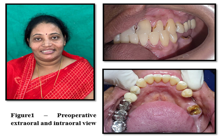
A 61-year-old female patient presented to the Department of Prosthodontics with the chief complaint of missing teeth in the lower left posterior region of the mandible for the past six months (Figure 1). The teeth had been lost due to extensive caries, leading to difficulty in mastication on the affected side, occasional food lodgment, and discomfort while chewing hard foods. The patient had no relevant systemic illness or history of parafunctional habits and expressed a desire for functional and esthetic rehabilitation using a fixed implant-supported prosthesis
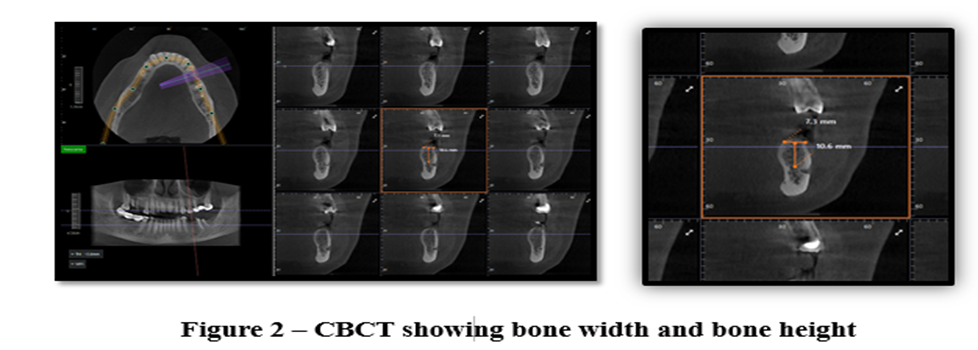
Clinical examination revealed an edentulous span in the 35 region with adequate keratinized mucosa and satisfactory interarch space. Radiographic assessment was performed using cone-beam computed tomography (CBCT), which showed a bone width of 7.9 mm and a vertical bone height of 10.6 mm, indicating sufficient bone availability for implant placement without the need for augmentation procedures (Figure 2).
Based on the available bone dimensions and the manufacturer’s protocol, a Zimmer Biomet Trabecular Metal™ porous tantalum implant measuring 4.5 × 10 mm was selected for rehabilitation1-4.
Prior to surgery, the patient underwent oral prophylaxis two weeks before the procedure, and was advised to use chlorhexidine (0.12%) mouth rinses twice daily for three days preoperatively. Prophylactic antibiotic therapy consisting of amoxicillin 1 g was administered one hour before implant placement. Under local anesthesia, a crestal incision was given in the 35 region, followed by full-thickness flap reflection to expose the alveolar crest (Figure 3,4).
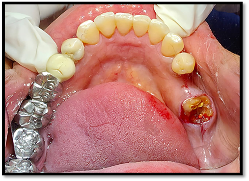
Figure 3 – Crestal incision
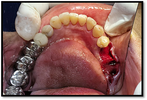
Figure 4 – Flap reflection
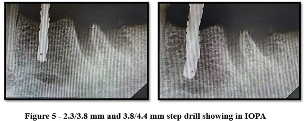
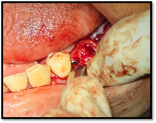
Figure 6 – Implant placement in 35 region
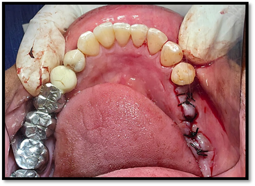
Figure 7 – Suture placement
After an uneventful healing period of two weeks, healing abutments were placed, and the soft tissue was allowed to mature. An impression was then made using impression coping to record the implant position and emergence profile accurately. A definitive prosthesis was fabricated and delivered after confirming proper fit, occlusion, and esthetics. Postoperative clinical and radiographic evaluation revealed successful osseointegration of the implant, stable peri-implant soft tissue, and improved functional and esthetic outcomes (Figure 8,9). The patient expressed satisfaction with the treatment, reporting restored chewing efficiency and comfort.
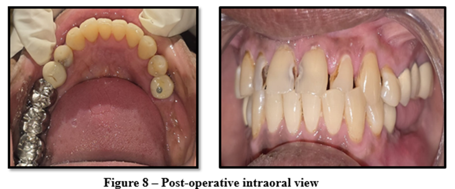
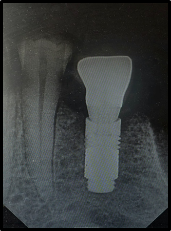
Figure 9 – Post-operative intraoral view
DISCUSSION:
Porous tantalum implants have gained increasing attention in implant dentistry because of their unique ability to achieve osseoincorporation, where bone grows both on and within the implant structure1,2. This biological advantage is particularly beneficial in posterior mandibular regions and in cases with reduced bone density4,5. The present case demonstrated excellent clinical and radiographic outcomes following the placement of a Zimmer Biomet Trabecular Metal™ porous tantalum implant in the 35 region, showing rapid healing, high primary stability, and favorable prosthetic results. These findings are consistent with earlier clinical case reports and studies that highlight the effectiveness of porous tantalum implants in challenging implant scenarios.
Lingam and Balasubramanian reported a similar successful mandibular rehabilitation using porous tantalum implants, emphasizing their superior osteoconductive behavior and long-term stability in compromised bone1. Likewise, Bencharit et al. described clinical applications of trabecular metal implants and found enhanced bone-implant contact and faster integration compared to traditional titanium implants2. Their observations align closely with the results obtained in this case, where the implant achieved predictable osseointegration and stable peri-implant soft tissue.
Several other case reports have demonstrated the advantage of porous tantalum in sites with poor bone quality or where grafting procedures were performed. Paganias et al. documented excellent osseous integration of porous tantalum implants in orthopedic and dental applications, concluding that the porous architecture supports rapid vascularization and bone regeneration. In a comparative clinical case study, Piglionico et al. observed significantly greater mineralized bone matrix formation around porous tantalum implants compared to titanium implants, reinforcing the material’s osteoinductive potential4. Similarly, Wang et al. reported superior osteogenesis and implant stability when using porous tantalum scaffolds fabricated through 3D-printing techniques, suggesting their suitability for load-bearing implant sites5.
From a biomechanical perspective, porous tantalum has an elastic modulus close to that of natural bone, reducing stress shielding and enhancing load distribution. This feature has been reported in multiple clinical case reports, including those involving sinus lift and ridge augmentation procedures, where tantalum implants showed improved clinical performance and secondary stability. The present case supports these observations, as the implant remained stable throughout the follow-up period and provided satisfactory functional and esthetic outcomes for the patient.
Overall, the clinical success of this case is in agreement with previously published reports that establish porous tantalum as a reliable and biologically superior implant material, particularly for posterior mandibular rehabilitation and compromised bone conditions. These consistent outcomes across different case reports and studies strengthen the evidence that porous tantalum implants can serve as a predictable and advanced alternative to conventional titanium implant systems.
CONCLUSION:
The successful outcome of this case demonstrates the strong clinical potential of porous tantalum implants as a next-generation biomaterial for implant rehabilitation. Owing to its trabecular architecture and high porosity, porous tantalum closely mimics cancellous bone and promotes rapid osseoincorporation, vascularization, and enhanced implant stability. In the present case, the implant showed excellent primary and secondary stability, favorable peri-implant soft-tissue adaptation, and a predictable prosthetic outcome with improved mastication and esthetics8,9. Compared with conventional titanium implants, porous tantalum offers biomechanical advantages such as reduced stress shielding and improved load distribution, making it particularly suitable for posterior mandibular and compromised bone situations. Although long-term clinical studies are still required, this case supports existing evidence that porous tantalum implants provide a reliable and biologically superior alternative for modern implant dentistry6,7.
REFERENCES:
1. Lingam MA, Balasubramanian I. Tantalum: A transmogrifying material in dental implant. SRM J Res Dent Sci. 2021;12(3):141–145.
2. Bencharit S, Byrd WC, Altarawneh S, Hosseini B, Leong A, Reside G, et al. Development and applications of porous tantalum trabecular metal-enhanced titanium dental implants. Clin Implant Dent Relat Res. 2014;16:817–826.
3. Paganias CG, Tsakotos GA, Koutsostathis SD, Macheras GA. Osseous integration in porous tantalum implants. Indian J Orthop. 2012;46(5):505–513.
4. Piglionico S, Bousquet J, Fatima N, Renaud M, Collart-Dutilleul PY, Bousquet P. Porous tantalum vs titanium implants: Enhanced mineralized matrix formation after stem cell proliferation and differentiation. J Clin Med. 2020;9(11):3657.
5. Wang H, Su K, Su L, Liang P, Ji P, Wang C. Comparison of 3D-printed porous tantalum and titanium scaffolds on osteointegration and osteogenesis. Mater Sci Eng C. 2019;104:109908.
6. Levine BR, Sporer S, Poggie RA, Della Valle CJ, Jacobs JJ. Experimental and clinical performance of porous tantalum in orthopedic applications. Biomaterials. 2006;27(27):4671–4681.
7. Zardiackas LD, Parsell DE, Dillon LD, Mitchell DW, Nunnery LA, Poggie R. Structure, metallurgy, and mechanical properties of a porous tantalum biomaterial. J Biomed Mater Res. 2001;58(2):180–187.
8. Sagomonyants KB, Hakim-Zargar M, Jhaveri A, Aronow MS, Gronowicz G. Porous tantalum stimulates the proliferation and osteogenesis of osteoblasts. J Biomed Mater Res A. 2011;97(4):489–496.
9. Karageorgiou V, Kaplan D. Porosity of 3D biomaterial scaffolds and osteogenesis. Biomaterials. 2005;26(27):5474–5491.
10. Arabnejad S, Burnett TL, Claeyssens F, et al. High-strength porous biomaterials for load-bearing orthopedic implants. Acta Biomater. 2017;56:212–224.