Clinico Mycological Profile And Dermoscopy In Dermatophytosis In A Tertiary Care Centre In North Kerala: A Cross Sectional Observational Study
- Amrutha Madai puthiyaveedu1 , MBBS,MD, Assistant Professor of Dermatology, KMCT Medical College, Kozhikode,Kerala-673602
- . Aishwaria Suresh , MBBS,MD,DNB ,MNAMS, Associate Professor of Dermatology, KMCT Medical College, Kozhikode,Kerala-673602
- Nasaha T.K3 , MBBS,MD,DNB, Assistant Professor of Dermatology, KMCT Medical College, Kozhikode,Kerala-673602
Article Information:
Abstract:
Background: Superficial mycotic infections affect 20–25% of the global population, with dermatophytes being the most common agents. In India, chronic dermatophytosis has risen sharply due to overcrowding, poor affordability, improper antifungal use, drug resistance, and widespread steroid abuse, leading to chronicity and relapse. Diagnosis becomes difficult in partially treated cases or when superimposed on other dermatoses, while KOH mounts and cultures show low positivity (40–58%) and require laboratory expertise, dermoscopy offers a rapid, noninvasive, and accessible adjunct for identifying dermatophytosis, especially in resource-limited settings. This study aims to evaluate dermoscopy’s usefulness and correlate findings with causative organisms to guide therapy. Aim: 1) assessing the dermoscopic patterns across various types of dermatophytosis, 2) identifying the fungal species isolated from cultures. Materials And Method: This six-month cross-sectional study included 60 mycologically proven dermatophytosis patients. KOH- or culture-positive individuals of all ages were enrolled. Skin scrapings underwent KOH microscopy and fungal culture. Dermoscopic images were taken using a Derm Lite DL4. Results: Male-to-female ratio was 2.2:1 and majority (43.3%) was in the age group15- 35 years. Tinea corporis was the most common type followed by Tinea cruris and 36 % of our patients had multiple sites involved. 90% was KOH positive .Fungal culture yielded dermatophytes in 41.7% cases. Dermoscopy patterns most commonly observed were superficial scales (98.3%), scales along creases (86.7%), collarette scales (76.7%), diffuse erythema (60%), brown black dots in 40%, red brown dots (35%) and perifollicular scales (30%). A significant association with p value 0.007 was observed with the dermoscopic findings of collarette of scales, scaling along creases and brown black dots. Conclusion: Dermatophytosis commonly affects the younger adult population in South India, with Tinea corporis and cruris being the most common presentation and Trichophyton mentagrophytes being the most common etiology. There is a rising incidence of Microsporum infections . Dermoscopy is no substitute for mycological study, but rather it complements it with consistent features. Hair involvement on dermoscopy may help predict the need for systemic antifungal treatment. This study reveals significant association between the type of organism and dermoscopic findings of collarette of scales, scaling along creases and brown black dots.
Keywords:
Article :
INTRODUCTION:
The prevalence of superficial mycotic infection worldwide is 20%-25%, of which dermatophytes are the most common agents , and in the last few years, we have seen a steep rise in the cases of chronic dermatophytic infections in Indian subcontinent (1). Dermatophytoses are currently a menace in India because of overcrowding, unaffordability and uncontrollable topical steroid abuse, improper use of antifungals and hence poor response and drug resistance. Also these results in chronicity, recurrence and relapse (2).They pose a diagnostic challenge to the treating dermatologist in circumstances such as partial treatment or fungus invasion on to other dermatoses, such as psoriasis or allergic contact dermatitis. This compels the dermatologists to seek investigations such as potassium hydroxide mount and fungal culture. However, positive yield in these tests is surprisingly low ranging from 40% to 58% in various studies(3). These conventional mycological examinations are also rather complex, time-consuming, and require trained personnel and mycological tools.
Dermoscopy can serve as a valuable adjunct in the noninvasive identification and differential diagnosis of several common dermatoses by enhancing the visibility of both surface structures and underlying characteristics that are not discernible to the naked eye(4).
In the lack of sufficient laboratory resources, dermoscopy serves as a complementary instrument to clinical diagnosis. Dermoscopy facilitates the prompt initiation of treatment, hence reducing the threat of dermatophytosis (5).
There are only few studies on dermoscopy of dermatophytosis(5).This study was carried out with the intention to evaluate the usefulness of dermoscopy in the diagnosis of dermatophytosis, co- relate the causative organism with the clinical appearance and dermoscopy findings and hence tailor the antifungals accordingly.
MATERIALS AND METHODS :
This was a cross-sectional study done over a period of 6 months from May 2025 to October 2025 on 60 consecutive patients with mycologically proven dermatophytosis in the out patient department of Dermatology in a tertiary care centre in North Kerala.
Study was conducted after receiving institutional research and ethical clearance. The primary aim of this study was to investigate the clinicomycological profile and dermoscopic patterns observed in dermatophytosis patients. Our objectives included: 1) assessing the dermoscopic patterns across various types of dermatophytosis, and 2) identifying the fungal species isolated from cultures.
We included all patients who are KOH smear positive or fungal culture positive, of all age groups and both sexes. Patients unwilling to participate in the study were excluded. For analytical purposes, lesions were categorized as chronic if duration of lesions were more than 6 months.
Mycological examination involved collecting skin scrapings from the active, peripheral edge of lesions, and from the roof of any vesicles or pustules present. Before collection, the affected area was thoroughly cleaned with alcohol to minimize surface contaminants. Samples were obtained using a sterile scalpel blade and placed into sterile plastic bottles. Laboratory procedures included direct microscopic examination with 10% KOH and fungal culture on Sabouraud's Dextrose Agar slopes containing chloramphenicol and cycloheximide for primary isolation. Identification was based on gross morphological features and microscopy using Lactophenol Cotton Blue (LPCB) mounts. Special tests, such as urease and hair perforation tests, were performed when necessary for definitive identification. Additionally, clinical photographs and dermoscopic images were captured after obtaining proper informed consent from each patient. A Dermalite DL4 dermoscope with 10x magnification was used, utilizing both polarized and non-polarized modes. Dermoscopic photographs were taken with a phone camera attached via an adaptor.
Statistical analysis: Data were entered in Microsoft office excel and analysis was done using SPSS software. Categorical variables were presented as frequency and percentage. Chi -square test /Fisher’s exact test was used for the statistical analysis and p value of <0.05 will be considered as statistically significant.
RESULTS:
Out of the 60 patients, male-to-female ratio was 2.2:1. Majority (43.3%) was in the age group 15- 35 years and had a duration of disease < 6 months ( 61.7%).Chronic infection lasting for >6 months were seen in 38.3% cases. Tinea corporis was the most common type observed, followed by Tinea cruris and 36 % of our patients had multiple sites involved (Table 1). There was relapse in 3.3% and recurrence was seen in 13.3% patients.65% of patients had no other co-morbidities, while 15% had atopy and 10% had diabetes mellitus. About 45% of patients had used topical antifungals before consulting here and 31.7% had used topical steroids.
All the patients included in the study were either KOH or culture positive and out of them 90% was KOH positive and showed thin hyaline branched septae. Fungal culture yielded dermatophytes in 41.7% cases.The fungal species most commonly isolated on culture was Trichophyton Mentagrophytes in 25%,(Fig 2) followed by Trichophyton Rubrum(Fig 3) in 8.3%,Microsporum Canis 5% (Fig 4) and Microsporum Gypseum 3.3%(Fig 5).Organism was not isolated in culture in 58.3%. Patients with Microsporum Canis infection had history of contact with pets,showed small sized /coin sized annular lesions and collarette of scales .
Dermoscopy was performed in all these cases and the most common patterns observed was superficial scales (98.3%),scales along creases (86.7%),collarette scales (76.7%),diffuse erythema (60%),brown black dots in 40%, red brown dots (35%) and perifollicular scales(30%) .Other patterns observed were telengiectasia (10%),comma hairs,unusual bends and dotted vessels in 5% cases each, follicular micropustules, non follicular micropustules, broken hairs, white hairs(Fig 6), hypo pigmentation in 3.3% each and black dots and morse code hair in 1.7%(Fig 7) . Long term use of topical steroids were seen in 31.7 % patients, which might be the cause for telangiectasia and hypopigmentation . We also noticed an unusual finding of white translucent hair in the lesional skin in about 3.3% patients.( (Table 8).A significant association with p value .007 was observed with the dermoscopic findings of collarette of scales,scaling along creases and brown black dots.( (Table 9)
[Table -1]: AGE DISTRIBUTION AND TYPE OF DERMATOPHYTOSIS
|
Clinical Diagnosis |
Age |
p value |
|||
|
<5 years |
5-15 years |
15-35 years |
>35 years |
||
|
T.CORPORIS T.CRURIS EXTENSIVE DERMATOPHYTOSIS T.MANUUM T.INCOGNITO T.PEDIS T.FACEAI |
0 1 0 0 0 0 0 |
10 5 0 0 0 0 0 |
16 16 3 1 0 1 4 |
15 6 4 1 2 0 2 |
0.151 0.079 0.477 0.911 0.311 0.722 0.534 |
[Table-8]: DERMOSCOPIC FINDINGS IN DERMATOPHYTOSIS
|
Dermoscopic finding |
Overall (n = 60) |
|
|
Frequency |
Percentage |
|
|
Superficial Scale Collarete of Scale Scaling Along Creases Perifollicular Scales Diffse Erythema Dotted Vessels Red Brown Dots Brown Black Dots Follicular Micropustules Non Follicular Micropustules Broken Hair Black Dots Morsecode Hair Telengiectasia Comma Hair Unusual Bends White Hair Hypopigmentation |
59 49 49 19 35 3 19 21 2 1 2 1 1 6 3 3 2 2 |
98.3 81.7 81.7 31.7 58.3 5 31.7 35 3.3 1.7 3.3 1.7 1.7 10 5 5 3.3 3.3 |
[Table -9]: ORGANISM ISOLATED IN CULTURE AND DERMOSCOPIC FINDINGS
|
Dermoscopic finding |
Organism in culture |
p value |
|||
|
Trichophyton Mentagrophytes (n=15) |
Trichophyton Rubrum (n=5) |
MicrosporumCanis (n=3) |
MicrosporumGypseum (n=2) |
|
|
|
Superficial Scale Collarette of Scale Scaling Along Creases Perifollicular Scales Diffuse Erythema Dotted Vessels Red Brown Dots Brown Black Dots Follicular Micropustules Non Follicular Micropustules Broken Hair Black Dots Morsecode Hair Telengiectasia Comma Hair Unusual Bends White Hair Hypopigmentation |
15 (100%) 15 (100%) 13 (86.7%) 8 (53.3%) 9 (60%) 1 (6.7%) 5 (33.3%) 3 (20%) 0(0%) 1(6.7%)
1(6.7%) 0(0%) 1(6.7%) 2 (13.3%) 1(6.7%) 1(6.7%) 1(6.7%) 1(6.7%) |
5 (100%) 5 (100%) 5 (100%) 1(20%) 2 (40%) 0 (0%) 1(20%) 3 (60%) 0 (0%) 0 (0%)
0 (0%) 0 (0%) 0 (0%) 0 (0%) 0 (0%) 0 (0%) 0 (0%) 0 (0%) |
3 (100%) 3 (100%) 3 (100%) 0 (0%) 3 (100%) 0 (0%) 0 (0%) 0 (0%) 0 (0%) 0 (0%)
0 (0%) 0 (0%) 0 (0%) 0 (0%) 0 (0%) 0 (0%) 0 (0%) 0 (0%) |
2 (100%) 1 (50%) 0 (0%) 1 (50%) 2 (100%) 0 (0%) 0 (0%) 1 (50%) 0 (0%) 0 (0%)
0 (0%) 1 (50%) 0 (0%) 0 (0%) 0 (0%) 0 (0%) 0 (0%) 0 (0%) |
--- 0.007 0.007 0.258 0.244 0.875 0.505 0.198 ---- 0.875
0.875 0.007 0.875 0.694 0.875 0.875 0.875 0.875 |
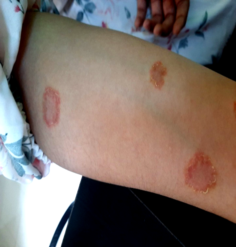
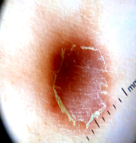
[Fig 4]: a) Clinical picture of Dermatophytosis caused by Microsporum Canis ; b) collarette of scales,superficial scales and scales along creases seen through dermoscopy
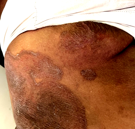
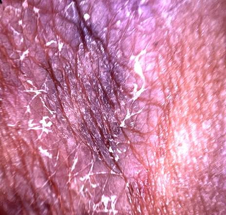
[Fig-5]: a) Clinical picture of Dermatophytosis caused by Microsporum Gypsium ; b) dermoscopic picture of Microsporum Gypsium showing superficial scales and scaling along creases
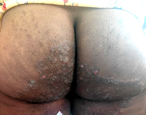
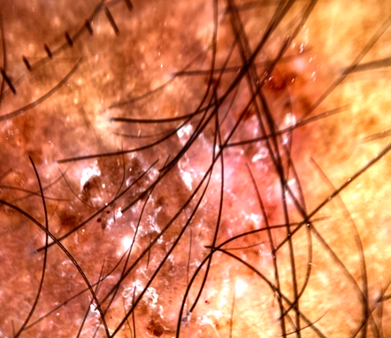
[Fig-2]: a) Clinical picture of dermatophytosis caused by Trichophyton mentagrophyte ; b) dermoscopic image showing redbrown dots, erythema and superficial scaling
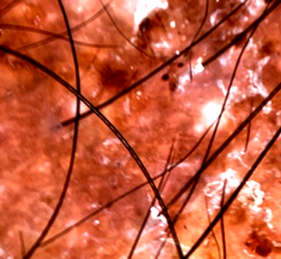
[Fig-7]: Dermoscopic image showing Morsecode hair
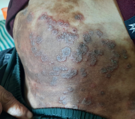
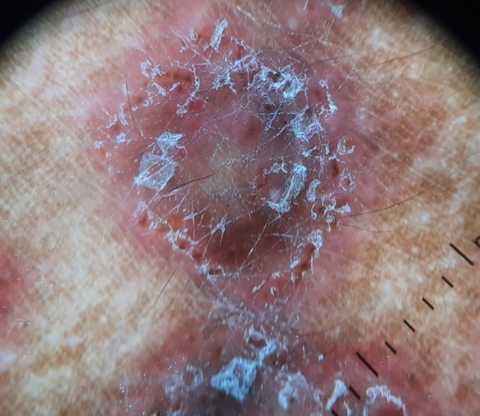
[Fig-3] : a) Clinical picture of Trichophyton Rubrum b) dermoscopic image showing red brown dots,scaling along creases ,and superficial scales .
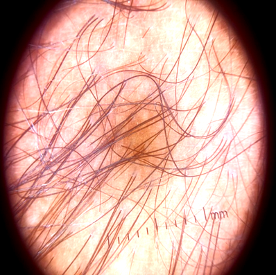
[Fig-6 ]: Dermoscopic image showing white hair
DISCUSSION:
The diagnosis of cutaneous fungal infection is usually made clinically with direct microscopic examination and fungal cultures wherever required; but are time-consuming, and require trained personnel and mycological tools and hence dermoscopy may serve as a valuable adjunct.(4) Tinea infections pose a diagnostic challenge to treating dermatologist during partial treatment, steroid abuse or fungus invasion on other dermatoses (5)
Our study showed a male preponderance as in previous studies(6).Kerala has a warm and humid climate almost round the year. According to Panda et al., dermatophytosis is a predominantly tropical dermatosis(7) so high humidity along with increased outdoor activity exposes males to an environment favourable for the growth of the fungus (8).
In this study, maximum number of cases of dermatophytosis were noted in the age group of 15- 35 years. Similar peak in the age group 20-30 yrs was observed by Singh et al , Janardan et al., Bhagra et al., Agarwal et al, and Hanumanthappa et al. However, Bindu et al. observed highest peak in second decade (9). Possible explanation for increase in incidence in this age group is that most of them stay in shared accommodations with lack of proper personal hygiene and sharing of fomites (7). 36 % of our patients had multiple sites involved with Tinea corporis and Tinea cruris being the most common forms. This is similar to previous studies by Mahajan et al (10) and Singh et al (8) where Tinea Corporis and Tinea Cruris were the most common forms observed.
Chronic infection lasting for >6 months were seen in 38.3% cases.There was relapse in 3.3% and recurrence was seen in 13.3% patients. This was similar to that observed by Saha et al with chronic (34.2%) and recurrent (12.6%) patients.
65% of patients had no other co-morbidities, while 15% had atopy and 10% had diabetes mellitus. Atopy and diabetes mellitus were the most common systemic associations in previous studies also(11). Vineeta et al(12) observed diabetes in 2.6%in first episode and in chronic cases diabetes was associated in 11% .
In our study, 31.7% cases were using over-the-counter (OTC) medications containing steroids without proper consultation. Many authors have observed relatively higher percentage of cases using OTCs e.g., Vineeta et al(12). in 63% cases, Dabas et al(13). in 77.94% cases and Mahajan et al(10). in 70.6% cases while it was lower in the study by Singh et al(8) 21.7%. Long term use of topical steroids were seen in 31.7 % patients, which might be the cause for telangiectasia and hypopigmentation .
Percentage of cases using topical antifungal preparation in our study were 45% compared to Singh et al(8) 23.1%, whereas it was 5.7%, 47%, and 7.35% in the studies by Mahajan et al., Vineeta et al. and Dabas et al respectively. Thus, most studies including ours highlight the improper and rampant use of steroid containing preparations (easy availability, low cost, early relief from inflammatory symptoms like itching) to be a major contributory factor for resurgence of dermatophytosis in recent times. These medications suppress the host cell mediated immunity (CMI) resulting in persistence and chronicity of the disease despite treatment(14). 90% of our patients were KOH positive ;this was similar to that observed by Singh et al(8 )97.7% cases and close to the observation made by Janardhan et al(15).
Fungal culture yielded dermatophytes in 41.7% of our patients.This is similar to previous studies from India( 9),(12),(14) .The fungal species most commonly isolated on culture was Trichophyton Mentagrophytes in 25%,followed by Trichophyton Rubrum in 8.3%. Singh et al(8), Agarwal et al (16) and Mahajan et al (10) also observed similar results. But previous studies from South India, have depicted T. rubrum as the commonest organism in contrast to our study which could indicate a changing prevalence of organism in South India also(9),(15),(17),(18).
The current epidemics over different geographical regions of the world are due to T. Mentagrophytes complex and M. Canis(19) . Nenoff et al. did mycological and molecular analysis of 201 patients from various parts of our country and found T. mentagrophytes as the predominant isolate and they also identified a new Indian genotype of this organism (T. Mentagrophytes ITS type VIII) responsible for the present epidemic of dermatophytosis(20).
Due to reasons unexplored, the prevalence of dermatophytosis due to M. canis is less prevalent in India (0.76–4.5%)(19). In our study Microsporum Canis was seen in 5% which is slightly higher than previous studies. In India, very few people used to keep pets but there is a rising trend now probably due to better economic status. There are lots of rodents, stray dogs and cats and monkeys, which could be possibly associated as a source or the agents for the spread of T. Mentagrophytes and Microsporum Canis infection. Also, many people dry their clothes and bed linen/bedding outside their houses, and there is every possibility of the clothes and linen getting contaminated either directly or indirectly from the environment(19). Microsporum Gypseum was seen in culture in 3.3% patients. Microsporum Gypseum is a geophilic dermatophyte and a rare cause of dermatophytosis in humans and can affect glabrous skin(21) .
On dermoscopy superficial scales were seen in 98.3%,scales along creases in 86.7%, and collarete scales in 76.7% which is similar to previous studies (2),(3).Other dermascopic features observed were diffuse erythema (60%),brown black dots in 40%,red brown dots (35%) and perifollicular scales(30%), telengiectasia(10%),comma hairs,unusual bends and dotted vessels in 5% cases each, follicular micropustules, non follicular micropustules, broken hairs, white hairs, hypo pigmentation in 3.3% each and black dots in 1.7%.
We noticed an unusual finding of white translucent hair in the lesional skin in about 3.3% patients,possibly due to massive fungal invasion or due to topical steroid use.Ankad et al (3 ) also recorded two patients showing hypopigmented terminal hairs. Bhat et al had also observed a new dermoscopic feature, consisting of translucent, easily deformable hairs that look weakened and transparent, and shows bends and proposed it likely due to massive fungal invasion involving the whole hair shaft (4). Glomez-Moyano et al. observed scaly, broken, translucent hairs and morse code hairs in a 57-year-old man with tinea incognito and provided a correlation of this dermoscopic finding with direct microscopy(22) .
A new criterion to start systemic antifungal therapy in tinea of vellus hair skin has been described the observation of parasitized vellus hairs on direct examination. Dermoscopic examination can predict from the outset which cases of tinea of vellus hair skin will respond poorly or even not respond at all to topical treatment alone. (4)
CONCLUSION:
Dermatophytosis commonly affects the younger adult population in South India,with Tinea Corporis and Cruris being the most common presentation and Trichophyton Mentagrophytes being the most common etiology.There is a rising incidence of Microsporum infections and patients with Microsporum Canis infection had small sized /coin sized annular lesions which showed collarette of scales. Dermoscopy is no substitute for mycological study, but rather it complements it with consistent features like superficial scales and scaling along creases. Hair involvement on dermoscopy may help predict the need for systemic antifungal treatment. This study reveals significant association between the type of organism and dermoscopic findings of collarette of scales, scaling along creases and brown black dots.
BIBLIOGRAPHY:
1. Kaul S, Yadav S, Dogra S. Treatment of dermatophytosis in elderly, children, and pregnant women. Indian Dermatol Online J. 2017;8(5):310.
2. Theunuo R, Ramesh V. Dermoscopic Patterns in Dermatophytosis with an Emphasis on Consistent Observation. Indian Dermatol Online J [Internet]. 2024 Sep 30
3. Ankad B, Mukherjee S, Nikam B, Reshme A, Sakhare P, Mural P. Dermoscopic characterization of dermatophytosis: A preliminary observation. Indian Dermatol Online J. 2020;11(2):202.
4. Bhat Y, Keen A, Hassan I, Latif I, Bashir S. Can dermoscopy serve as a diagnostic tool in dermatophytosis? A pilot study. Indian Dermatol Online J. 2019;10(5):530.
5. A Study to Establish Correlation between Dermoscopic and Clinical Findings of Dermatophytosis. Indian J Public Health Res Dev [Internet]. 2020 Mar 23
6. Verma SB, Panda S, Nenoff P, Singal A, Rudramuruthy SM, Uhrlass S, et al. The unprecedented epidemic-like scenario of dermatophytosis in India: I. Epidemiology, risk factors and clinical features. Indian J Dermatol Venereol Leprol. 2021 Mar 23;87:154–75.
7. Panda S, Verma S. The menace of dermatophytosis in India: The evidence that we need. Indian J DermatolVenereolLeprol. 2017;83:281–4.
8. Singh BSTP, Tripathy T, Kar BR, Ray A. Clinicomycological Study of Dermatophytosis in a Tertiary Care Hospital in Eastern India: A Cross-sectional Study. Indian Dermatol Online J. 2019 Sep 26;11(1):46-50.
9. Bindu V, Pavithran K. Clinico-mycological study of dermatophytosis in Calicut. Indian J Dermatol Venereol Leprol. 2002;68:259–61
10. Mahajan S, Tilak R, Kaushal SK, Mishra RN, Pandey SS. Clinico-mycological study of dermatophytic infections and their sensitivity to antifungal drugs in a tertiary care center. Indian J Dermatol Venereol Leprol. 2017;83:436–40.
11. Sentamilselvi G, Kamalam A, Ajithadas K, Janaki C, Thambiah AS. Scenario of chronic dermatophytosis: an Indian study. Mycopathologia. 1997-1998;140(3):129-35.
12. Vineetha M, Sheeja S, Celine MI, Sadeep MS, Palackal S, Shanimole PE, Das SS. Profile of Dermatophytosis in a Tertiary Care Center. Indian J Dermatol. 2018 Nov-Dec;63(6):490-495.
13. Dabas R, Janney MS, Subramaniyan R, Arora S, Lal VS, Donaparthi N. Use of over-the-counter topical medications in dermatophytosis: A cross-sectional, single-center, pilot study from a tertiary care hospital. Indian J Drugs Dermatol. 2018;4:13–7.
14. Saha, Indraneel; Podder, Indrashis1,; Chowdhury, S.N.1; Bhattacharya, Susmita. Clinico-Mycological Profile of Treatment-Naïve, Chronic, Recurrent and Steroid-Modified Dermatophytosis at a Tertiary Care Centre in Eastern India: An Institution-Based Cross-SectionalStudy. Indian Dermatology Online Journal 12(5):p 714-721, Sep–Oct 2021.
15. Janardhan B, Vani G. Clinico mycological study of dermatophytosis. Int J Res Med Sci. 2017;5:31–9.
16. Agarwal US, Saran J, Agarwal P. Clinico mycological study of dermatophytes in a tertiary care centre in Northwest India. Indian J Dermatol Venereol Leprol 2014;80:194
17. Surendran KA, Bhat RM, Boloor R, Nandakishore B, Sukumar D. A clinical and mycological study of dermatophytic infections Indian J Dermatol. 2014;59:262–7
18. Manjunath M, Koppad M, Dadapeer S. Clinicomycological study of Dermatomycosis in a tertiary care hospital Indian J Microbiol Res. 2016;3:190–3
19. Thakur R, Kalsi AS. Outbreaks And Epidemics Of Superficial Dermatophytosis Due To Trichophyton mentagrophytes Complex And Microsporum canis: Global And Indian Scenario. Clin Cosmet Investig Dermatol. 2019 Dec 11;12:887-893.
20. Nenoff P, Verma SB, Vasani R, Burmester A, Hipler UC, Wittig F, et al. The current Indian epidemic of superficial dermatophytosis due to Trichophyton mentagrophytes–a molecular study. Mycoses 2018.
21. Tobeigei FH, Joseph MR, Al-Hakami A, Hamid ME. Microsporum gypseum Infection Among Two Related Families With a Zoonotic Aspect: A Prospective Case Series. Cureus. 2023 Dec 31;15(12):e51402. doi: 10.7759/cureus.51402. PMID: 38292972; PMCID: PMC10826859.
22. Gomez-Moyano E, Crespo-Erchiga V, Martinez Pilar L, Martinez Garcıa S. Correlation between dermoscopy and direct microscopy of morse code hairs in tinea incognito J Am Acad Dermatol. 2016;74:e7–