The Effect of Bromelain on Cytoplasmic Vacuolization and Myofibril Loss in Cardiac Muscle Cells of Doxorubicin-Administered Rattus norvegicus Wistar Strain Rats
- Kiki Mario , Departement of surgery, Faculty of Medicine, Airlangga University/ dr. Soetomo Hospital, Surabaya, Indonesia
- Asdi Wihandono , Division of Oncology Surgery, Departement of surgery, Faculty of Medicine, Airlangga University/ dr. Soetomo Hospital, Surabaya, Indonesia
- Marjono Dwi Wibowo , Division of Head and Neck Surgery, Departement of surgery, Faculty of Medicine, Airlangga University/ dr. Soetomo Hospital, Surabaya, Indonesia
Article Information:
Abstract:
Background: Breast cancer is the most prevalent and often fatal malignancy among women, affecting both developed and developing countries. Doxorubicin has risk of cardiotoxicity, particularly cardiomyopathy, and increases with improved patient prognosis. Bromelain offers extensive beneficial properties, such as treating cardiovascular disorders. Objective: This study was conducted to determine the histologic effect of bromelaine on myofibril loss and vacuolization of cardiac muscle. Methods: This study employed an experimental design with a posttest-only control group using in vivo Wistar rats (Rattus norvegicus) as experimental animals. Results: Doxorubicin administration resulted in statistically significant cardiac damage when compared to the negative control group [Mean (SD) P1 vs P2: 0.70 (0.308) vs 1.66 (0.313), p-value = 0.001]. Bromelain administration significantly reduced the damage score compared to doxorubicin alone [Mean(SD) P2 vs P3: 0.70 (0.308) vs 1.16 (0.270), p-value = 0.027]. Conclusion: There was a statistically significant difference observed in the effect of bromelain on myofibril loss and vacuolization within the cardiac muscle tissue of Wistar rats administered doxorubicin.
Keywords:
Article :
INTRODUCTION:
Breast cancer is the most prevalent and often fatal malignancy among women, affecting both developed and developing countries. According to the 2018 GLOBOCAN estimates by the International Agency for Research on Cancer (IARC), there were approximately 2.1 million new breast cancer cases worldwide (11.6% of all cancers), resulting in 626,679 deaths (15% of all cancer deaths).[1] In Indonesia, the prevalence of breast cancer reached 0.5 per 1000 women (Ministry of Health of the Republic of Indonesia, 2015). Data from Dr. Soetomo Hospital revealed that 77% of patients presented with advanced-stage disease. Cancer Research UK reported that between 2002 and 2006, the survival rate for patients diagnosed at an early stage was 90%, while for those with advanced-stage disease, it was only 15%. Metastasis is the primary cause of increased mortality and morbidity in breast carcinoma. According to PERABOI 2020, breast cancer treatment modalities include surgery, chemotherapy, hormonal therapy, targeted therapy, immunotherapy, and supportive therapy.[2,3,4]
Doxorubicin, a commonly used chemotherapeutic agent, significantly improves the prognosis and survival rate of breast cancer patients.[3-10] Despite its advantages, the risk of cardiotoxicity, particularly cardiomyopathy, increases with improved patient prognosis. Studies have even reported that as patients enter long-term survivorship (generally > 5 years post-diagnosis), cardiovascular mortality rates can equal or even exceed those of their primary disease.[11-19] Consequently, the risk of doxorubicin-induced cardiomyopathy often limits treatment continuation. Doxorubicin administration triggers changes in cardiac muscle cell structure, leading to impaired cardiac function, cardiomyopathy, and heart failure.[20-28] Observed damage includes myofibril loss and cytoplasmic vacuolization, which can occur separately or concurrently after doxorubicin administration.[29] The level of doxorubicin toxicity is dose-dependent, generally increasing when the administered dose exceeds 400 mg/m² – 700 mg/m². Currently, the precise mechanism of doxorubicin-induced cardiotoxicity (DIC) remains unclear. It is most likely caused by myocardial injury leading to left ventricular dysfunction.[20,30]
In recent years, research has focused on preventing and managing drug-induced cardiotoxicity. Various approaches to reduce the risk of cardiac dysfunction have been explored, ranging from doxorubicin dose limitation to the use of cardioprotective agents. However, there is currently no universally agreed-upon solution to prevent or treat doxorubicin-induced cardiotoxicity. On the other hand, natural plant-derived compounds have recently gained popularity due to their perceived minimal side effects, non-toxicity, affordability, and easy accessibility. Bromelain, for example, is a combination of several thiol endopeptidases derived from the fruit peel, stem, and/or roots of pineapple (Ananas comosus). Bromelain contains various compounds, including thiol endopeptidases, as well as protease inhibitors, glucosidase, cellulase, and escarase. Bromelain offers extensive beneficial properties, such as treating cardiovascular disorders, fibrinolytic, anti-edema, antithrombotic, and anti-inflammatory effects, making it useful for treating burns, blood clots, inflammation, enhancing antibiotic effectiveness, and even cancer.[31-35] Bromelain inhibits thrombus formation, reduces platelet aggregation and blood viscosity, lowers blood pressure, increases total antioxidant capacity (TAC) and oxygen utility capacity, prevents reduced flow in interstitial edema, enhances cardiac microcirculation, and maintains cardiac muscle tissue, all of which are beneficial for human cardiovascular health.[34,36-37]
METHOD:
This study employed an experimental design with a posttest-only control group using in vivo Wistar rats (Rattus norvegicus) as experimental animals. The interventions administered were: (1) doxorubicin administration and (2) doxorubicin administration combined with bromelain, with observations conducted over a 14-day period.
This study was conducted at the Faculty of Veterinary Medicine Laboratory, Universitas Airlangga, Surabaya, and further histopathological examinations were performed at the Department of Anatomy-Histology, Faculty of Medicine, Universitas Airlangga. The research was scheduled for March 2024.
Doxorubicin HCl at a dose of 15 mg/kg body weight was administered intraperitoneally to each rat in the doxorubicin-receiving groups. The doxorubicin was given three times a week (every other day) for 14 days.[35] Bromelain, at a volume of 6.2 mL/day, was administered once daily via oral gavage or pipette 30 minutes prior to doxorubicin HCl administration.[38]
Cardiac muscle cell samples from the rats were collected and then stained with hematoxylin and eosin (H&E). Stained sections were observed under a light microscope at 400x magnification at the Department of Anatomy-Histology, Faculty of Medicine, Universitas Airlangga. Diffuse vacuolization and flaccid cardiac muscle cell myofibril loss were recorded as percentages per field of view. Vacuolization observed with H&E staining included interstitial edema and intracellular swelling.[39]
For statistical analysis, unpaired numerical comparative analysis between two groups was performed using an independent samples t-test. If the data distribution was not normal, the Mann-Whitney U test was used.
RESULTS AND DISCUSSION :
Vacuolization and myofibril loss in the study specimens were assessed using histopathological examination. After collection, specimens underwent histological preparation followed by Hematoxylin-Eosin (H&E) staining. Each specimen was then evaluated for its degree of vacuolization and myofibril loss.
Analysis of the results indicated that the doxorubicin-treated group exhibited a higher degree of vacuolization and/or myofibril loss compared to the control group. In the group treated with doxorubicin and bromelain extract, an increase in vacuolization and/or myofibril loss was still observed when compared to the control group. However, this increase was less pronounced than in the doxorubicin-only group.
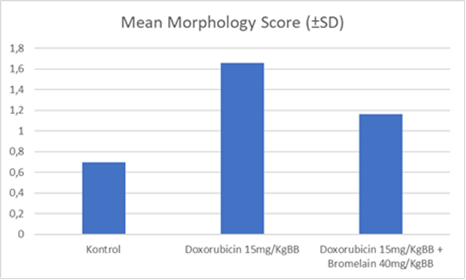
Figure 1. Graphs of Mean Morphology Score for Each Group
From the graph above, the doxorubicin group shows the highest mean morphological score of 1.66 (0.313), indicating greater damage to cardiomyocytes in this group compared to the others.
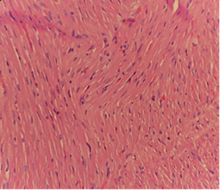
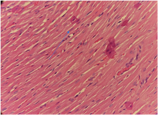
Figure 2. Histopathological Image with Score of 0 (left) and score of 1 (right)
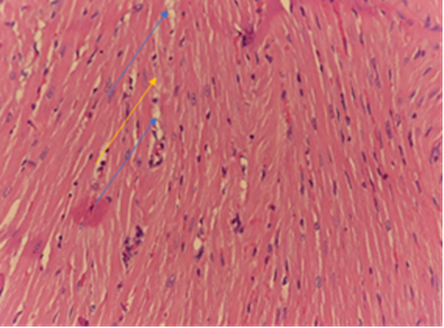
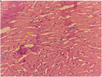
Figure 3. Histopathological Image with Score of 1.5 (left) and score of 2 (right)
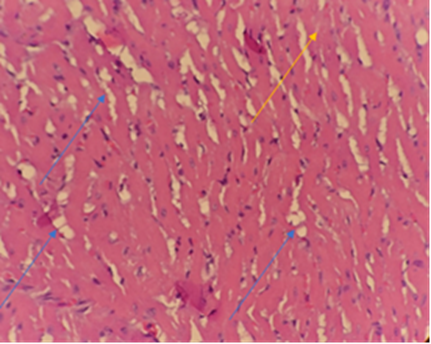
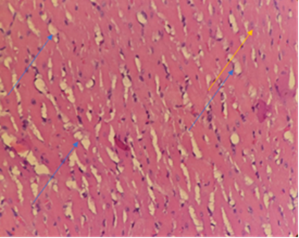
Figure 4. Histopathological Image with Score of 2.5 (left) and score of 3 (right)
Table 1. Independent Samples T-Test
|
Group Comparison |
n |
Mean (±SD) |
p-value |
|
P1 |
5 |
0,70 (0,308) |
0,001 |
|
P2 |
5 |
1,66 (0,313) |
|
|
P1 |
5 |
0,70 (0,308) |
0,036 |
|
P3 |
5 |
1,16 (0,270) |
|
|
P2 |
5 |
1,66 (0,313) |
0,027 |
|
P3 |
5 |
1,16 (0,270) |
In this study, independent samples t-tests were conducted to compare the mean morphological scores of cardiomyocyte damage across the different sample groups. These tests revealed significant differences in cardiomyocyte damage among the groups.
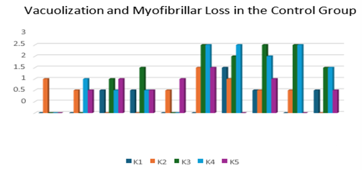
Figure 5. Bar Chart of Vacuolization and Myofibrillar Loss in the Control Group
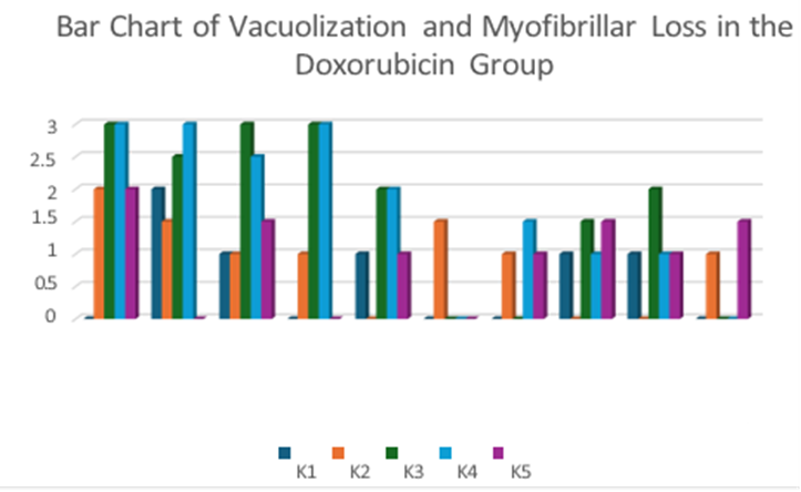
Figure 6. Bar Chart of Vacuolization and Myofibrillar Loss in the Doxorubicin Group
According to figure 5 and 6, doxorubicin administration resulted in statistically significant cardiac damage when compared to the negative control group [Mean (SD) P1 vs P2: 0.70 (0.308) vs 1.66 (0.313), p-value = 0.001]. This indicates that doxorubicin significantly increased cardiomyocyte injury.
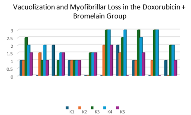
Figure 7. Bar Chart of Vacuolization and Myofibrillar Loss in the Doxorubicin + Bromelain Group
While bromelain administration did not completely prevent damage, it is presented in figure 7 that there was still a significant difference compared to the normal control group, although the extent of damage was less severe than in the doxorubicin-only group [Mean(SD) P1 vs P3: 0.70 (0.308) vs 1.16 (0.270), p-value = 0.036].
Crucially, bromelain administration significantly reduced the damage score compared to doxorubicin alone [Mean(SD) P2 vs P3: 0.70 (0.308) vs 1.16 (0.270), p-value = 0.027]. This finding suggests that bromelain provides a protective effect against doxorubicin-induced cardiomyocyte damage, even though it may not fully prevent all damage caused by doxorubicin administration.
Analysis of vacuolization and myofibril loss across the groups revealed statistically significant differences among all three, despite variations in percentage values based on the field of view within the control group. These findings support the study's hypothesis: the doxorubicin and bromelain group exhibited cardioprotective effects regarding vacuolization and myofibril loss compared to the doxorubicin-only group.
This study also found significant vacuolization and myofibril loss in myocardial tissue treated with doxorubicin. This aligns with observed damage, leading to cardiomyopathy in patients receiving doxorubicin even for short periods, which, if continued, can result in severe cardiac muscle damage.[29]
The cardiac muscle damage observed with doxorubicin use is a direct result of the drug's induction, causing extensive fibrosis and the presence of scattered cardiomyocytes with vacuolar degeneration. Cardiomyocytes containing vacuoles are typically found in fibrotic areas of the heart (known as "Adria cells"). Fibrosis is a predominant feature in cardiac tissue experiencing acute damage.[29] This is consistent with the findings in this study, where the doxorubicin-only group showed both vacuole formation and increased myofibril loss.[40]
Our analysis of vacuolization and myofibril loss across the experimental groups showed statistically significant differences among all three. This was observed despite some variations in percentage values within the control group's fields of view. These findings strongly support our study's hypothesis: the group treated with doxorubicin and bromelain demonstrated cardioprotective effects against vacuolization and myofibril loss when compared to the group that received doxorubicin alone.
Furthermore, we found significant vacuolization and myofibril loss in myocardial tissue exposed to doxorubicin. This aligns with previously observed damage, which can lead to cardiomyopathy in patients receiving doxorubicin, even for short durations. Prolonged exposure can result in severe and persistent damage to cardiac muscle.[29]
The cardiac muscle damage seen with doxorubicin use is directly induced by the drug itself. This damage manifests as extensive fibrosis and the presence of scattered cardiomyocytes with vacuolar degeneration. These vacuolated cardiomyocytes, often referred to as "Adria cells," are typically found in fibrotic regions of the heart. Fibrosis is a predominant feature in cardiac tissue experiencing acute damage.[29] Our findings are consistent with this, as the doxorubicin-only group exhibited both noticeable vacuole formation and increased myofibril loss.
CONCLUSION:
There was a statistically significant difference observed in the effect of bromelain on myofibril loss and vacuolization within the cardiac muscle tissue of Wistar rats administered doxorubicin
BIBLIOGRAPHY:
1. Sung, H., Ferlay, J., Siegel, R. L., Laversanne, M., Soerjomataram, I., Jemal, A., & Bray, F. (2021). Global Cancer Statistics 2020: GLOBOCAN Estimates of Incidence and Mortality Worldwide for 36 Cancers in 185 Countries. CA: A Cancer Journal for Clinicians, 71(3), 209–249. https://doi.org/10.3322/caac.21660
2. PERABOI. (2020). Panduan Penatalaksanaan Kanker Solid PERABOI 2010.
3. Cardoso, F., Costa, A., Senkus, E., Aapro, M., André, F., Barrios, C. H., Bergh, J., Bhattacharyya, G., Biganzoli, L., Cardoso, M. J., Carey, L., Corneliussen-James, D., Curigliano, G., Dieras, V., El Saghir, N., Eniu, A., Fallowfield, L., Fenech, D., Francis, P., … Winer, E. (2017). 3rd ESO–ESMO International Consensus Guidelines for Advanced Breast Cancer (ABC 3). Annals of Oncology, 28(1), 16–33. https://doi.org/10.1093/annonc/mdw544
4. Sutantio, J. A., Ishardyanto, H., & Herman, E. (2013). Hubungan Antara Kadar Vitamin D Darah Dan Wanita Pasca Menopause Dengan Locally Advanced Breast Cancer Di Rsud Dr . Soetomo Surabaya the Relationship Between Vitamin D Serum Level and Clinical Response of Neoadjuvant Chemotherapy on Post Menopausal Women .Hikisz, P., & Bernasinska-Slomczewska, J. (2021). Beneficial Properties of Bromelain. Nutrients, 13(12). https://doi.org/10.3390/nu1312431
5. Gupta, S. K., Garg, A., Bär, C., Chatterjee, S., Foinquinos, A., Milting, H., Streckfus-Bomeke, K., Fiedler, J., & Thum, T. (2018). Quaking inhibits doksorubisin-mediated cardiotoxicity through regulation of cardiac circular RNA expression short communication. Circulation Research, 122(2), 246–254. https://doi.org/10.1161/CIRCRESAHA.117.311335
6. Angsutararux, P., Luanpitpong, S., & Issaragrisil, S. (2015). Chemotherapy-Induced Cardiotoxicity: Overview of the Roles of Oxidative Stress. Oxidative Medicine and Cellular Longevity, 2015. https://doi.org/10.1155/2015/795602
7. Deavall, D. G., Martin, E. A., Horner, J. M., & Roberts, R. (2012). Drug-induced oxidative stress and toxicity. Journal of Toxicology, 2012. https://doi.org/10.1155/2012/645460
8. Kong, C. Y., Guo, Z., Song, P., Zhang, X., Yuan, Y. P., Teng, T., Yan, L., & Tang, Q. Z. (2022). Underlying the Mechanisms of Doksorubisin-Induced Acute Cardiotoxicity: Oxidative Stress and Cell Death. International Journal of Biological Sciences, 18(2), 760. https://doi.org/10.7150/IJBS.65258
9. Lyu, Y. L., Kerrigan, J. E., Lin, C.-P., Azarova, A. M., Tsai, Y.-C., Ban, Y., & Liu, L. F. (2007). Topoisomerase IIβ–Mediated DNA Double-Strand Breaks: Implications in Doksorubisin Cardiotoxicity and Prevention by Dexrazoxane. Cancer Research, 67(18), 8839–8846. https://doi.org/10.1158/0008-5472.CAN-07-1649
10. Lenneman, C. G., & Sawyer, D. B. (2016). Cardio-Oncology: An Update on Cardiotoxicity of Cancer-Related Treatment. Circulation Research, 118(6), 1008–1020. https://doi.org/10.1161/CIRCRESAHA.115.303633
11. Mu, H., Liu, H., Zhang, J., Huang, J., Zhu, C., Lu, Y., Shi, Y., & Wang, Y. (2019). Ursolic acid prevents doksorubisin‐induced cardiac toxicity in mice through eNOS activation and inhibition of eNOS uncoupling. Journal of Cellular and Molecular Medicine, 23(3), 2174. https://doi.org/10.1111/JCMM.14130
12. Rivankar, S. (2014). An overview of doksorubisin formulations in cancer therapy. Journal of Cancer Research and Therapeutics, 10(4), 853–858. https://doi.org/10.4103/0973-1482.139267
13. Sangweni, N. F., Gabuza, K., Huisamen, B., Mabasa, L., van Vuuren, D., & Johnson, R. (2022). Molecular insights into the pathophysiology of doksorubisin-induced cardiotoxicity: a graphical representation. Archives of Toxicology, 96(6), 1541–1550. https://doi.org/10.1007/S00204-022-03262-W/FIGURES/6
14. Sheibani, M., Azizi, Y., Shayan, M., Nezamoleslami, S., Eslami, F., Farjoo, M. H., & Dehpour, A. R. (2022). Doksorubisin-Induced Cardiotoxicity: An Overview on Pre-clinical Therapeutic Approaches. Cardiovascular Toxicology, 22(4), 292–310. https://doi.org/10.1007/S12012-022-09721-1/FIGURES/3
15. Sheibani, M., Nezamoleslami, S., Faghir-Ghanesefat, H., Emami, A. hossein, & Dehpour, A. R. (2020). Cardioprotective effects of dapsone against doksorubisin-induced cardiotoxicity in rats. Cancer Chemotherapy and Pharmacology, 85(3), 563–571. https://doi.org/10.1007/s00280-019-04019-6
16. Yarmohmmadi, F., Rahimi, N., Faghir-Ghanesefat, H., Javadian, N., Abdollahi, A., Pasalar, P., Jazayeri, F., Ejtemaeemehr, S., & Dehpour, A. R. (2017). Protective effects of agmatine on doksorubisin-induced chronic cardiotoxicity in rat. European Journal of Pharmacology, 796, 39–44. https://doi.org/10.1016/j.ejphar.2016.12.022
17. Zhang, L., Jiang, Y. H., Fan, C., Zhang, Q., Jiang, Y. H., Li, Y., & Xue, Y. T. (2021). MCC950 attenuates doksorubisin-induced myocardial injury in vivo and in vitro by inhibiting NLRP3-mediated pyroptosis. Biomedicine & Pharmacotherapy, 143, 112133. https://doi.org/10.1016/J.BIOPHA.2021.112133
18. Zhang, Y. Y., Meng, C., Zhang, X. M., Yuan, C. H., Wen, M. Da, Chen, Z., Dong, D. C., Gao, Y. H., Liu, C., & Zhang, Z. (2015). Ophiopogonin D attenuates doksorubisin-induced autophagic cell death by relieving mitochondrial damage in vitro and in vivo. The Journal of Pharmacology and Experimental Therapeutics, 352(1), 166–174. https://doi.org/10.1124/JPET.114.219261
19. Abdel-Qadir, H., Austin, P. C., Lee, D. S., Amir, E., Tu, J. V., Thavendiranathan, P., Fung, K., & Anderson, G. M. (2017). A Population-Based Study of Cardiovascular Mortality Following Early-Stage Breast Cancer. JAMA Cardiology, 2(1), 88–93. https://doi.org/10.1001/JAMACARDIO.2016.3841
20. Rawat, P. S., Jaiswal, A., Khurana, A., Bhatti, J. S., & Navik, U. (2021a). Doksorubisin-induced cardiotoxicity: An update on the molecular mechanism and novel therapeutic strategies for effective management. In Biomedicine and Pharmacotherapy (Vol. 139). Elsevier Masson s.r.l. https://doi.org/10.1016/j.biopha.2021.111708
21. Rawat, P. S., Jaiswal, A., Khurana, A., Bhatti, J. S., & Navik, U. (2021b). Doksorubisin-induced cardiotoxicity: An update on the molecular mechanism and novel therapeutic strategies for effective management. Biomedicine & Pharmacotherapy, 139, 111708. https://doi.org/10.1016/J.BIOPHA.2021.111708
22. D’Oria, R., Schipani, R., Leonardini, A., Natalicchio, A., Perrini, S., Cignarelli, A., Laviola, L., & Giorgino, F. (2020). The Role of Oxidative Stress in Cardiac Disease: From Physiological Response to Injury Factor. Oxidative Medicine and Cellular Longevity, 2020. https://doi.org/10.1155/2020/5732956
23. Geisberg, C. A., Abdallah, W. M., da Silva, M., Silverstein, C., Smith, H. M., Abramson, V., Mayer, I., Means-Powell, J., Freehardt, D., White, B., Lenihan, D., & Sawyer, D. B. (2013). Circulating Neuregulin During the Transition From Stage A to Stage B/C Heart Failure in a Breast Cancer Cohort. Journal of Cardiac Failure, 19(1), 10–15. https://doi.org/10.1016/j.cardfail.2012.11.006
24. McGowan, J. V., Chung, R., Maulik, A., Piotrowska, I., Walker, J. M., & Yellon, D. M. (2017). Anthracycline Chemotherapy and Cardiotoxicity. Cardiovascular Drugs and Therapy, 31(1), 63–75. https://doi.org/10.1007/s10557-016-6711-0
25. Russo, M., Bono, E., & Ghigo, A. (2021). The Interplay Between Autophagy and Senescence in Anthracycline Cardiotoxicity. Current Heart Failure Reports, 18(4), 180–190. https://doi.org/10.1007/S11897-021-00519-W
26. Smuder, A. J., Kavazis, A. N., Min, K., & Powers, S. K. (2013). Doksorubisin-induced markers of myocardial autophagic signaling in sedentary and exercise trained animals. Journal of Applied Physiology (Bethesda, Md. : 1985), 115(2), 176–185. https://doi.org/10.1152/JAPPLPHYSIOL.00924.2012
27. van der Zanden, S. Y., Qiao, X., & Neefjes, J. (2021). New insights into the activities and toxicities of the old anticancer drug doksorubisin. In FEBS Journal (Vol. 288, Issue 21, pp. 6095–6111). John Wiley and Sons Inc. https://doi.org/10.1111/febs.15583
28. Tran, B., & Bedard, P. L. (2011). Luminal-B breast cancer and novel therapeutic targets. Breast Cancer Research, 13(6), 221. https://doi.org/10.1186/bcr2904
29. Takemura, G., & Fujiwara, H. (2007). Doxorubicin-Induced Cardiomyopathy. From the Cardiotoxic Mechanisms to Management. Progress in Cardiovascular Diseases, 49(5), 330–352. https://doi.org/10.1016/j.pcad.2006.10.002
30. Renu, K., V.G., A., Tirupathi, T. P., & Arunachalam, S. (2018). Molecular mechanism of doksorubisin-induced cardiomyopathy – An update. Volume 818, Pages 241 - 253, 818, 241–253. https://doi.org/10.1016/j.ejphar.2017.10.043
31. Chandwani, N. D., Maurya, N., Nikhade, P., & Chandwani, J. (2022). Comparative evaluation of antimicrobial efficacy of calcium hydroxide, triple antibiotic paste and bromelain against Enterococcus faecalis: An In Vitro study. Journal of Conservative Dentistry : JCD, 25(1), 63–67. https://doi.org/10.4103/JCD.JCD_461_21
32. Agrawal, P., Nikhade, P., Patel, A., Mankar, N., & Sedani, S. (2022). Bromelain: A Potent Phytomedicine. Cureus, 14(8). https://doi.org/10.7759/CUREUS.27876
33. Hikisz, P., & Bernasinska-Slomczewska, J. (2021). Beneficial Properties of Bromelain. Nutrients, 13(12). https://doi.org/10.3390/nu13124313
34. Pekas, E. J., Shin, J., Headid, R. J., Son, W. M., Layec, G., Yadav, S. K., Scott, S. D., & Park, S. Y. (2021). Combined anthocyanins and bromelain supplement improves endothelial function and skeletal muscle oxygenation status in adults: a double-blind placebo-controlled randomised crossover clinical trial. The British Journal of Nutrition, 125(2), 161–171. https://doi.org/10.1017/S0007114520002548
35. Varilla, C., Marcone, M., Paiva, L., & Baptista, J. (2021b). Bromelain, a Group of Pineapple Proteolytic Complex Enzymes (Bromelain) and Their Possible Therapeutic and Clinical Effects. A Summary. Foods, 10(10), 2249. https://doi.org/10.3390/foods10102249
36. Altmann, K.-H. (2009). Cancer Chemotherapy: Basic Science to the Clinic. By Rachel Airley. ChemMedChem, 4(12), 2123–2124. https://doi.org/10.1002/cmdc.200900323
37. Kajarabille, N., & Latunde-Dada, G. O. (2019). Programmed Cell-Death by Ferroptosis: Antioxidants as Mitigators. International Journal of Molecular Sciences 2019, Vol. 20, Page 4968, 20(19), 4968. https://doi.org/10.3390/IJMS20194968
38. Violita, R. E., & Probosari, E. (2015). Pengaruh Pemberian Sari Batang Nanas (Bromelain) Terhadap Jumlah Limfosit Tikus Wistar Yang Diberi Paparan Asap Rokok. http://ejournal-s1.undip.ac.id/index.php/jnc
39. Cha, M. J., Seo, J. W., Kim, H. J., Kim, M. K., Yoon, H. S., Jo, S. W., Oh, S., & Chang, J. H. (2021). Early changes in rat heart after high-dose irradiation: Implications for antiarrhythmic effects of cardiac radioablation. Journal of the American Heart Association, 10(6). https://doi.org/10.1161/JAHA.120.019072
40. Kajarabille, N., & Latunde-Dada, G. O. (2019). Programmed Cell-Death by Ferroptosis: Antioxidants as Mitigators. International Journal of Molecular Sciences 2019, Vol. 20, Page 4968, 20(19), 4968.