A Comparative Evaluation of Bone Changes Following Immediate Dental Implant Placement with Autogenous Bone Graft at Different Intervals: An In Vivo (Original Research) Study
- Vipul Yadav , Associate Professor, Department of Public Health Dentistry, Post Graduate Institute of Dental Sciences, Rohtak, Haryana, India (Corresponding Author)
- Devesh Thakur , MDS (PG 3rd Yr), Department of Prosthodontics and Crown and Bridge, Kalinga Institute of Dental Sciences, Bhubaneswar, Odisha, India
- Arati Hoskhande , Assistant Professor, Department of Prosthodontics and Crown and Bridge, SB Patil Institute for Dental Sciences and Research, Bidar, Karnataka, India
- Ali Hasan4 , Reader, Department of Oral and Maxillofacial Surgery, Hazaribagh Dental College, Demotad, Jharkhand, India
- Shweta Mishra , Associate Professor, Department of Oral Pathology and Microbiology, Maharana Pratap Dental College, Kanpur, Uttar Pradesh, India
- Ujjwal Kunal , PG Student (1st Yr), Department of Oral Pathology and Microbiology, Rama Dental College, Kanpur, Uttar Pradesh, India
Article Information:
Abstract:
Aim: This study evaluates bone changes following immediate dental implant placement with autogenous bone graft at different intervals. Materials and Methods: A total of 30 patients with a root stump in the right mandibular first molar sought tooth replacement options. Of these, 20 patients chose immediate implant placement with an implant-supported prosthesis. Inclusion criteria included adults aged 25 to 50, absence of acute infection, favourable soft tissue, and a history of atraumatic extraction. Exclusions were for smokers, individuals with uncontrolled mental health issues, systemic diseases, or pregnancy. After informed consent, a clinical examination and cone-beam computed tomography (CBCT) were conducted to assess suitability for implantation. Patients received a chlorhexidine rinse, an inferior alveolar nerve block, and a careful extraction of the root stump. A dental implant was placed, and an autogenous bone graft filled the void from the extraction. The mucoperiosteal flap was sutured to promote healing. Two months later, a healing abutment was installed, and after three months, a final prosthetic crown was placed. The study included 20 patients divided into two groups: Group 1 (10 patients) received immediate implant placement with bone grafting, while Group 2 (10 patients) did not. Bone changes were evaluated at (3, 6, and 9) month intervals to assess the outcomes of the techniques used. Statistical Analysis and Results: This study evaluates 20 patients with root stumps in the right mandibular first molar who sought immediate implant placement for restoration. The group consisted of 11 males and 9 females, divided into two: Group 1 received immediate implant with autogenous bone grafting (10 patients), while Group 2 had implants placed without grafting (10 patients). Bone changes were assessed at 3, 6, and 9 months. After 3 months, 2 patients in Group 1 showed no radiolucent changes, compared to 7 in Group 2. At 6 months, only 1 patient in Group 1 had stable outcomes, while Group 2 had 3. By 9 months, all patients in Group 1 showed no changes, while 2 in Group 2 were stable. A one-way ANOVA analysis revealed significant differences between the groups, suggesting further research on grafting techniques' impact on long-term implant success and gender differences in root resorption among orthodontic patients. Conclusion: This study concluded that autogenous grafting is vital for large bone defects, minimising ridge collapse and preserving bone density over time compared to untreated gaps. Immediate grafting following tooth extraction significantly reduces bone loss, maintains ridge aesthetics, and enhances implant success rates.
Keywords:
Article :
INTRODUCTION:
Dental implants represent a significant advancement in restorative dentistry, serving as artificial tooth roots crafted from biocompatible titanium. These sophisticated devices are surgically embedded into the jawbone, providing a robust and enduring foundation for a range of replacement options, including crowns, bridges, and dentures. Beyond their functional role in restoring essential activities such as chewing, speaking, and smiling, dental implants also play a pivotal role in enhancing a patient's self-esteem and overall quality of life. Importantly, they contribute to maintaining jawbone health by preventing bone loss, thereby preserving the structural integrity and positioning of adjacent natural teeth.1,2 The implant process unfolds in several critical phases to ensure optimal outcomes. Initially, a thorough assessment and treatment planning phase is conducted, where the dental professional evaluates the patient’s oral health and maps out a tailored approach. The first step involves the precise surgical placement of the implant into the jawbone. This is followed by a vital period known as osseointegration, during which the titanium implant gradually fuses with the surrounding bone tissue. This biologic integration is essential for establishing a stable and secure connection, which ultimately supports the prosthetic tooth effectively. Once osseointegration is successfully achieved, the next phase involves attaching the prosthetic tooth to the implant, culminating in a seamless restoration that closely mimics the appearance and function of natural dentition.3,4 A cornerstone of the dental implant procedure is the use of Cone Beam Computed Tomography (CBCT), an innovative imaging technology that provides detailed three-dimensional views of the jaw structure. This cutting-edge imaging allows dental professionals to assess both the quality and quantity of available bone with remarkable precision. Through CBCT, practitioners can visualise vital anatomical features, plan the optimal positioning of the implant, and anticipate any potential complications related to underlying anatomical structures. By offering such comprehensive insights, CBCT significantly enhances the safety, predictability, and overall success of implant surgeries, ultimately improving long-term outcomes for patients.5,6 Timing plays a critical role in implant placement strategies. Immediate placement of the implant following tooth extraction comes with distinct advantages, including expedited treatment timelines and enhanced aesthetic results. This approach minimises the gap between tooth loss and restoration, fostering faster rehabilitation for the patient. On the contrary, delayed placement also has noteworthy benefits, particularly in allowing sufficient time for bone regeneration and reducing the risk of infection. This strategy enables the jawbone to heal appropriately before the introduction of the implant, contributing to improved long-term success rates. Both techniques, immediate and delayed placement,can achieve high success outcomes, dependent on the individual patient’s oral health, bone quality, and the specifics of their treatment plan.8,9 For patients facing significant bone volume loss, dental bone grafting is often a necessary procedure to create a stable environment for implant placement. A variety of grafting materials can be utilised, including autografts (where bone is harvested from the patient’s own body) and allografts (bone sourced from a donor). Among these options, autogenous bone grafts are widely acknowledged as the "gold standard" due to their superior healing properties and elimination of rejection risks. This grafting technique is particularly crucial in regions experiencing substantial bone loss, as it not only facilitates successful implant placement but also enhances the likelihood of favourable long-term outcomes by promoting healthy bone regeneration.10-12 The present study meticulously examined the changes in bone structure following immediate dental implant placement combined with autogenous bone grafting. By analysing the effects of this intervention at various postoperative intervals, the research aims to yield valuable insights that could inform optimal treatment protocols and improve patient outcomes. In doing so, the study aspires to contribute to the evolution of dental implant techniques and enhance the quality of care provided to individuals seeking restorative dental solutions.
MATERIALS AND METHODS:
A total of 30 patients presented with the primary complaint of a root stump in the right mandibular first molar and were seeking options for tooth replacement. Among these, 20 patients expressed a preference for immediate implant placement, accompanied by an implant-supported prosthesis. The inclusion criteria for this study encompassed adults aged between 25 and 50 years, incorporating both male and female participants, all of whom had a root stump in the mandibular first molar. Additional prerequisites included the absence of any acute infection, favorable soft tissue conditions, and a documented history of atraumatic tooth extraction. On the other hand, patients were excluded from participation if they exhibited a smoking habit, endured uncontrolled mental health issues, had systemic diseases that could interfere with healing, or were pregnant.After obtaining informed consent from all participants, a comprehensive clinical examination was performed, supplemented by cone-beam computed tomography (CBCT). This imaging modality was critical for assessing the anatomical suitability for implant placement, allowing for meticulous planning and evaluation of bone quality and quantity. To ensure optimal hygiene throughout the surgical procedure, each patient underwent a chlorhexidine mouthwash rinse, reinforcing our commitment to maintaining a sterile environment.An inferior alveolar nerve block was then administered to effectively anesthetize the area, minimizing discomfort for the patient during the procedure. A precise incision was made using a 15-scalpel blade, allowing for the careful elevation of a mucoperiosteal flap that provided necessary access to the underlying bone structure. The root stump of the mandibular first molar was meticulously extracted, taking great care to preserve adjacent tissues.Once the surgical site was adequately prepared, a dental implant was strategically placed within the cortical layer of the bone. To address any void resulting from the root stump removal, the site was enhanced with an autogenous bone graft, which was harvested directly from the patient. This approach aimed to improve the stability and integration of the implant within the bony environment.After the implant placement, the mucoperiosteal flap was delicately repositioned, and sutures were applied to promote healing and maintain stability throughout the recovery phase. Two months post-implant placement, a follow-up appointment was scheduled where a healing abutment was installed, facilitating the next steps in the prosthetic restoration process.At the three-month mark following the initial implant surgery, patients received their final prosthetic crown, which was supported by the newly placed implant. To ensure a comprehensive understanding of the biological response in the surrounding bone, CBCT evaluations were conducted at three-month intervals (specifically at 3 months, 6 months, and 9 months) following the initial placement. The study included a total of 20 patients who were divided into two distinct groups for thorough analysis. Group 1 consisted of 10 patients who underwent immediate implant placement supplemented with autogenous bone grafting, while Group 2 included 10 patients who received immediate implant placement without the addition of bone grafting. Bone changes were systematically evaluated at designated intervals, providing essential insights into the effectiveness and outcomes associated with various immediate dental implant placement techniques and the use of autogenous bone grafts. Ultimately, this study aims to deepen our understanding of bone alterations following such procedures, thereby contributing to enhanced practices and outcomes in dental implantology.
Statistical Analysis and Results
In this study, we employed the SPSS software version 29.0 to carry out all our statistical analyses with precision and reliability. To thoroughly evaluate our findings, we implemented the chi-square test, a robust statistical method renowned for its effectiveness in comparing proportions across various groups. This analytical approach allowed us to perform a comprehensive examination of categorical data, facilitating the identification of trends and relationships within the dataset. By using the chi-square test, we were able to uncover significant insights that illuminate the underlying patterns and associations, further enriching our understanding of the data at hand.
RESULTS :
This study focuses on a cohort of 20 patients, all of whom presented with root stumps in their right mandibular first molar, indicating a requirement for dental intervention. These patients actively sought immediate implant placement, preferring the option of an implant-supported prosthesis to restore function and aesthetics. The gender distribution within this group was relatively balanced, consisting of 11 males and 9 females, which could provide a more comprehensive analysis of treatment outcomes across genders. Table 1 presents a detailed statistical overview of the participating patients, categorized by age and gender, offering insights into the demographic characteristics of the cohort. Additionally, Graph 1 visually illustrates the demographic distribution and associated characteristics, enabling a clearer understanding of the population under study. To facilitate a comparative analysis of treatment outcomes, the 20 patients were systematically divided into two distinct groups. Group 1 comprised 10 patients who underwent immediate implant placement supplemented by autogenous bone grafting. This technique involved using the patient’s own bone tissue, which is often believed to enhance healing and integration of the implant. In contrast, Group 2 consisted of 10 patients who received immediate implants without any grafting material, providing a baseline for evaluating the efficacy of the grafting technique. The evaluation of bone changes following implant placement was performed meticulously at pre-established intervals of 3 months, 6 months, and 9 months. This longitudinal approach aimed to assess the effectiveness of the different implant techniques and the potential advantages conferred by using autogenous bone grafts. In Table 2, the findings for Group 1 (n=10) are outlined, with an emphasis on monitoring changes in the radiolucent area around the implants at three-month intervals. The statistical analysis utilized the Pearson Chi-Square test to determine the significance of the observed outcomes. Notably, after the initial 3-month period, no radiolucent changes were detected in 2 individuals, suggesting a potential stability of the bone status during this critical early timeframe. Conversely, for Group 2 (n=10), Table 3 details the patients who underwent immediate implant placement without autogenous grafts. Their progress was similarly evaluated at three-month intervals using the Pearson Chi-Square test for statistical analysis. Among this group, 7 patients demonstrated no changes in the radiolucent area after 3 months, highlighting some level of success, yet also pointing to the variations in bone response without grafting. As the evaluation continued, Table 4 addresses the findings for Group 1 at the six-month mark, where the same assessment methodologies were applied. At this interval, only 1 patient showed no changes in the radiolucent area, hinting at a possible improvement in bone integration and suggesting that the grafting technique may yield favorable outcomes over time. In contrast, Table 5 outlines the results for Group 2 at the same six-month mark, revealing that 3 patients exhibited no changes, which underscores the necessity for further research into the effectiveness of bone grafting in immediate implant placements. Table 6 further focuses on Group 1 for the nine-month evaluation, with results indicating that none of the patients in this group experienced any change in the radiolucent area. This finding suggests a favourable long-term outcome associated with the autogenous grafting technique. Meanwhile, Table 7 summarizes the findings for Group 2 after the nine-month period, where 2 patients showed no changes, reaffirming the previously observed trends. Finally, Table 8 provides an overall statistical estimation among all studied groups through a one-way ANOVA analysis, facilitating a comprehensive evaluation of the differences in outcomes between the two methodologies employed. The analysis not only enhances our understanding of immediate implant placement strategies but also sheds light on gender differences related to root resorption among orthodontic patients, suggesting a need for further exploration in this area. This comprehensive approach to the study offers valuable insights into effective dental practices and the implications of grafting techniques on long-term implant success.
Table 1: Age & gender based statistical description of contributing patients
|
Age Group (Yrs) |
Male |
Female |
Total |
P value |
|
25-30 |
3 |
2 |
5 |
0.01* |
|
31-35 |
3 |
1 |
4 |
0.50 |
|
36-40 |
1 |
1 |
2 |
0.02* |
|
41-45 |
2 |
3 |
5 |
0.60 |
|
46-50 |
2 |
2 |
4 |
0.40 |
|
Total |
11 |
9 |
20 |
*Significant |
|
*p<0.05 significant |
||||
Graph 1: Patients' demographic distribution and associated details
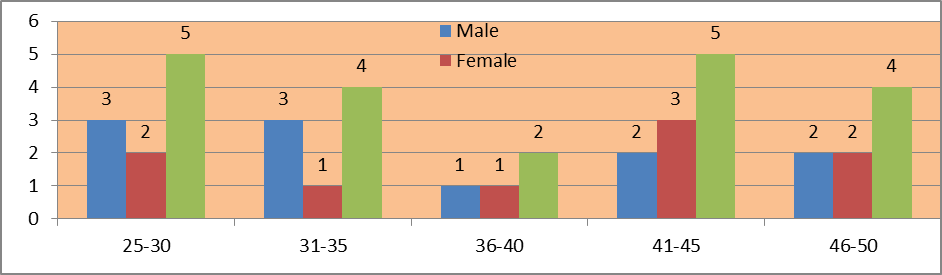
Table 2: Group 1 (n=10) consisted of patients who underwent immediate implant placement with autogenous bone grafts. Their condition was evaluated at three-month intervals. The Pearson Chi-Square test was used to assess the significance of the outcomes. This approach enhances our understanding of gender differences in root resorption among orthodontic patients
|
Criteria to evaluate bone changes |
N |
Mean |
Std. Dev. |
Std. Error |
95% CI |
Pearson Chi-Square Value |
df |
p value |
|
Bone remodelling &resorption |
2 |
1.08 |
1.06 |
1.05 |
1.04 |
1.03 |
1.05 |
2.0 |
|
Bone density & quality |
3 |
1.12 |
1.08 |
1.16 |
1.15 |
1.12 |
1.02 |
0.2* |
|
Marginal bone loss |
2 |
1.08 |
1.06 |
1.05 |
1.04 |
1.03 |
1.05 |
2.0 |
|
Peri-implant gap |
1 |
1.05 |
1.03 |
1.02 |
1.01 |
1.03 |
1.04 |
0.01* |
|
No change within the radiolucent area |
2 |
1.08 |
1.06 |
1.05 |
1.04 |
1.03 |
1.05 |
2.0 |
|
*p<0.05 significant |
||||||||
Table 3: Group 2 (n=10) consisted of patients who underwent immediate implant placement without autogenous bone grafts. Their condition was evaluated at three-month intervals. The Pearson Chi-Square test was used to assess the significance of the outcomes. This approach enhances our understanding of gender differences in root resorption among orthodontic patients
|
Criteria to evaluate bone changes |
N |
Mean |
Std. Dev. |
Std. Error |
95% CI |
Pearson Chi-Square Value |
df |
p value |
|
Bone remodelling &resorption |
0 |
- |
- |
- |
- |
- |
- |
- |
|
Bone density & quality |
0 |
- |
- |
- |
- |
- |
- |
- |
|
Marginal bone loss |
2 |
1.08 |
1.06 |
1.05 |
1.04 |
1.03 |
1.05 |
2.0 |
|
Peri-implant gap |
1 |
1.05 |
1.03 |
1.02 |
1.01 |
1.03 |
1.04 |
0.01* |
|
No change within the radiolucent area |
7 |
1.24 |
1.18 |
1.26 |
1.22 |
1.19 |
1.07 |
0.3* |
|
*p<0.05 significant |
||||||||
Table 4: Group 1 (n=10) consisted of patients who underwent immediate implant placement with autogenous bone grafts. Their condition was evaluated at six-month intervals. The Pearson Chi-Square test was used to assess the significance of the outcomes. This approach enhances our understanding of gender differences in root resorption among orthodontic patients
|
Criteria to evaluate bone changes |
N |
Mean |
Std. Dev. |
Std. Error |
95% CI |
Pearson Chi-Square Value |
df |
p value |
|
Bone remodelling &resorption |
1 |
1.05 |
1.03 |
1.02 |
1.01 |
1.03 |
1.04 |
0.01* |
|
Bone density & quality |
3 |
1.12 |
1.08 |
1.16 |
1.15 |
1.12 |
1.02 |
0.2* |
|
Marginal bone loss |
3 |
1.12 |
1.08 |
1.16 |
1.15 |
1.12 |
1.02 |
0.2* |
|
Peri-implant gap |
2 |
1.08 |
1.06 |
1.05 |
1.04 |
1.03 |
1.05 |
2.0 |
|
No change within the radiolucent area |
1 |
1.05 |
1.03 |
1.02 |
1.01 |
1.03 |
1.04 |
0.01* |
|
*p<0.05 significant |
||||||||
Table 5: Group 2 (n=10) consisted of patients who underwent immediate implant placement without autogenous bone grafts. Their condition was evaluated at six-month intervals. The Pearson Chi-Square test was used to assess the significance of the outcomes. This approach enhances our understanding of gender differences in root resorption among orthodontic patients
|
Criteria to evaluate bone changes |
N |
Mean |
Std. Dev. |
Std. Error |
95% CI |
Pearson Chi-Square Value |
df |
p value |
|
Bone remodelling &resorption |
1 |
1.05 |
1.03 |
1.02 |
1.01 |
1.03 |
1.04 |
0.01* |
|
Bone density & quality |
2 |
1.08 |
1.06 |
1.05 |
1.04 |
1.03 |
1.05 |
2.0 |
|
Marginal bone loss |
1 |
1.05 |
1.03 |
1.02 |
1.01 |
1.03 |
1.04 |
0.01* |
|
Peri-implant gap |
3 |
1.12 |
1.08 |
1.16 |
1.15 |
1.12 |
1.02 |
0.2* |
|
No change within the radiolucent area |
3 |
1.12 |
1.08 |
1.16 |
1.15 |
1.12 |
1.02 |
0.2* |
|
*p<0.05 significant |
||||||||
Table 6: Group 1 (n=10) consisted of patients who underwent immediate implant placement with autogenous bone grafts. Their condition was evaluated at nine-month intervals. The Pearson Chi-Square test was used to assess the significance of the outcomes. This approach enhances our understanding of gender differences in root resorption among orthodontic patients
|
Criteria to evaluate bone changes |
N |
Mean |
Std. Dev. |
Std. Error |
95% CI |
Pearson Chi-Square Value |
df |
p value |
|
Bone remodelling &resorption |
2 |
1.08 |
1.06 |
1.05 |
1.04 |
1.03 |
1.05 |
2.0 |
|
Bone density & quality |
3 |
1.12 |
1.08 |
1.16 |
1.15 |
1.12 |
1.02 |
0.2* |
|
Marginal bone loss |
3 |
1.12 |
1.08 |
1.16 |
1.15 |
1.12 |
1.02 |
0.2* |
|
Peri-implant gap |
2 |
1.08 |
1.06 |
1.05 |
1.04 |
1.03 |
1.05 |
2.0 |
|
No change within the radiolucent area |
0 |
- |
- |
- |
- |
- |
- |
- |
|
*p<0.05 significant |
||||||||
Table 7: Group 2 (n=10) consisted of patients who underwent immediate implant placement without autogenous bone grafts. Their condition was evaluated at nine-month intervals. The Pearson Chi-Square test was used to assess the significance of the outcomes. This approach enhances our understanding of gender differences in root resorption among orthodontic patients
|
Criteria to evaluate bone changes |
N |
Mean |
Std. Dev. |
Std. Error |
95% CI |
Pearson Chi-Square Value |
df |
p value |
|
Bone remodelling &resorption |
2 |
1.08 |
1.06 |
1.05 |
1.04 |
1.03 |
1.05 |
2.0 |
|
Bone density & quality |
2 |
1.08 |
1.06 |
1.05 |
1.04 |
1.03 |
1.05 |
2.0 |
|
Marginal bone loss |
1 |
1.05 |
1.03 |
1.02 |
1.01 |
1.03 |
1.04 |
0.01* |
|
Peri-implant gap |
3 |
1.12 |
1.08 |
1.16 |
1.15 |
1.12 |
1.02 |
0.2* |
|
No change within the radiolucent area |
2 |
1.08 |
1.06 |
1.05 |
1.04 |
1.03 |
1.05 |
2.0 |
|
*p<0.05 significant |
||||||||
Table 8: Estimation amongst all studied groups using one-way ANOVA
|
Variables |
Degree of Freedom |
Sum of Squares ∑ |
Mean Sum of Squares m∑ |
F |
Level of Sig. (p) |
|
Between Groups |
5 |
2.243 |
2.283 |
1.7 |
0.01* |
|
Within Groups |
17 |
2.194 |
2.456 |
_ |
|
|
Cumulative |
126.20 |
6.343 |
*p<0.05 significant |
||
DISCUSSION:
Iyer S et al. reviewed in their study that dental implants are a highly advanced solution for replacing missing teeth, offering numerous functional and aesthetic benefits that enhance a patient's quality of life. At its foundation, the dental implant consists of a durable titanium screw that is precisely inserted into the jawbone, mimicking the natural role of a tooth root. This innovative design provides exceptional stability and support for the artificial tooth or crown, forming a resilient foundation for long-term dental restoration. The titanium used in these implants is biocompatible, allowing it to integrate seamlessly with the living bone through a process known as osseointegration. This pivotal concepthas revolutionised dental practices worldwide, significantly improving the lives of countless individuals facing tooth loss by restoring their ability to eat, speak, and smile confidently.13,14 Wilson GM et al showed in their study that in the realm of modern dentistry, Cone Beam Computed Tomography (CBCT) has emerged as a crucial tool for treatment assessment and planning. This cutting-edge imaging technology generates intricate three-dimensional images of dental structures, enabling practitioners to visualize complex anatomical relationships with unparalleled accuracy. The depth of detail provided by CBCT not only facilitates the development of accurate and customized treatment plans but also enhances communication with patients regarding their dental health and potential treatment outcomes. Engaging patients in discussions about the importance of CBCT imaging empowers them with a comprehensive understanding of their unique dental conditions. This is particularly vital in cases where procedures like bone augmentation may be necessary. By fostering a proactive environment, dental professionals can secure informed consent and create a collaborative atmosphere where patients feel engaged and invested in their treatment journey.15,16 Qin R et al included in their study immediate implant placement is an innovative technique in the field of implant dentistry, allowing practitioners to insert an implant directly into the extraction site immediately following tooth removal. This approach offers multiple advantages, including the preservation of natural bone contours and the aesthetics of the gums, thereby maintaining a pleasing appearance during the healing period. In contrast, the delayed placement method necessitates the extraction site to heal sufficiently before the implant can be placed, which can result in a more stable and secure foundation for the new tooth. However, this method can prolong the overall treatment timeline and may require additional visits to the dental office, which is an important consideration for both patients and clinicians.17,18 Donker VJ et al reviewed in their study that to effectively tackle bony defects encountered during surgical procedures, bone grafts have emerged as the gold standard in regenerative dentistry. A variety of materials are available for use in bone grafting, each with distinct properties and applications. These materials include autografts, which are derived from the patient’s own body; allografts, sourced from human donors; xenografts, obtained from animal tissues; and alloplasts, which are synthetic grafting materials designed to mimic the properties of natural bone.19,20 Zuhr O et al showed in their study that among these options, autogenous bone grafts are particularly revered for their superior effectiveness. These grafts not only stimulate the growth of new bone but also integrate seamlessly with the existing bone tissue. This integration promotes a more natural healing response, which is crucial for achieving long-term stability and functionality in the treated area. The biological compatibility of autografts typically leads to a lower rate of rejection and complications compared to other graft types.21Tuncay KS et al reviewed in their study that however, the use of autografts is not without its challenges. One significant drawback is the potential for morbidity at the donor site, where additional surgical trauma can occur. This risk emphasizes the critical need for careful consideration in selecting the most appropriate grafting material, especially in complex procedures such as sinus lifts or maxillary augmentations, where precise anatomical management is essential. Ultimately, the success of these interventions is contingent upon a combination of the materials chosen and the surgical techniques employed. Surgeons must adopt a tailored and individualized approach in restorative dental medicine to ensure optimal outcomes for their patients. By combining the right grafting material with meticulous surgical planning and execution, practitioners can significantly enhance healing and functionality, thereby meeting the specific needs of each patient.22,23
CONCLUSION:
In the context of this study's limitations, the authors thoroughly investigated the changes in bone structure following the immediate placement of dental implants using autogenous bone grafts at various intervals. The findings revealed that autogenous bone grafting is essential for addressing significant bone defects, particularly those characterized by large bone loss. This approach effectively minimizes the risk of severe ridge collapse, providing superior preservation and density of the bone over time when compared to scenarios where the gap is left untreated.By opting for immediate grafting with the patient’s own bone following tooth extraction, we can significantly curb bone loss, uphold the aesthetics of the ridge, and enhance the long-term success rates of implants. This technique stands in stark contrast to delayed approaches or situations where no grafting is performed. The insights derived from this study pave the way for improved treatment protocols in challenging clinical scenarios, ultimately leading to more effective clinical practices and therapeutic interventions in the future.
BIBLIOGRAPHY:
1. Yeh YT, Chiou LL, Chen HH, Lin GH, Kao RT, Curtis DA. Influence of Dental Implant Diameters on Prosthesis Complications: A Systematic Review and Meta-analysis. Int J Oral Maxillofac Implants. 2025 Sep 26;40(5):529-546.
2. Rushinek H, Cohen A, Casap N, Alterman M. The Effect of Implant-Associated Factors on the Long-Term Outcomes of Dental Implants. Oral MaxillofacSurg Clin North Am. 2025 Feb;37(1):51-63.
3. Sukegawa S, Yoshii K, Hara T, Tanaka F, Taki Y, Inoue Y, Yamashita K, Nakai F, Nakai Y, Miyazaki R, Ishihama T, Miyake M. Optimizing dental implant identification using deep learning leveraging artificial data. Sci Rep. 2025 Jan 29;15(1):3724.
4. Häggman-Henrikson B, Ali D, Aljamal M, Chrcanovic BR. Bruxism and dental implants: A systematic review and meta-analysis. J Oral Rehabil. 2024 Jan;51(1):202-217.
5. Slagter KW, Meijer HJA, Hentenaar DFM, Vissink A, Raghoebar GM. Immediate single-tooth implant placement with simultaneous bone augmentation versus delayed implant placement after alveolar ridge preservation in bony defect sites in the esthetic region: A 5-year randomized controlled trial. J Periodontol. 2021 Dec;92(12):1738-1748.
6. Pauwels R, Araki K, Siewerdsen JH, Thongvigitmanee SS. Technical aspects of dental CBCT: state of the art. DentomaxillofacRadiol. 2015;44(1):20140224.
7. Yepes JF, Al-Sabbagh M. Use of cone-beam computed tomography in early detection of implant failure. Dent Clin North Am. 2015 Jan;59(1):41-56.
8. Patel R, Ucer C, Wright S, Khan RS. Differences in Dental Implant Survival between Immediate vs. Delayed Placement: A Systematic Review and Meta-Analysis. Dent J (Basel). 2023 Sep 15;11(9):218.
9. Riachi E, Juodzbalys G, Maciuliene D. Clinical Outcomes of Immediate, Early, and Delayed Implant Placement in the Esthetic Zone: A Systematic Review and Meta-analysis. Int J Oral Maxillofac Implants. 2024 Oct 16;39(5):157-173.
10. Sghaireen MG, Shrivastava D, Alnusayri MO, Alahmari AD, Aldajani AM, Srivastava KC, Alam MK. Bone Grafts in Dental Implant Management: A Narrative Review. Curr Pediatr Rev. 2022;19(1):15-20.
11. Baldwin P, Li DJ, Auston DA, Mir HS, Yoon RS, Koval KJ. Autograft, Allograft, and Bone Graft Substitutes: Clinical Evidence and Indications for Use in the Setting of Orthopaedic Trauma Surgery. J Orthop Trauma. 2019 Apr;33(4):203-213.
12. Miron RJ. Optimized bone grafting. Periodontol 2000. 2024 Feb;94(1):143-160.
13. Iyer S, Annamma LM, Hassaan Rasheed M, Ahmed N. Dental Implant Failures: Part 2. Dent Clin North Am. 2026 Jan;70(1):191-208.
14. Yan Y, Sharma P, Suvan J, Blanco C, Linares A, Leira Y, Blanco J, D'Aiuto F. Dental-implant inflamed surface area: A quantification and simulation study. J Periodontol. 2025 Sep;96(9):994-1003.
15. Wilson GM, McGregor JC, Gibson G, Jurasic MM, Evans CT, Suda KJ. Factors associated with dental implant loss/complications in the Veterans Health Administration, 2015-2019. J Public Health Dent. 2023 Sep;83(4):408-412.
16. Alkharrat AR, Schmitter M, Rues S, Rammelsberg P. Fracture behavior of all-ceramic, implant-supported, and tooth-implant-supported fixed dental prostheses. Clin Oral Investig. 2018 May;22(4):1663-1673.
17. Qin R, Chen Y, Han C, Wu D, Yu F, He D. Immediate Implant Placement With or Without Immediate Provisionalization in the Maxillary Esthetic Zone: A Systematic Review and Meta-analysis. Int J Oral Maxillofac Implants. 2023 May, Jun;38(3):422-434c.
18. Omami G, Al Yafi F. Should Cone Beam Computed Tomography Be Routinely Obtained in Implant Planning? Dent Clin North Am. 2019 Jul;63(3):363-379.
19. Donker VJ, Raghoebar GM, Slagter KW, Hentenaar DFM, Vissink A, Meijer HJA. Immediate implant placement with immediate or delayed provisionalization in the maxillary aesthetic zone: A 10-year randomized trial. J Clin Periodontol. 2024 Jun;51(6):722-732.
20. Pardal-Peláez B, Flores-Fraile J, Pardal-Refoyo JL, Montero J. Implant loss and crestal bone loss in immediate versus delayed load in edentulous mandibles: A systematic review and meta-analysis. J Prosthet Dent. 2021 Mar;125(3):437-444
21. Zuhr O, Bäumer D, Hürzeler M. The addition of soft tissue replacement grafts in plastic periodontal and implant surgery: critical elements in design and execution. J Clin Periodontol. 2014 Apr;41 Suppl 15:S123-42.
22. Tuncay KS, Günbey E, Şimşek G, Acar B, Karasen RM. Comparison of autogeneous and homogeneous nasal dorsal onlay grafts in an experimental design. Eur Arch Otorhinolaryngol. 2015 Mar;272(3):619-25.
23. Laiamnuay P, Chaiyasamut T, Zhang R, Seriwatanachai D, Kretapirom K. Comparable efficacy of autogenous tooth-derived grafts and xenografts in post-extraction osseous defects: a split-mouth randomized trial. Clin Oral Investig. 2025 Jul 23;29(8):388.