Evaluation Of Litchi Chinensis Extracts For Anticancer Effects Through Cellular And Molecular Mechanisms: A Sytematic Review
- Ruvanthika A.V.K , Undergraduate (Bachelor of Dental Surgery), SRM Dental College, Ramapuram, Bharathi Salai, Chennai, TN, India.
- Sindhu R , MDS, Senior Lecturer, Department of Public Health Dentistry, SRM Dental College, Ramapuram, Bharathi Salai, Chennai, TN, India
- Muthulakshmi S , Post Graduate, Department of Public Health Dentistry, SRM Dental College, Ramapuram, Bharathi Salai, Chennai, TN, India
- Prabu D , MDS, Ph.D, Professor and HOD, Department of Public Health Dentistry, SRM Dental College, Ramapuram, Bharathi Salai, Chennai, TN, India
- Raj mohan M , MDS, Reader, Department of Public Health Dentistry, SRM Dental College, Ramapuram, Bharathi Salai, Chennai, TN, India
- Dinesh Dhamodhar , MDS, Reader, Department of Public Health Dentistry, SRM Dental College, Ramapuram, Bharathi Salai, Chennai, TN, India
- Lubna fathima , MDS, Senior Lecturer, Department of Public Health Dentistry, SRM Dental College, Ramapuram, Bharathi Salai, Chennai, TN, India
Article Information:
Abstract:
Background: Litchi chinensisis a fruit rich in flavonoids and procyanidins that possesses strong potential.preclinical experiment demonstrate its extracts inhibits the proliferation of cancer cells,induce apotopis,modulate key pathways .This review presents facts supporting its uses as natural anticancer drug. Aim: To conduct a systematic analysis of preclinical evidence regarding the anticancer efficacy of litchi chinensis extracts from several plant components. Materials And Methods: In vitro and in vivo studies on litchi extracts from seeds, pericarp, mesocarp, and pulp were explored. The assessment was targeted towards anticancer parametes including cell proflieration, induction of apoptosis, cell cycle arrest, tumour inhibition,and immunomodulatory effects.Techniques to find in vitro and in vivo research on anticancer properties, a systematic review was conducted using Pubmed and Google scholar. apoptopsis ,cell profileration, and tumour suppression were among the outcomes of studies. key process and pathways were identified by summarizing data. Results: Litchi chinensis extracts showed strong anticancer activity against various cancer type.Total flavonoids of litchi and litchi derived procyanidins exhibited strong inhibition of tumour growth in animal models with low toxicity.The effects were mediated through the modulation of signaling pathways. LPC also modulated gut microbiota composition, indicating the participation of gut – immune axis in its anticancer effects. Conclusion: By inhibiting cell proflieration, inducing apoptosis, arresting the cell cycle, and modulating key molecular pathways, seed, pericarp, pulp extracts of litchi chinensis have potent anticancer activity. their lower toxicities and immunomodulatory and antimetastatic activities highlight their promise as safe,multitargeted therapeutics for development of novel cancer therapies.
Keywords:
Article :
INTRODUCTION:
Litchi chinensis of the sapindaceae family is a native Chinese tree species that is well known for its sweet,juicy fruits.Due to its comforting for warm climates, it is extensively cultivated worldwide, particularly in tropical and subtropical regions1.The discovery of new drugs and the expanding field of nutraceuticals still heavily rely on plants, especially those employed in traditional medicine. Herbal treatments such as homeopathy and Ayurveda have long been used to cure varios aliments and physiological problems.One well-known example is Litchi chinensis, which has a long history of traditional Indian medical applications. Epicatechin, procyanidins, leucocyanidin, cyanidin glycoside, malvidin glycoside, saponins, and other bioactive substances2 .
Litchi has the ability to prevent cancer. The anticancer potential of Litchi Fruit Pericarp (LFP) extract, which is abundant in polyphenolic chemicals and has shown impressive antioxidative activities, is being investigated in this study. Building on its potential,this study utilise both in vivo and in vitro models to assess the anticancer of activity of LFP extract on human cancer cases 3 .The pro-oxidant feature of polyphenols works against the cell’s metabolic process. Both apoptosis and inhibit cell proliferation may be involved. Research also revealed that numerous additional impacts of polyphenols had been noted, such as the decreases in enzymes like telomerase and lipoxygenase. The most prevalent flavonoid derivatives with simple structures To put it another way, the chemical structures of several phenolic chemicals that are frequently found in coffee and fruits4.Consuming foods and beverages high in polyphenols (such as catechins, flavones, and antocyanines) has been linked to a lower incidence of cancer, according to epidemiological and animal research. A number of molecular theories have been put out to explain this effect.The second most frequent cancers in women were stomach(4.9%),prostate (7.3%),colorectum (9.6%),breast (11.6%) Also , the main cause of cancer death was lung cancer5.The most common cancers in males and women,respectively, were lung and breast cancer.Herbal extracts can reduce clinical symptoms, increase a patient’s chances of survival, and enhance their quality of life when used in conjunction with traditional anticancer treatments6.
Almost 2.5 million new lung cancer cases,one in eight of all cancer globally(12.4% of total cancer),occurred in 2022 7.With an anticipated 1.8 million deaths (18.7%), lung cancer was also the most common cause of cancer-related mortality. Colorectal (9.3%), liver (7.8%), female breast (6.9%), and stomach (6.8%) cancers were next in line. The most common cancers in males and women, respectively, were lung and breast cancer8.Autophagy determines the tumour host interaction by facilitating stress adaptation qand suppression of activation of innate and adaptive imuune responses. Furthermore, especially in a microenvironment with limited nutrients, autophagy might encourage a cross-talk between the tumor and the stroma, which can stimulate tumor growth9.Both in vitro and in vivo, the n-butyl alcohol extract of Litchi seeds showed anti-prostate cancer activities, and the most prevalent chemical components were flavonoid compounds10.Colorectal cancer (CRC) has now emerged as the leading cancer type in Taiwan as the diet of the Taiwanese people has been westernized in recent decades Though there is a prominent present and past emphasis .In Taiwan on traditional Chinese medicine, little effort Has gone into studying the efficacy of natural plants or Herbs as new bioactive compounds against colorectal Cancer11.litchi has the capability of enhancing metabolic health, for it is found to have numerous helpful substances with has antioxidant, anti-inflammatory and tumor-preventive properties It was already demonstrated in alcohol-fed mice that the phenolic extract of litchi pulp might protect against Liver damage by alleviating gut microbiota dysbiosis and intestinal barrier dysfunction12.
Procyanidins are considered to be prebiotic that is susceptible to degradation by gut microbiota and metabolized into different phenolic procyanidins function as an immunomodulator through control of gut microbiota in the treatment of colon cancer13.Anthocyanins from pericarp of Litchi have been reported to suppress linoleic acid oxidation very strongly and demonstrate dose-dependent free radical scavenging activity toward DPPH radical Epicatechin proanthocyanidin B2, proanthocyanidin B4, and the ethyl acetate fraction of Litchi pericarp were was discovered to have much stronger stimulatory activities on splenocyte proliferation compared to that of rutin,while exhibited lower cytotoxicities to human breast cancer cell MCF-7 and human embryolic lung fibroblasts than paclitaxel14.
In the past ten years, the scientists were interested in litchi seed and its active constituents for anti-tumor activity Chen and colleagues administered litchi seed water extract or granules to mouse xenograft of mouse Ehrlich ascites tumor cells, sarcoma S180 tumor cells and liver tumor cells and discovered the reduced tumours15.We conclude that Numerous pharmacological studies have shown that extracts and components from various parts of the Litchi plant (including seeds, peels, pulps, and flowers) display anticancer properties by influencing processes such as cell proliferation, apoptosis, autophagy, metastasis, sensitivity to chemotherapy and radiotherapy, stemness, metabolism, angiogenesis, and immune response through multiple targeting mechanisms16.This systematic review aims to unveil the preclinical evidence on anticancer potential of litchi chinensis extract derived from different plant parts focusing on their effects on cell proflieration, apoptosis induction, cell cycle regulation, tumour inhibition,and immunomodulatory mechanisms.
MATERIALS AND METHODS:
This systematic review was carried out to explore the anticancer potential of Litchi chinensis extracts obtained from the seed, pericarp, pulp, and mesocarp. The review focused on preclinical in vitro and in vivo studies that examined how these extracts influence cell proliferation, apoptosis, tumor suppression, and molecular signaling pathways. The review process followed the PRISMA (Preferred Reporting Items for Systematic Reviews and Meta-Analyses, 2020) guidelines to ensure accuracy and transparency in study selection and reporting.
Eligibility Criteria
Inclusion Criteria
· Original in vitro or in vivo studies published between 2000 and 2024.
· Full-text, peer-reviewed articles available in English.
· Studies assessing Litchi chinensis extracts (seed, pericarp, or pulp) for anticancer effects such as apoptosis, cell-cycle arrest, or tumor growth inhibition.
Exclusion Criteria
· Reviews, case reports, or meta-analyses.
· Human clinical studies or research without experimental data.
· Studies lacking anticancer outcomes or using herbal mixtures not specific to Litchi chinensis.
Search Strategy
Relevant literature was gathered from PubMed, Google Scholar, ScienceDirect, and ResearchGate using keywords such as:
“Litchi chinensis” AND (“anticancer” OR “antitumor” OR “flavonoids” OR “procyanidins” OR “apoptosis” OR “tumor suppression”).
Reference lists from selected studies were also reviewed to identify any additional research articles.
Study Selection
Initially, 300 studies were retrieved through database searches. After removing duplicates, 179 articles were screened based on titles and abstracts. Of these, 34 full-text studies were reviewed in detail, and finally, 5 studies met all the inclusion criteria and were included in this review.
RESULTS:
This review identified five preclinical studies demonstrating that Litchi chinensis extracts exhibit strong anticancer activity. Compounds such as Total Flavonoids of Litchi Seed (TFLS) and Litchi Procyanidins (LPC) effectively reduced tumor cell growth, induced apoptosis, and regulated signaling pathways like AKT/mTOR, NF-κB, and MAPK. These extracts also showed low toxicity in animal models, suggesting their potential as safe, natural anticancer agents.
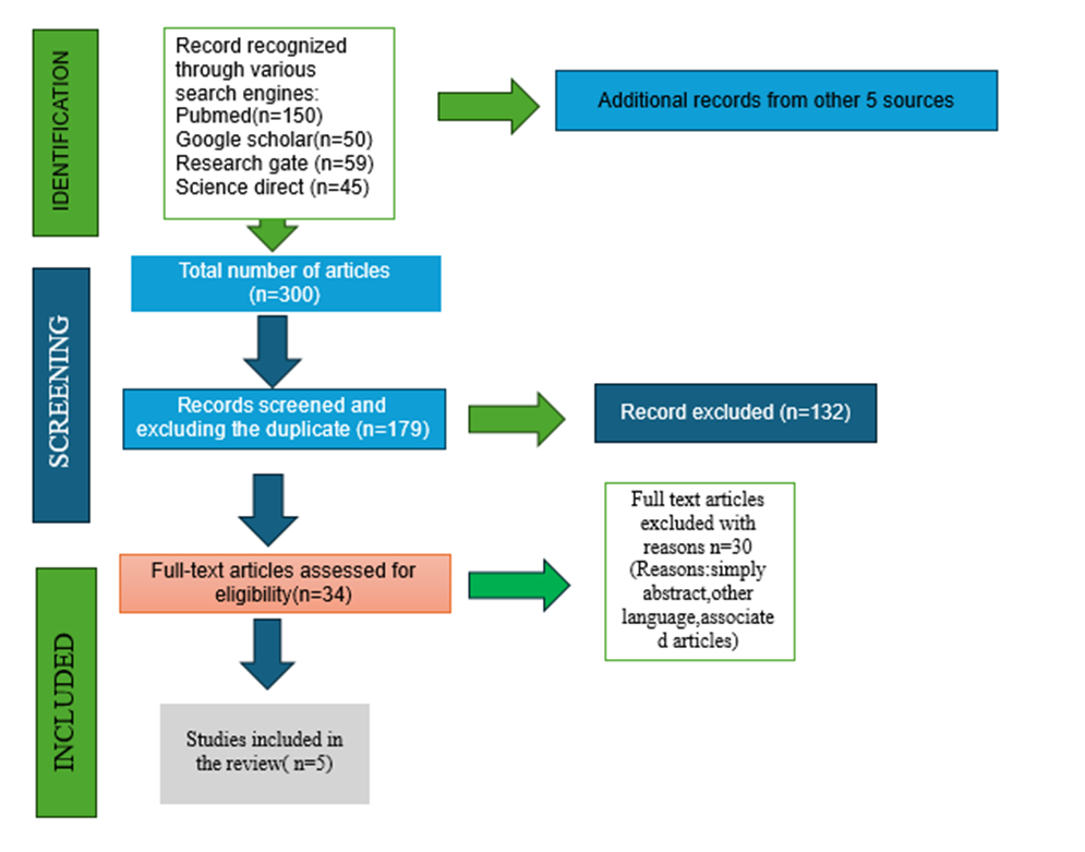
Figure 1 illustrates the PRISMA flow diagram, showing the number of studies identified, screened, excluded, and included in the final analysis and shows how studies were carefully selected for this review.out of 300 records identified from multiple data bases, 179 were screened.Following eligibility assessment , 5 studies were finally included in systematic review17.
TABLE 1: OVERVIEW OF INCLUDED PRECLINICAL STUDIES EVALUATING ANTICANCER EFFECTS OF LITCHI CHINENSIS EXTRACT
|
S.NO |
AUTHOR NAME /YEAR |
STUDY DESIGN |
CELL PROLIFERATION |
FLOW CYTOMETRY/CELL CYCLE ANALYSIS /APOTOPSIS |
WESTERN BLOTTING ANALYSIS/ IMUUNOBLOTTING |
|
1 |
Ming chang et. al6 2021 |
1.laboratory study 2.animal study |
CELL PROLIFERATION 1. PC3 and DU145 cells were treated with TFLS, and cell viability was assessed using MTS assay, with IC50 values calculated from absorbance readings. 2.NA (i)cell line 1. 32 male nude mice were injected with PC3-luc cells and treated with TFLS (40/80mg/kg) daily, with body weight and tumor size monitored. (ii)acute toxicity 2. TFLS acute toxicity was evaluated in Kunming mice at 18g/kg, assessing behavior, body weight, and organ weight.
|
FLOW CYTOMETRY PC3 and DU145 cells were subjected to TFLS treatment and subsequently examined for apoptosis and cell cycle distribution using flow cytometry.
|
WESTERNBLOTTING Protein detection was achieved using Western blot analysis with SDS-PAGE and ECL chemiluminescence
|
|
Yuan yao et.al18 2022 |
laboratory study |
CELL PROFILERATION The cell viability of NCM460, CT26, and HCT116 cells treated with LPC was examined using the MTT test, which measures formazan production at 570 nm.
|
FLOW CYTOMETRY Flow cytometry was used to examine immune cell markers CD3, CD4, and CD8 in spleens and tumors, with an Accuri C6 cytometer and FlowJo software.
|
WESTERN BLOT ANALYSIS Western blot analysis was performed on lung tissue proteins to detect and quantify expression of β-tubulin, E-cadherin, vimentin, and PCNA using the Odyssey system.
|
|
|
3 |
Sonia Emanuele et. al8 2018 |
laboratory study |
NA |
FLOW CYTOMETRY PI staining and flow cytometry assessed cell death through PI uptake and cell cycle distribution via DNA fragmentation analysis. |
WESTERN BLOT ANALYSIS Western blotting analyzed protein expression, with γ-tubulin as a loading control, and band intensity quantified and normalized accordingly. |
|
4 |
Yuan-chiang chung et.al19 2017 |
laboratory study
|
CELL PROFILERATION Cells were treated with LCSE (0-150 μg/mL) for 24 hours, and cell counts were performed using a hemocytometer with trypan blue staining. |
CELL CYCLE ANALYSIS APOPTOSIS |
IMMUNOBLOTING Western blotting was performed by separating proteins via SDS-PAGE, transferring them to PVDF membranes, and detecting them using primary and secondary antibodies with enhanced chemiluminescence. |
|
5 |
Chih-ping hsu et. al11 2012 |
Laboratory study |
CELL PROFILERATION Cell proliferation was evaluated in SW480 and Colo320DM cells treated with LCSP (0-150 μg/mL) for 24 hours, with cell counts determined using a hemocytometer and trypan blue staining. |
CELL CYCLE ANALYSIS APOPTOSIS
|
IMMUNOBLOTTING Western blotting was performed on CRC cell lysates, with proteins separated by SDS-PAGE, transferred to PVDF membranes, and detected using primary and secondary antibodies with enhanced chemiluminescence. |
Table 1 shows the key characteristics of the preclinical studies included and presents detailed information on extract used(seed, pericarp, pulp, or mesocarp).table also outlines the specific assays and analytical methods employed to assess anticancer parameters such as cell proliferation, cell cycle arrest, apoptopsis and immunoblotting techniques like western blot, flow cytometry used to evaluate the pathway and protein expression. These data provide a detailed review of technique and experimental frameworks that support anticancer characteristics.
TABLE 2: CHARACTERISTIC PRIMARY OUTCOME AND RESULTS OF STUDIES INCLUDED IN THIS REVIEW
|
S.NO |
AUTHOR NAME / YEAR |
FINDINGS |
EFFECT MEASURE |
CONCLUSION |
|
Ming chang et .al6 2021 |
CELL PROFILERATION TFLS effectively suppressed prostate cancer cell growth, reducing viability and colony formation in both PC3 and DU145 cells in a dose- and time-dependent manner. FLOW CYTOMETRY Flow cytometry analysis revealed TFLS induces apoptosis in prostate cancer cells (PC3 and DU145), with increased apoptotic rates (31.10% and 26.23%, respectively), supported by TUNEL assay confirmation. WESTERNBLOT ANALYSIS TFLS treatment altered apoptosis-related protein expression, upregulating cleaved PARP and Bax while downregulating Bcl-2 in PC3 and DU145 cells, indicating apoptosis induction. ANIMAL STUDY CELL LINE TFLS inhibited tumor growth in a PC3-luc xenograft model, reducing tumor weight and volume without affecting body weight. ACUTE TOXICITY TFLS exhibited anti-cancer properties by targeting the AKT pathway, inhibiting key phosphorylation events without showing in vivo toxicity. |
CELL PROLIFERATION TFLS demonstrated potent anti-cancer activity, suppressing prostate cancer cell growth and viability in a dose- and time-dependent manner, with significant inhibition of colony formation.
FLOW CYTOMETRY TFLS proved to be an effective measure in inducing apoptosis in prostate cancer cells (PC3 and DU145), as evidenced by increased apoptotic rates (31.10% and 26.23%) in flow cytometry analysis, further confirmed by TUNEL assay.
WESTERNBLOT ANALYSIS TFLS induced apoptosis in PC3 and DU145 cells by upregulating cleaved PARP and Bax and downregulating Bcl-2.
ANIMAL STUDY CELL LINE TFLS showed strong antitumour activity, reducing PC3 xenograft tumour growth by 20.87% and 44.6% at 40 and 80 mg/kg, respectively. ACUTE TOXICITY TFLS significantly inhibited prostate cancer cell growth and tumor progression with low toxicity, suggesting its potential as an effective antitumor agent.
|
CELL PROLIFERATION TFLS inhibited PC3 and DU145 cell growth in a dose- and time-dependent manner.
FLOW CYTOMETRY TFLS showed strong anti-cancer effects by inducing apoptosis in PC3 and DU145 cells, as confirmed by flow cytometry and TUNEL assays, highlighting its therapeutic potential.
WESTERN BLOT ANALYSIS TFLS induced apoptosis in prostate cancer cells by upregulating cleaved PARP and Bax while downregulating Bcl-2, without altering cell cycle progression. ANIMAL STUDY CELL LINE TFLS shows strong anticancer effects, reducing tumor size while maintaining body weight. ACUTE TOXICITY TFLS was safe and well tolerated, with a high LD50 (>18 g/kg) and no toxicity, supporting its potential for safe use.
|
|
|
2 |
2022 |
CELL PROLIFERATION FLOW CYTOMETRY WESTERN BLOT ANALYSIS |
CELL PROLIFERATION FLOW CYTOMETRY WESTERN BLOT ANALYSIS |
CELL PROLIFERATION FLOW CYTOMETRY WESTERN BLOT ANALYSIS |
|
3 |
2018 |
FLOW CYTOMETRY WESTERN BLOTTING ANALYSIS |
FLOW CYTOMETRY WESTERN BLOTTING ANALYSIS |
FLOW CYTOMETRY WESTERN BLOTTING ANALYSIS |
|
4 |
2017 |
CELL PROLIFERATION CELL CYCLE ANALYSIS APOPTOSIS IMMUNOBLOTTING |
CELL PROLIFERATION CELL CYCLE ANALYSIS APOPTOSIS IMMUNOBLOTTING |
CELL PROLIFERATION CELL CYCLE ANALYSIS APOPTOSIS IMMUNOBLOTTING |
|
5 |
Chih-ping hsu et.al11 2012 |
CELL PROLIFERATION CELL CYCLE ANALYSIS APOPTOSIS IMMUNOBLOTTING
|
CELL PROLIFERATION CELL CYCLE ANALYSIS APOPTOSIS IMMUNOBLOTTING |
CELL PROLIFERATION CELL CYCLE ANALYSIS APOPTOSIS IMMUNOBLOTTING |
Table 2 explains the key results from the studies included in the review.it outlines the how different extracts of litchi chinensis such as seed,pulp,pericarp,mesocarp, were found to slow down the cancer cell growth ,trigger the cell death, and block the cell cycle and also summarizes the changes in important molecular markers and signaling pathways and findings from the animal studies included showing reduced tumor growth and minimal toxicity.extracts shows strong anticancer activity through several biological mechanisms.
TABLE 3: RISK OF BIAS ASSESSMENT
|
Author name |
RANDOMISATION |
ALLOCATION CONCEALMENT |
COMPARSION GROUP |
CONFOUNDING |
EXPERIMENTAL CONDITIONS |
BLINDING |
COMPLETE OUT COME DATA |
EXPOSURE CHARACTERISTIC |
OUTCOME ASSESSMENT |
OUTCOME REPORTING |
NO OTHER THREATS |
|
chang M et al., (TFLS,prostate cancer and xenograft ) |
|
|
|
|
|
|
|
|
|
|
|
|
Yao Y et al.,2023(LPC, Colon cancer,gut microbiota) |
|
|
|
|
|
|
|
|
|
|
|
|
Emanuele S et al.,2018(sicilian litchi,HT-29 cells |
|
|
|
|
|
|
|
|
|
|
|
|
Chung Y -C et al.,2017(LCSE, NSCLC A549&H661)
|
|
|
|
|
|
|
|
|
|
|
|
|
Hsu c-p et al.,2012 (LCSP,colorectal SW480\colo320DM) |
|
|
|
|
|
|
|
|
|
|
|
Table 3 shows the bias analysis of all included studies.it is categorized as probably high risk, probably low risk, definitely high risk, definitely low risk20,21
Yellow colour indicates probably high rish,where bias was likely but results were not clearly reported.
orange colour indicates definitely high risk of bias analysis due to major methodological flaws such as lack of bing and randomisation.
Green colour indicates definitely low risk of bias reflecting strong methodology and transparent reporting.
Blue colour signifies definitely low risk shows minor limitations with minimal impact on validity.
DISCUSSION:
This systematic review explains the strong evidence for anticancer activities of litchi chinensis extracts prepared from seeds, pericarp,pulp,and mesocarp of the plants is summarized .Across multiple preclinical studies, litchi-derived compounds demonstrated broad-spectrum anticancer activity against various type of cancers, such as lung,breast,colon,and prostate cancers. litchi chinensis has been traditionally used in china,india,Vietnam,Indonesia for medicine, as research shown from phytochemical studies, the fruit is reported to be in phenolics,triterpenes,sterols,flavonoids,and other bioactive compounds .Notably, Extracts and isolated compounds of litchi have been found to posses a number of health benefits ,such as antioxidant, anticancer ,antiviral, antimicrobial,anti-obesity activites22. Epicatechin, proanthocyanidin B2, and B4 were isolated and refined by researchers from the litchi pericarp.
These compounds enhanced mouse immune cells and exerted significant immunomodulatory activity. Besides, they possess anti-breast cancer properties and were less toxic than the widely used chemotherapy drug paclitaxel. The study further highlights the potential medicinal benefits of litchi-derived chemicals.23.The pericarp and seeds of litchi and longan fruits account for approximately 30% of the dry weight of the fruit and are rich in bioactive compounds. The extracts have anti-inflammatory, anti-cancer, and antioxidant properties, among other health benefits. Valorization of these by-products for their pharmacological value might contribute significantly to improving human health with increasing fruit production, and this aspect deserves further studies.24. Extracts and compounds from various parts of the Litchi fruit (seeds, peels, pulp, and flowers) have which have shown anti-cancer properties by inhibiting cancer cell proliferation, inducing cell death, and interfering with processes related to cancerdemonstrated anti-cancer properties, including inhibiting cancer cell growth, inducing cell death, and impacting cancer-related processes25.Litchi fruit pericarp (LFP) extract, rich in polyphenolic compounds, shows potent antioxidant activity. This study investigates LFP extract's anti-cancer effects on human breast cancer cells in vitro and in vivo, examining its impact on cell viability, colony formation, DNA synthesis, and gene expression to understand its mechanism of action26.
This study showed that extracts from Sicilian litchi fruit have anti-tumor effects on colon cancer cells (HT29) by decreasing cell viability and proliferation, causing cell cycle arrest and caspase-dependent cell death, and initiating autophagy. The extracts also altered important proteins linked to autophagy including ATG1/ULK1, beclin-1, LC3-II, and p62, highlighting the potential therapeutic applications of litchi extracts in cancer treatment27.Litchi seed extract (LCSP), rich in polyphenols, demonstrated anti-cancer effects on colorectal cancer cells by inducing apoptotic cell death, arresting the cell cycle in the G2/M phase, and regulating key proteins such as cyclins, Bax/Bcl-2 ratio, and caspase 3, suggesting its potential as a chemopreventive agent for colorectal cancer28. Litchi chinensis exhibits various bioactive properties, including anti-cancer effects in its fruit and seeds, antihyperglycemic, antihyperlipidemic, antiplatelet, and antiviral activities in its seeds, and a high flavonoid content in its fruit that may be effective against breast cancer, highlighting its potential health benefits29. Procyanidin C1, a polyphenol derived from seed extract, was discovered to have anti-tumor actions in colorectal cancer (CRC) cells and to inhibit miR-501-3p. Previous research demonstrated PCC1's potential anti-tumor effects in various cancers, including breast, melanoma, liver, and lung cancer, by inducing apoptosis, inhibiting cell growth, and modulating key signaling pathways.30.
TFLS and LPC show potent anticancer activity via distinct mechanisms. By blocking the AKT pathway and controlling Bcl-2, Bax, and PARP, TFLS causes dose- and time-dependent growth inhibition in prostate cancer. LPC from Litchi chinensis inhibits colon cancer proliferation and metastasis by modulating EMT markers (E-cadherin, vimentin, PCNA) and gut microbiota–mediated immune responses. Flow cytometry and western blot confirm TFLS-induced apoptosis and LPC’s suppression of metastatic signaling. Both reduced tumor growth in vivo with minimal toxicity. Combining TFLS (apoptosis inducer) and LPC (immune, anti-metastatic modulator) may yield synergistic, multi-targeted cancer therapy6,18 Litchi extracts show strong anticancer effects via distinct mechanisms. LCSE inhibited NSCLC cell proliferation with low toxicity, induced G1/G2-M cell cycle arrest, and triggered mitochondrial apoptosis through caspase and Bcl-2 regulation. It also downregulated EGFR, Akt, and Erk-1/2 pathways. In HT29 colon cancer cells, 150 µg/mL litchi extract altered cell cycle distribution and reduced PCNA expression, confirming suppressed proliferation. Overall, LCSE showed multi-targeted effects involving apoptosis and signaling inhibition, while the HT29 extract mainly affected proliferation and cell cycle control, highlighting the therapeutic potential of litchi-derived compounds8,19.Litchi exhibits promising anticancer effects through multiple mechanisms, yet more standardized and clinically focused studies are required to validate its efficacy and safety for therapeutic use.
Ming Chang et.al,20216 Demonstrated that Litchi chinensis (TFLS) flavonoid extracts inhibited prostate cancer cell growth in PC3 and DU145 lines efficiently and in a dose-dependent manner. TUNEL and flow cytometry assays confirmed TFLS-induced apoptosis, which was associated with decreased Bcl-2 expression and enhanced expression of cleaved PARP and Bax. High anticancer activity and low toxicity were suggested by in vivo experiments showing reduced growth of tumors with no alteration of body weight. TFLS was shown to mainly suppress the AKT signaling pathway in producing anticancer effects, which would make it a safe option for prostate cancer treatment.Yuan Yao et.al, 202218Found that LPC isolated from Litchi chinensis seeds inhibited the growth of colon cancer cells and tumour dissemination but with minor risk. Since disturbance suppressed immune response and activity, its anticancer effect relied on gut microbiota. LPC upregulated E-cadherin and decreased PCNA and vimentin levels, indicating enhanced suppression of tumours and metastasis.
Sonia Emanuele et.al,20188 Demonstrated that Litchi chinensis extracts possessed strong anti-proliferative effects against colon cancer cells (HT29). The extracts induced DNA fragmentation and altered the cell cycle distribution, which demonstrated cell death and cell growth arrest, as determined by flow cytometry. Further, a significant reduction in PCNA expression was established by Western blot analysis verifying the ability of the extracts to inhibit cell proliferation and increase anticancer activity.Yuan-Chiang Chung et.al,201719Found that Litchi chinensis seed extract (LCSE) caused minimal toxicity to normal lung cells but effectively inhibited the proliferation and colony formation of non-small cell lung cancer (NSCLC) cells. LCSE was shown, through cell cycle studies, to change significant regulatory proteins and induce G1 arrest in A549 cells and G2/M arrest in NCI-H661 cells. Apoptosis research confirmed that LCSE triggered caspase and induced changes in Bcl-2 family proteins to trigger mitochondrial-dependent apoptosis. Cyclins D1, A, and B1 were found to be downregulated in immunoblotting data, which correlated with cell cycle arrest and reduced proliferation. The findings show the huge potential of LCSE as a multi-targeted, selective anticancer agent in the treatment of lung cancer.
Chih-Ping Hsu et.al,201211 Demonstrated that Litchi chinensis seed polyphenol (LCSP), which is rich in flavonoids, tannins, and polyphenols, inhibited the growth of colorectal cancer (CRC) cells efficiently and dose-dependently, with SW480 cells being the most sensitive. LCSP induced G2/M phase arrest by downregulating cyclins D1, A, and B1, based on cell cycle research. Increased annexin V-positive cells, cleaved PARP, and increased Bax:Bcl-2 ratio were markers of mitochondrial-mediated cell death, which were confirmed by apoptosis studies. The results were confirmed by immunoblotting, which indicated reduced levels of cell cycle proteins that are associated with G2/M arrest. All of these findings indicate that LCSP suppresses the proliferation of CRC cells by targeting the apoptotic and cell cycle progression pathways.
Noor JJ et al., 202531 demonstrated that natural phytochemicals such as gingerol suppress cancer-promoting signaling pathways while enhancing tumor-suppressor activity. In contrast, contaminants identified in traditionally dried fish were shown to activate molecular pathways associated with chronic inflammation, DNA damage, and carcinogenesis. These findings highlight how dietary components can distinctly modulate oncogenic versus protective cellular mechanisms. In a similar manner, polyphenol-rich extracts from Litchi chinensis including flavonoids, procyanidins, and anthocyanins exert anticancer effects by inhibiting proliferative signaling pathways (AKT, EGFR, MAPK), inducing apoptosis, and regulating immune and autophagy-related responses. Unlike carcinogenic dietary exposures that shift cellular signaling toward tumor promotion, litchi-derived bioactives promote a molecular environment favoring growth suppression and programmed cell death. This comparison underscores the broader concept that dietary choices can influence cancer risk through pathway-specific mechanisms and reinforces the potential role of Litchi chinensis as a protective, nutraceutical candidate in cancer prevention and adjunct therapy.
The risk of bias analysis revealed some methological variations among studies.most of the included studies showed probably low to moderate risk of bias,primarly due to incomplete reporting of randomization procedures ,allocation concealment,and blinding methods.A few studies were definitely high risk where experimental details such as blinding of outcome assessors or random sample allocation ere not clearly described.these limitations could potentially influence the outcome measurements or introduce selection bias.several studies demonstrated definitely low risk of bias shows strong methodological design,reproducible results,and transparent reporting of outcome.This review highlights that litchi chinensis fruit, already well known for its therapeutic properties,has immense potential to act against cancer.its pulp,pericarp,and seeds have all been reported to posses extracts with anti tumour activity,reducing the profileration of cancer cells and inducing apoptosis.In particular,its high phytochemical composition,mainly flavonoids,procyanidins, and polyphenol,is related to these actions.Extract have been found to posses potent anticancer efficiacy in animal and in vitro models,coupled with low toxicity, raising hopes may be safer and natural. Despite encouraging results,little is known about its safety,metabolism,and bioavailability in humans,with most research being confined to preclinical models.future development should include well planned clinical trials,improved delivery systems,including nanoparticles formulations, and studies on combination of litchi chemicals with various chemotherapeutic drugs that are currently available.these may pave the way for developing sustainable, affordable,and effective therapeutic options for prevention and treatment of cancer.
CONCLUSION:
Extracts of Litchi chinensis from the seed, pericarp, and pulp demonstrate notable anticancer activity by suppressing cell proliferation, promoting apoptosis, inducing cell cycle arrest, and regulating pathways such as AKT/mTOR, NF-κB, and MAPK. They also show immunomodulatory and anti-metastatic effects with low toxicity in preclinical studies. These results highlight litchi-derived compounds as safe, multi-targeted agents with strong potential for cancer therapy.The bioactive compounds particularly flavonoids,procyanidins,and poly phenol are responsible for these effects and exhibit low toxicity in animal models,supporting their potential as safe.The current evidence is largely limited to invitro and animal studies.further research involving human trials,pharmacokinetic profiling ,long term safety evaluation is essential.overall,litchi chinensis represents a potent source of natural potent source of natural compounds that contribute to the development of innovative,effective and less toxic cancer therapies in the future.
CONFLICTS OF INTEREST:There are no conflicts of interest.
BIBLIOGRAPHY:
1. Kilari EK, Putta S. Biological and Phytopharmacological Descriptions of Litchi Chinensis. Pharmacogn Rev. 2016 Jan-Jun;10(19):60-5. doi: 10.4103/0973-7847.176548. PMID: 27041875; PMCID: PMC4791990.
2. Gontier E, Boussouel N, Terrasse C, Jannoyer M, Ménard M, Thomasset B, Bourgaud F. Litchi chinensis fatty acid diversity: occurrence of the unusual cyclopropanoic fatty acids. Biochem Soc Trans. 2000 Dec;28(6):578-80. PMID: 11171131.
3. Wang X, Yuan S, Wang J, Lin P, Liu G, Lu Y, Zhang J, Wang W, Wei Y. Anticancer activity of litchi fruit pericarp extract against human breast cancer in vitro and in vivo. Toxicol Appl Pharmacol. 2006 Sep 1;215(2):168-78. doi: 10.1016/j.taap.2006.02.004. Epub 2006 Mar 23. PMID: 16563451.
4. Abbas M, Saeed F, Anjum FM, Afzaal M, Tufail T, Bashir MS, Ishtiaq A, Hussain S, Suleria HAR. Natural polyphenols: An overview. International Journal of Food Properties [Internet]. 2016 Oct 3;20(8):1689–1699. Available from: https://doi.org/10.1080/10942912.2016.1220393
5. Naasani I, Oh-Hashi F, Oh-Hara T, Feng WY, Johnston J, Chan K, Tsuruo T. Blocking telomerase by dietary polyphenols is a major mechanism for limiting the growth of human cancer cells in vitro and in vivo. Cancer Res. 2003 Feb 15;63(4):824-30. PMID: 12591733.
6. Chang M, Zhu D, Chen Y, Zhang W, Liu X, Li XL, Cheng Z, Su Z, Zhang J, Lu Y, Guo H. Total Flavonoids of Litchi Seed Attenuate Prostate Cancer Progression Via Inhibiting AKT/mTOR and NF-kB Signaling Pathways. Front Pharmacol. 2021 Sep 23;12:758219. doi: 10.3389/fphar.2021.758219. PMID: 34630125; PMCID: PMC8495171.
7. Bray F, Laversanne M, Sung H, Ferlay J, Siegel RL, Soerjomataram I, Jemal A. Global cancer statistics 2022: GLOBOCAN estimates of incidence and mortality worldwide for 36 cancers in 185 countries. CA Cancer J Clin. 2024 May-Jun;74(3):229-263. doi: 10.3322/caac.21834. Epub 2024 Apr 4. PMID: 38572751.
8. Emanuele S, Notaro A, Palumbo Piccionello A, Maggio A, Lauricella M, D'Anneo A, Cernigliaro C, Calvaruso G, Giuliano M. Sicilian Litchi Fruit Extracts Induce Autophagy versus Apoptosis Switch in Human Colon Cancer Cells. Nutrients. 2018 Oct 12;10(10):1490. doi: 10.3390/nu10101490. PMID: 30322062; PMCID: PMC6213492.
9. Chang M, Zhu D, Chen Y, Zhang W, Liu X, Li XL, Cheng Z, Su Z, Zhang J, Lu Y, Guo H. Total Flavonoids of Litchi Seed Attenuate Prostate Cancer Progression Via Inhibiting AKT/mTOR and NF-kB Signaling Pathways. Front Pharmacol. 2021 Sep 23;12:758219. doi: 10.3389/fphar.2021.758219. PMID: 34630125; PMCID: PMC8495171.
10. Chang M, Zhu D, Chen Y, Zhang W, Liu X, Li XL, Cheng Z, Su Z, Zhang J, Lu Y, Guo H. Total Flavonoids of Litchi Seed Attenuate Prostate Cancer Progression Via Inhibiting AKT/mTOR and NF-kB Signaling Pathways. Front Pharmacol. 2021 Sep 23;12:758219. doi: 10.3389/fphar.2021.758219. PMID: 34630125; PMCID: PMC8495171.
11. Hsu CP, Lin CC, Huang CC, Lin YH, Chou JC, Tsia YT, Su JR, Chung YC. Induction of apoptosis and cell cycle arrest in human colorectal carcinoma by Litchi seed extract. J Biomed Biotechnol. 2012;2012:341479. doi: 10.1155/2012/341479. Epub 2012 Oct 3. PMID: 23093841; PMCID: PMC3470890.
12. Jinato T, Chayanupatkul M, Dissayabutra T, Chutaputti A, Tangkijvanich P, Chuaypen N. Litchi-Derived Polyphenol Alleviates Liver Steatosis and Gut Dysbiosis in Patients with Non-Alcoholic Fatty Liver Disease: A Randomized Double-Blinded, Placebo-Controlled Study. Nutrients. 2022 Jul 16;14(14):2921. doi: 10.3390/nu14142921. PMID: 35889878; PMCID: PMC9319370.
13. Yao Y, Feng S, Li X, Liu T, Ye S, Ma L, Man S. Litchi procyanidins inhibit colon cancer proliferation and metastasis by triggering gut-lung axis immunotherapy. Cell Death Dis. 2023 Feb 11;14(2):109. doi: 10.1038/s41419-022-05482-5. PMID: 36774343; PMCID: PMC9922286.
14. Gong Y, Fang F, Zhang X, Liu B, Luo H, Li Z, Zhang X, Zhang Z, Pang X. B Type and Complex A/B Type Epicatechin Trimers Isolated from Litchi pericarp Aqueous Extract Show High Antioxidant and Anticancer Activity. Int J Mol Sci. 2018 Jan 19;19(1):301. doi: 10.3390/ijms19010301. PMID: 29351247; PMCID: PMC5796246.
15. Lin CC, Chung YC, Hsu CP. Anti-cancer potential of litchi seed extract. World Journal of Experimental Medicine [Internet]. 2013 Jan 1;3(4):56. Available from: https://doi.org/10.5493/wjem.v3.i4.56.
16. Cao S, Han Y, Li Q, Chen Y, Zhu D, Su Z, Guo H. Mapping Pharmacological Network of Multi-Targeting Litchi Ingredients in cancer therapeutics. Frontiers in Pharmacology [Internet]. 2020 Apr 24;11:451. Available from: https://doi.org/10.3389/fphar.2020.00451.
17. Page MJ, McKenzie JE, Bossuyt PM, Boutron I, Hoffmann TC, Mulrow CD, Shamseer L, Tetzlaff JM, Akl EA, Brennan SE, Chou R, Glanville J, Grimshaw JM, Hróbjartsson A, Lalu MM, Li T, Loder EW, Mayo-Wilson E, McDonald S, McGuinness LA, Stewart LA, Thomas J, Tricco AC, Welch VA, Whiting P, Moher D. The PRISMA 2020 statement: an updated guideline for reporting systematic reviews. BMJ [Internet]. 2021 Mar 29;372:n71. Available from: https://doi.org/10.1136/bmj.n71.
18. Yao Y, Feng S, Li X, Liu T, Ye S, Ma L, Man S. Litchi procyanidins inhibit colon cancer proliferation and metastasis by triggering gut-lung axis immunotherapy. Cell Death and Disease [Internet]. 2023 Feb 11;14(2):109. Available from: https://doi.org/10.1038/s41419-022-05482-5.
19. Chung YC, Chen CH, Tsai YT, Lin CC, Chou JC, Kao TY, Huang CC, Cheng CH, Hsu CP. Litchi seed extract inhibits epidermal growth factor receptor signaling and growth of Two Non-small cell lung carcinoma cells. BMC Complementary and Alternative Medicine [Internet]. 2017 Jan 5;17(1):16. Available from: https://doi.org/10.1186/s12906-016-1541-y.
20. Puljak L. Adequate and complete reporting of Cochrane risk of bias tool. Pain. 2019 Apr;160(4):984. doi: 10.1097/j.pain.0000000000001469. PMID: 30889116.
21. Handbook for Conducting a Literature-Based Health Assessment Using OHAT Approach for Systematic Review and Evidence Integration (2nd ed.). (2019) National Institute of Environmental Health Sciences. (Original work published 2015).
22. Ibrahim SRM, Mohamed GA. Litchi chinensis: medicinal uses, phytochemistry, and pharmacology. Journal of Ethnopharmacology [Internet]. 2015 Sep 3;174:492–513. Available from: https://doi.org/10.1016/j.jep.2015.08.054.
23. Zhao M, Yang B, Wang J, Liu Y, Yu L, Jiang Y. Immunomodulatory and anticancer activities of flavonoids extracted from litchi (Litchi chinensis Sonn.) pericarp. International Immunopharmacology [Internet]. 2006 Oct 6;7(2):162–166. Available from: https://doi.org/10.1016/j.intimp.2006.09.003.
24. Zhu XR, Wang H, Sun J, Yang B, Duan XW, Jiang YM. Pericarp and seed of litchi and longan fruits: constituent, extraction, bioactive activity, and potential utilization. Journal of Zhejiang University SCIENCE B [Internet]. 2019 May 17;20(6):503–512. Available from: https://doi.org/10.1631/jzus.b1900161.
25. Cao S, Han Y, Li Q, Chen Y, Zhu D, Su Z, Guo H. Mapping Pharmacological Network of Multi-Targeting Litchi Ingredients in cancer therapeutics. Frontiers in Pharmacology [Internet]. 2020 Apr 24;11:451. Available from: https://doi.org/10.3389/fphar.2020.00451.
26. Wang X, Yuan S, Wang J, Lin P, Liu G, Lu Y, Zhang J, Wang W, Wei Y. Anticancer activity of litchi fruit pericarp extract against human breast cancer in vitro and in vivo. Toxicol Appl Pharmacol. 2006 Sep 1;215(2):168-78. doi: 10.1016/j.taap.2006.02.004. Epub 2006 Mar 23. PMID: 16563451.
27. Emanuele S, Notaro A, Piccionello AP, Maggio A, Lauricella M, D’Anneo A, Cernigliaro C, Calvaruso G, Giuliano M. Sicilian Litchi Fruit Extracts Induce Autophagy versus Apoptosis Switch in Human Colon Cancer Cells. Nutrients [Internet]. 2018 Oct 12;10(10):1490. Available from: https://doi.org/10.3390/nu10101490.
28. Emanuele S, Notaro A, Piccionello AP, Maggio A, Lauricella M, D’Anneo A, Cernigliaro C, Calvaruso G, Giuliano M. Sicilian Litchi Fruit Extracts Induce Autophagy versus Apoptosis Switch in Human Colon Cancer Cells. Nutrients [Internet]. 2018 Oct 12;10(10):1490. Available from: https://doi.org/10.3390/nu10101490.
29. Bhat RS, Al-Daihan S. Antimicrobial activity of Litchi chinensis and Nephelium lappaceum aqueous seed extracts against some pathogenic bacterial strains. Journal of King Saud University - Science [Internet]. 2013 Jun 6;26(1):79–82. Available from: https://doi.org/10.1016/j.jksus.2013.05.007.
30. Lv JL, Tan YJ, Ren YS, Ma R, Wang X, Wang SY, Liu WQ, Zheng QS, Yao JC, Tian J, Li J. Procyanidin C1 inhibits tumor growth and metastasis in colon cancer via modulating miR-501-3p/HIGD1A axis. Journal of Advanced Research [Internet]. 2023 Jul 20;60:215–231. Available from: https://doi.org/10.1016/j.jare.2023.07.007.
31. Noor JJ, Sindhu R, Jothi AB, Prabu D, Mohan MR, Dhamodhar D, Fathima L, Haripriya R. Modulatory effects of gingerol in cancer cell growth through activation and suppression of signal pathways in cancer cell growth Systemic review. Journal of Pharmacy and Bioallied Sciences [Internet]. 2024 Dec 1;16(Suppl 5):S4314–S4319. Available from: https://doi.org/10.4103/jpbs.jpbs_1001_24.