Unveiling a Cysteine Protease Isolated from the Latex of Wrightia tinctoria: Purification and Biochemical Characterization
- Rashmi D , School of Sciences, Department of Biochemistry, Maharani Cluster University, India. Palace Road, Bangalore, Karnataka, India
- Nagesh Babu R , Department of Biochemistry, Jnanasahyadri, Kuvempu University, Shankaraghatta-577451, Shivamogga, Karnataka, India
- Sathisha J. Gonchigar , Department of Biochemistry, Jnanasahyadri, Kuvempu University, Shankaraghatta-577451, Shivamogga, Karnataka, India
Article Information:
Abstract:
Background: Plant-derived cysteine proteases have emerged as a compelling subject of investigation, capturing scientific interest owing to their potential applications in diverse industries, including the food and biotechnology industries. In this study, for the first time a cysteine protease was isolated and purified from the latex of Wrightia tinctoria (WT), and its hemostatic potential was evaluated. The crude enzyme was purified using Sephadex G-75 gel filtration and diethyl-aminoethyl (DEAE) ion-exchange chromatography. The purified enzyme demonstrated optimal activity at pH 8.0 and 70 °C. Proteolytic activity was significantly inhibited by Hg2+, and Cu2+ ions. The N-terminal amino acid sequence of the purified WT, "KDFELPKSVPWRKKGAV," showed significant homology with proteases from Arabidopsis thaliana and Glycine max. The purified protease was characterized, and its thrombin-like (coagulant and fibrinogenolytic activity) and plasmin-like (blood and plasma clot lysis) activities were evaluated. The enzyme exhibited a specific activity of 87 IU/mg protein with azo-casein as a substrate, and its molecular weight was determined to be 27 kDa via sodium dodecyl sulfate polyacrylamide gel electrophoresis (SDS-PAGE). Thus, Wrightia tinctoria (WT), a cysteine protease, exhibits distinct fibrin and fibrinogenolytic properties, suggesting its potential for preventing or treating cardiovascular diseases and thrombosis-related diseases.
Keywords:
Article :
INTRODUCTION:
The entire study is graphically presented as follows:
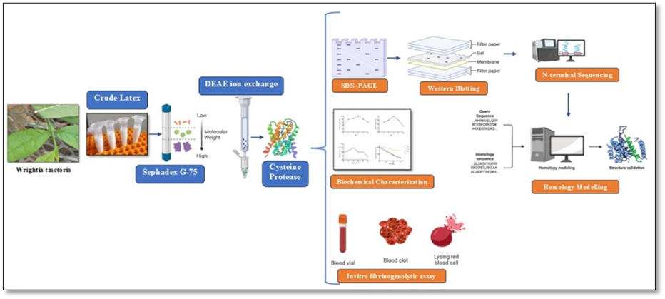
Proteolytic enzymes are intricately involved in numerous biological processes throughout the plant life cycle and play a pivotal role in plant growth and development (Liu et al., 2018). These proteases are integral to plant defense mechanisms, as they initiate a hypersensitive response to pathogen attacks and participate in innate immunity (Balakireva et al., 2018 and Molik et al., 2025). Although most commercial proteases are derived from microbial sources, plant proteases are becoming increasingly important for industrial applications due to their marked stability. Among these plant proteases, cysteine proteases, such as papain, bromelain, and ficin, are often used in different industries (González-Rábade et al., 2011). As the demand for industrial proteases increases, there is a growing need for the purification and characterization of proteases from different sources. Latex-producing plants are a valuable source of pharmacologically active compounds, including proteases. Latex, a complex fluid secreted by specialized cells in certain plants, is often found in plant families such as Euphorbiaceae, Apocynaceae, Moraceae, Papaveraceae and Asclepiadaceae. These families are distributed across ecosystems and produce latex as a defence mechanism (Molik et al., 2025). The latex of various plant species contains bioactive compounds, including proteins, enzymes, alkaloids, glycosides, cardenolides, terpenoids, furanocoumarins and starch (Konno et al., 2011). The presence of proteases in latex helps plants defend against parasites, herbivores, and pathogens by attacking invading organisms once the plant cell is lysed. These latexes exhibit pharmacological properties and are used in folk medicine. Latex from several plant species is involved in hemostasis, wound healing and pain relief. Screening of latex showed high proteolytic activity. Over 110 latices from different plant families contain at least one proteolytic enzyme. Most belong to the cysteine or serine endopeptidase family, and only one belongs to the aspartic endopeptidase family (Badgujar et al., 2014 and Mnif et al., 2015). Similarly, Plant latex is a rich source of proteolytic enzymes with potential biomedical applications, particularly in hemostasis. Among them, thrombin-like enzymes have garnered interest for their ability to mimic thrombin by catalyzing the conversion of fibrinogen to fibrin, facilitating clot formation (Selvaraja et al., 2025).
Therefore, the present study aimed to purify a novel cysteine protease from the latex of the plant Wrightia tinctoria (Roxb). and investigate its biochemical characteristics W. tinctoria belongs to the Apocynaceae family and is a small deciduous tree distributed in all parts of India. W. tinctoria is a medicinally important plant, and its extract exhibits antibacterial, antidandruff, and antipsoriasis activities (Tomar et al., 2008). It exudes copious amounts of latex upon wounding of the leaf or stem throughout the year, and there is no seasonal change in the protein levels or composition of latex. Thus, latex can be harvested at any time of the year for the purification of proteases.
MATERIALS AND METHODS:
Plant materials, latex collection, and preparation of crude enzyme source from Wrightia tinctoria (WT) were identified by Prof. Suresh Kumar C, School of Sciences, Department of Botany, Maharani Cluster University, India. Voucher specimens of each plant were deposited at the Herbarium, Department of Botany, Maharani Cluster University, India. Latex from each plant was collected separately in pre-chilled 1X PBS (phosphate buffer saline (PBS) microfuge tubes (1:1). The mixture was centrifuged at 13,000 rpm for 30 min to remove the gum and other insoluble materials from the supernatant. The resulting clear liquid phase, termed “crude latex,” was stored at 4 °C and used for enzyme purification.
Acetone Precipitation
The crude latex was subjected to acetone precipitation. Acetone fractions of 60-80 % (v/v) were collected by centrifugation at 13,000 rpm, and the precipitate obtained in each fraction was air-dried and resuspended in a minimal volume of 50 mM Tris-HCl buffer (pH 8.0).
Sephadex G-75 Gel Filtration
The acetone fraction (60-80%) was subjected to gel filtration using a Sephadex G-75 column (2.5 cm × 90 cm) equilibrated with buffer A (25 mM Tris-HCl, pH 8.0, containing Triton X-100 at 0.5%). Fractions of 1.5 ml were collected at a flow rate of 30 ml/h using the same buffer. The protein content (absorbance at 280 nm) and proteolytic activity of all fractions were determined, and the active fractions were pooled.
DEAE Anion-Exchange Chromatography
Active fractions from Sephadex G-75 gel filtration were applied to a DEAE column (2 cm × 10 cm) pre-equilibrated with a buffer solution (50 mM Tris-HCl, pH 8.0). After washing with the same buffer, bound proteins were eluted using a linear gradient of NaCl (0 to 1.0 M) in buffer B. Fractions of 1.0 ml were collected at 60 ml/h and analyzed for protease activity and protein concentrations. Fractions exhibiting protease activity were pooled and stored at −20 °C for further analysis.
Protein Concentration
Protein concentration was determined using the method described by Bradford (1976), using bovine serum albumin as a standard.
Cysteine protease activity assay
Proteolytic activity was determined using azocasein substrate, as described by modifying the method of (Sorokhaibam et al., 2015). A 0.5ml aliquot of the diluted enzyme was mixed with 0.5 ml of 100 mM Tris–HCl buffer (pH 8.0) containing 1 % (w/v) casein and incubated for 15 min at 70 °C. The reaction was stopped by adding 0.5 ml of 20 % (w/v) TCA. The mixture was allowed to stand at room temperature for 10 min and centrifuged at 13,000 rpm for 15 min to remove the precipitate. A blank was prepared similarly, using 0.5 ml of 100 mM Tris–HCl buffer (pH 8.0) instead of the enzyme. The absorbance of the supernatant was measured at 280 nm wavelength. A standard curve was generated using 0-50 mg/l tyrosine . One unit of protease activity was defined as the enzyme required to liberate 1 μg of tyrosine per minute under experimental conditions.
Zymography analysis
Zymography was performed as described by Garcia-Carreno et al., (1993). After native electrophoresis, the gel was submerged in 100 mM Tris–HCl buffer (pH 8.0) with 2.5% Triton X-100 for 30 min to remove the SDS. The gel was washed thrice with Tris–HCl buffer and immersed in 50 ml of 1% casein in Tris–HCl buffer (pH 8.0) for 10 min at 4°C. The plates were then incubated at 60°C for 20 min to develop the activity zone. The gel was stained with 0.25% Coomassie Brilliant Blue R-250 in 45% ethanol and 10% acetic acid and destained with 5% ethanol and 7.5% acetic acid. Clear zones on a blue background indicate protease activity.
SDS-PAGE and Western Blotting
The purified enzyme from DEAE anion-exchange chromatography was subjected to SDS-PAGE and Western blotting (Charnsa et al., 2025). Proteins (6 µg) were mixed with SDS loading buffer, boiled at 95 °C for 5 min, and separated on 12% SDS-PAGE gels under non-reducing and reducing conditions. Parallel acrylamide gels were resolved under identical conditions: one was stained with Coomassie protein stain, while the other underwent western blotting and transfer to a PVDF membrane. The PVDF band corresponding to the protease was excised, and the N-terminal amino acid sequence was determined by Edman degradation on an automated ABI Procise 494 (Applied Biosystems, Foster City, CA).
Effect of pH on Activity and Stability
The optimum pH for casein hydrolysis by the purified enzyme was studied over pH 5.0-10.0, at 60 °C for 15 min. To check pH stability, the enzyme was incubated for 60 min at 4 °C in different buffers, and residual proteolytic activities were determined under standard conditions.
Effect of Temperature on the Activity and Stability
The protease activity was tested at temperatures ranging from 30 to 90 °C for 15 min at pH 8.0, using casein as the substrate. Thermal stability was tested by incubating the enzyme at different temperatures for 1 h and measuring the residual activity under standard assay conditions. The non-heated enzyme was considered as the 100% control.
Effects of Metal Ions on Enzyme Activity
The influence of metal ions (5 mM) on protease activity was investigated by adding monovalent (K+ or Na+) and divalent (Ca2+, Mn2+, Zn2+, Cu2+, Ba2+, Mg2+, or Hg2+) ions to the reaction mixture. The proteolytic activity of the purified enzyme without additives was considered 100%.
Determination of Fibrinolytic Activity
The fibrinolytic activity of the purified protease was determined by modifying the method of (Astrup and Müllertz 1952) using a 1% agarose gel plate containing 0.2% fibrin. A mixture of 0.2 g agarose, 4 mL of 1% fibrin solution, and 16 mL of 0.1 M sodium phosphate buffer (pH 6.0) was warmed in a hot water bath. The clear suspension was poured into a petri dish (10 cm diameter) and allowed to set at room temperature. Three 0.5 cm diameter wells were punched in each plate. Two diluted protease samples containing 0.65 µg and 1.30 µg protein were loaded in two wells, while heat-inactivated protease (1.30 µg protein) served as a control in the third well. The gel plate was incubated at 37 °C for 6 h and then treated with 10% TCA. Fibrinolytic activity was confirmed by the presence of clear zones around the sample wells against an opaque background.
Thrombin-like activity
Recalcification time (Coagulant assay)
Fresh blood was mixed with 0.11 M trisodium citrate (9:1) and centrifuged for 15 min at 134×g. The supernatant was used as platelet-poor plasma (PPP). Enzyme extract (0.01 M Tris–HCl buffer, pH 7.4) was added to 0.3 mL of pre-warmed PPP (37 °C). The mixture was incubated for 1 min at 37 °C. To form a clot, 30 μL of 0.25 M calcium chloride was added. The time for noticeable clot appearance after the addition of calcium chloride was noted. Tris–HCl buffer was added instead of the enzyme in the control experiments (Condrea et al., 1983).
Plasmin-like activity
RESULTS AND DISCUSSION :
A new cysteine protease from the latex of Wrightia tinctoria (WT) was extracted and purified successively by gel filtration followed by DEAE cellulose anion exchange chromatography. The purification table is summarized (Table 1). Initially, the crude enzyme extract was acetone precipitated and then subjected to Sephadex G-75 gel filtration (Figure. 1A). Active fractions were pooled and loaded onto a DEAE cellulose chromatography column equilibrated with buffer B. Bound proteins were eluted with a linear gradient of NaCl concentrations ranging from 0 to 1.0 M. Protease activity appeared as a single peak (Fig. 1B). At the final purification step, the cysteine protease was purified eightfold, with a recovery of 12.8% and a specific activity of 28.3 U/mg using casein as a substrate.
Table 1. Purification of cysteine protease from Latex of W. tinctoria
|
Sl.No |
Purification steps |
Total protein (mg) |
Total activity (units)a |
Specific activity (units/mg) |
% recovery |
|
1 |
Crude Latex |
353.7 |
6960 |
19.7 |
100 |
|
2 |
Sephadex G-75 (Gel filtration) |
286.4 |
5817 |
20.3 |
83.5 |
|
3 |
DEAE ion exchange |
31 |
897 |
28.3 |
12.8 |
aDefinition of 1 unit: the amount of enzyme under the assay conditions described, resulting in an increase of 0.01 unit absorbance at 280 nm per minute of digestion. Casein was used as substrate.
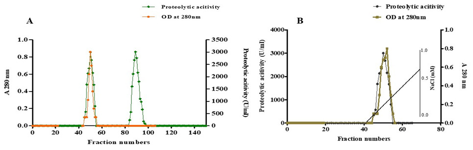
Fig. 1 A. Purification profile of plant protease from the latex of Wrightia tinctoria via gel filtration on a Sephadex G-75 column. The 60–80 % acetone precipitate was resuspended in 100 mM Tris–HCl buffer (pH 8.0) and applied to a 2.6 cm×90 cm column, equilibrated, and eluted with buffer A at a flow rate of 30 ml/h. Fractions collected from the column were assayed for protein content at 280 nm and protease activity. B. Elution profile of the cysteine protease from DEAE Cellulose. Active fractions from gel filtration G-75 were collected and applied to DEAE Cellulose equilibrated with buffer. The enzyme was eluted with a linear gradient of NaCl (0-1.0 M) in buffer at a rate of 70 ml/h
The purified cysteine protease showed a single band on SDS-PAGE, confirming that the enzyme was homogeneous and had a molecular weight of approximately 27 kDa, corresponding to that estimated by DEAE cellulose chromatography (Fig. 2A). The purity of the enzyme was also evaluated using zymogram activity staining with a clear band of casein hydrolysis, indicating the purity and homogeneity of the purified enzyme (Figure 2B). The obtained molecular weight was lower than those of other plant species investigated (Hashim et al., 2011 and Mnif et al., 2015). However, These enzymes typically have molecular weights ranging from 20 to 50 kDa, with those isolated from latex commonly falling within the 20-30 kDa range. Cysteine proteases are characterized by a cysteine residue at their catalytic site, which plays a pivotal role in the nucleophilic attack of the enzyme on peptide bonds (Sharma et al., 2018). Their catalytic mechanism involves the formation of a covalent enzyme–substrate complex, facilitated by the thiol (-SH) group of cysteine residue. However, cysteine proteases are highly sensitive to oxidation owing to the redox activity of the catalytic cysteine, leading to rapid inactivation upon exposure to air (Verma et al., 2016).
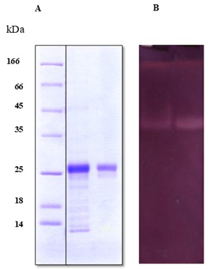
Fig. 2 A. SDS-PAGE of the purified cysteine protease from the latex of Wrightia tinctoria. Lane 1, standard protein marker of different molecular weights; lane 2; Sephadex G-75 column fraction, lane 3, purified enzyme from DEAE-Cellulose column. B Zymogram detection of the proteolytic activity of the purified enzyme from the latex of Wrightia tinctoria.
The N-terminal amino acid sequence of the cysteine protease was determined using the automated Edman method after SDS-PAGE and electroblotting. The initial sequence residues of the cysteine protease were determined to be “VPETVDWRSKGAV”. The obtained sequence showed sequence similarity with other plant proteases and the conserved consensus sequences VDWR and KGAV, as well as the proline residue located at position 2 of the N-terminal sequence (Figure 3). The N-terminal sequence of W. tinctoria cysteine proteinase was compared with those of some sequences in the database, and it shared 92 % identity with the cysteine proteinase from Arabidopsis thaliana and Glycine max, 76.92 % identity with papaya proteinase omega from Carica papaya (Reveil et al., 1993), 61.54 % identity with proteases isolated from Morrenia brachystephana Griseb (Vairo Cavalli et al., 2003), Funastrum clausum (Morcelle, et al., 2004), and Asclepias curassavica L. (Mnif et al., 2015), 53.85 % identity with endopeptidases from the latex of Morrenia odorata (Mnif et al., 2015), and 46.15 % identity with CMS1MS1-B from the latex of Carica candamarcensis (Teixeira et al., 2008). These results indicate that the purified W. tinctoria protease is a novel protease.
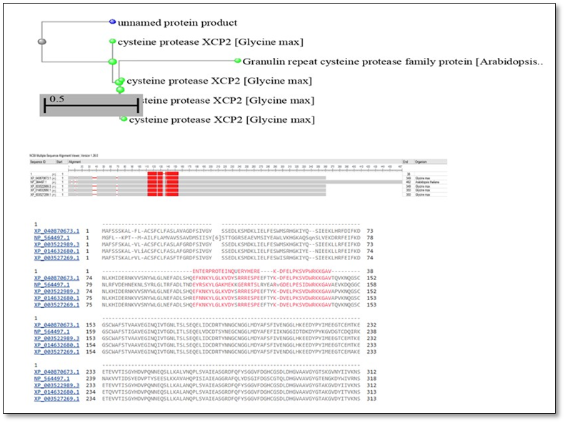
Fig. 4. Multiple sequence alignment of the N-terminal amino acid sequence of purified cysteine protease from the latex of Wrightia tinctoria with the sequences of other plant proteases
The effect of pH on the activity of the purified Wrightia tinctoria cysteine protease was studied using casein as a substrate over a pH range of 5.0–10.0 at 60 °C (Fig. 5A). The purified protease was highly active in the pH range of 6.0–9.0, with optimum activity at approximately pH 8.0. The relative activities at pH 6.0, 7.0, 9.0, and 10.0 were approximately 81, 94, 83, and 70 %, respectively, of that measured at pH 8.0.
Protease activity decreased significantly at pH 10.0 and was only 21 % of the maximum enzyme activity. The optimum pH of cysteine protease was similar to that reported for eumiliin from Euphorbia milii var. hislopii latex (Fonseca et al., 2010). The optimum activity of procerain B, a protease extracted from the latex of Calotropis procera, was in the pH range of 6.5–8.5 (Singh et al., 2010). The optimum pH of cysteine protease was higher than that of ginger rhizome, which showed maximum activity at pH 5.5 (Barakat et al., 2023). However, it was lower than that reported for asclepain, a cysteine peptidase of the Asclepias curassavica latex, which showed maximum activity within the pH range of 9.4–10.2. The cysteine peptidase VQ-VII, isolated from V. quercifolia latex, exhibited an optimal pH of 8.5. The pH stability profile of the cysteine protease (Fig. 3b) showed that after 1 h of incubation, the protease retained 51, 45, and 43 % of its original activity at pH 8.0, 7.0, and 9.0, respectively. However, at pH 6.0 and 12.0, the enzyme retained only 26 and 31 %, respectively (Ningthoujam et al., 2025) (Figure 5A)
Similarly, the purified protease displayed optimal activity at 50 °C (Fig. 5). For confirmation, the enzyme activity was measured over different time periods (0 to 80 min) at 50 °C using the caseinolytic standard assay. The results showed that the purified enzyme retained 100% of its activity when incubated with the substrate at 50 °C for 75 min, and a very slight decrease (5%) was observed at 89 min. Therefore, this temperature (50 °C) was selected as the standard temperature for all experiments. For thermal stability, the purified enzyme maintained 100% activity when incubated alone for 30 min at temperatures up to 40 °C; however, at 50 and 60 °C, the activity was decreased. In addition, 47% of the enzyme activity was still present at 70 °C. It can be concluded that the purified enzyme is a thermostable enzyme with optimal activity at 50 °C and pH 7 to 8. Herein, we also investigated the influence of various metal ions on enzyme activity. At 5 mM, Cu2+ and Hg2+ act as potent inhibitors of enzyme activity, whereas Ca2+ and Mg2+ have no impact on enzyme activity (Table 2). Similar results were reported for cysteine proteases from Fasciola gigantica and Triticum aestivum. Furthermore, the heavy metal ions Zn2+, Ni2+, Hg2+, and Cu2+ are effective enzyme inhibitors, whereas Ca2+ and Mg2+ have no impact on enzyme activity. Most of the tested heavy metal ions inhibited the cysteine proteases purified from Fasiola gigantica and Triticum aestivum. Hg2+ poisoning is commonly attributed to the strong interactions between Hg2+ and cysteine thiolate anions in cysteine proteases (Tomar et al., 2008 and Ningthoujam et al., 2025).
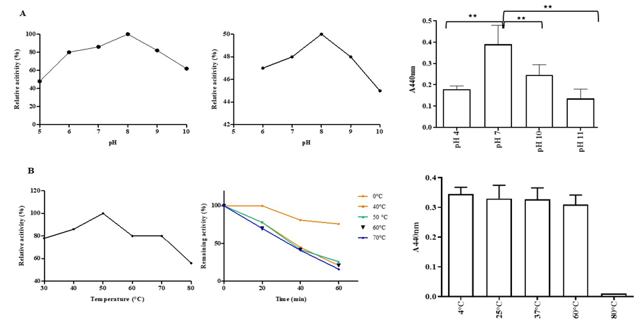
Fig.5 Effects of temperature (A) and pH (B) on the enzyme activity of WT cysteine protease. Enzyme activity was measured using azo-casein assays at 440 nm. (A) Cysteine protease activity was assessed after incubation at temperatures ranging from 4 to 80 °C. (B) Cysteine protease was analyzed by incubation at 37 °C for 30 min over a pH range of 4, 7, 10, and 11. The data shown are the mean ± S.D. of three independent experiments. The asterisk indicates the presence and level of significant differences (** p < 0.01) from other groups.
Table -2. Effects of various metal ions (5 mM) on the purified cysteine protease from the latex of Wrightia tinctoria
|
Metal ions |
Relative activity (%) |
|
Control |
100±0 |
|
Ca2+ |
51.22±0.52 |
|
Zn2+ |
7.35±0.1 |
|
Cu2+ |
0 |
|
Mg2+ |
22.67±0.98 |
|
Hg2+ |
0 |
|
Mn2+ |
12.2±1.06 |
|
Na+ |
60.48±0.14 |
|
K+ |
78.115±0.46 |
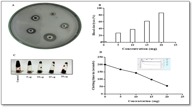
Fig. 6. Determination of the fibrinolytic activity of the purified protease.A; Fibrinolytic activity was determined using
the fibrin-agarose gel plate method, as described in the section on fibrinolytic activity by fibrin agarose gel plate method. Sample well 5, 10,15 and 20 µg purified protease; control well III, 1.3 µg heat-inactivated purified protease. B; Thrombolytic activity of purified cysteine protease from Wrightia tinctoria C; Representative image of blood clot lysis by cysteine protease from Wrightia tinctoria. D; Concentration-dependent recalcification time of purified cysteine protease from Wrightia tinctoria
In recent years, there has been growing scientific interest in the hemostatic potential of plant latexes, owing to their traditional medicinal use and bioactive enzymatic components (Urs et al., 2021, and Uday et al., 2017). Studies investigating the procoagulant and fibrin(ogen)olytic properties of latex-derived proteases have employed various analytical approaches to elucidate their mechanisms of action. These include in vitro hemostatic assays to evaluate clotting activity (Siritapetawee et al., 2020), electrophoretic techniques to assess substrate specificity and fibrinogen degradation patterns (Singh et al., 2021), and mass spectrometry-based analyses to characterize protease composition and post-translational modifications (Shivaprasad et al., 2010).
Several studies have highlighted the dual functionality of plant latex proteases, wherein the same enzymatic preparation can promote both clot formation and fibrin degradation. Proteases derived from plant latex often exhibit thrombin-like activity by rapidly degrading the Aα and Bβ chains of fibrinogen, leading to fibrin clot formation. However, upon prolonged incubation or at higher concentrations, these enzymes have also been observed to hydrolyze existing fibrin networks, indicating underlying fibrinolytic or plasmin-like activities (Kusuma et al., 2021). This functional duality is thought to arise from the co-existence of multiple enzyme classes within the latex, such as serine and cysteine proteases, each contributing differently to coagulation and fibrinolysis. Our results showed that the rate of clot formation varied with the protease concentration. The time taken for fibrin clot formation decreased with increasing concentrations. The recalcification time with 10 µg of protease was found to be 39 s ± 0.33, whereas the control value was 200 s in human platelet-poor plasma (PPP). The purified protease exhibited a 77.62% increase in clot-inducing ability (Fig 6 D). Similarly, the thrombin-like activity of the purified cysteine protease was observed as the hydrolysis of pure fibrinogen to form fibrin. The fibrinogen agarose plate assay revealed that the zone of precipitation by the protease was larger than that produced by 0.2 U of thrombin. The purified cysteine protease (2 µg protein) showed a zone of precipitation of 1.3 cm, suggesting significant enhancement in fibrinogenolytic activity after purification (Fig. 6 A and B). Ninety-six percentage of clot lysis was exhibited by 25 µg of cysteine protease. This resulted in a 89.86% enhancement compared to its cysteine protease performance (Figure 6C). The obtained results revealed the involvement of latex proteases in clot formation and the thrombolytic aspect of the wound healing process (Banu et al., 2017). Earlier reports with purified plant latex proteases, such as AMP48 (S. grantii), indicated that the susceptibility of fibrinogen subunits seems to follow Aα > Bβ >> γ chains. The pattern of fibrinogenolysis by another purified latex protease, hirtin (Euphorbia hirta), demonstrated its preferential specificity for hydrolysis of Aα and Bβ subunits and subsequent release of fibrinopeptides to assit fibrin clot formation (with mild γ subunit hydrolysis) (Singh et al., 2021).
CONCLUSION :
Plant latex proteases have emerged as promising candidates for exogenous hemostatic agents, primarily because of their thrombin-like and fibrinolytic activities. Thus, the latex of Wrightia tinctoria is a potential source of novel cysteine proteases with a molecular mass of 27 kDa. The purified protease acted on azocasein as its substrate, with an optimum pH of 6 and an optimum temperature of 50°C. The purified protease showed significant sequence similarity to existing proteases. In addition, the purified cysteine protease exhibited fibrinolytic activity. The results provide further insights into the potential of purified proteases from Wrightia tinctoria as therapeutic agents for industrial applications.
BIBLIOGRAPHY:
1. Badgujar, S. B. (2014). Evaluation of the hemostatic activity of latex from three Euphorbiaceae species. Journal of ethnopharmacology, 151(1), 733-739.
2. Balakireva AV, Zamyatnin AA. Indispensable role of proteases in plant innate immunity. Int J Mol Sci, 19 (2018) 629
3. Banu, S. H., Nagashree, S., Latha, B., & Chethankumar, M. (2017). Biochemical characterization of proteases isolated from the latex of Tabernaemontana divaricata L. and Carissa carandas L.: their role in hemostasis. J Pharmacogn Phytochem, 6(6), 06-09.
4. Barakat, A. Z., Abdel-Aty, A. M., Ibrahim, M. K., Salah, H. A., Hegazy, U. M., Azouz, R. A., ... & Mohamed, S. A. (2023). Purification and characterization of cysteine protease of Sarcocystis fusiformis from infected Egyptian water buffaloes. Scientific Reports, 13(1), 16123.
5. Bradford MM. A rapid and sensitive method for the quantitation of microgram quantities of protein utilizing the principle of protein‐dye binding. Anal Biochem. 1976;72:248‐254.
6. Charnsatabut, C.; Suwanchaikasem, P.; Rattanapisit, K.; Iksen, I.; Pongrakhananon, V.; Bulaon, C.J.I.; Phoolcharoen, W. Optimized expression of human interleukin-15 in Nicotiana benthamiana and in vitro assessment of its activity on human keratinocytes. Biotechnol. Rep. 2025, 46, e00889.
7. Doolittle, R.F. Clotting of Mammalian Fibrinogens by Papain: A Re-Examination. Biochemistry 2014, 53, 6687–6694.
8. Fonseca, K. C., Morais, N. C. G., Queiroz, M. R., Silva, M. C., Gomes, M. S., Costa, J. O., ... & Oliveira, F. (2010). Purification and biochemical characterization of Eumiliin from Euphorbia milii var. hislopii latex. Phytochemistry, 71(7), 708-715.
9. Garciacarreno, F. L., Dimes, L. E., & Haard, N. F. (1993). Substrate-gel electrophoresis for composition and molecular weight of proteinases or proteinaceous proteinase inhibitors. Analytical biochemistry, 214(1), 65-69.
10. Gonzalez-Rabade, N., Badillo-Corona, J. A., Aranda-Barradas, J. S., & del Carmen Oliver-Salvador, M. (2011). Production of plant proteases in vivo and in vitro—a review. Biotechnology advances, 29(6), 983-996.
11. Hahn B‐S, Cho SY, Ahn MY, Kim YS. Purification and characterization of a plasmin‐like protease from Tenodera sinensis (Chinese mantis). Insect Biochem Mol Biol. 2001;31(6): 573‐581
12. Hashim, M. M., Mingsheng, D., Iqbal, M. F., Xiaohong, C., Schrick, K., Cordova, C., ... & Fujioka, S. (2011). PROTEIN BIOCHEMISTRY AND PROTEOMICS. Phytochemistry, 72(6), 431-434.
13. Kembhavi, A. A., Kulkarni, A., & Pant, A. (1993). Salt-tolerant and thermostable alkaline protease from Bacillus subtilis NCIM No. 64. Applied biochemistry and biotechnology, 38(1), 83-92.
14. Kusuma, C.G.; Gubbiveeranna, V.; Sumachirayu, C.K.; Bhavana, S.; Ravikumar, H.; Nagaraju, S. Thrombin- and Plasmin-like and Platelet-Aggregation-Inducing Activities of Plumeria alba L. Latex: Action of Cysteine Protease. J. Ethnopharmacol. 2021, 273, 114000.
15. Liu H, Hu M, Wang Q, Cheng L & Zhang Z, Role of papainlike cysteine proteases in plant development. Front Plant Sci, 9 (2018) 717
16. Mnif, I. H., Siala, R., Nasri, R., Mhamdi, S., Nasri, M., & Kamoun, A. S. (2015). A cysteine protease isolated from the latex of Ficus microcarpa: purification and biochemical characterization. Applied biochemistry and biotechnology, 175(3), 1732-1744.
17. Mohamed, S. A., Fahmy, A. S., Mohamed, T. M., & Hamdy, S. M. (2005). Proteases in egg, miracidium and adult of Fasciola gigantica. Characterization of serine and cysteine proteases from adult. Comparative Biochemistry and Physiology Part B: Biochemistry and Molecular Biology, 142(2), 192-200.
18. Molik, Z. A., Ajayi, T. O., Ogunniyi, Q. A., Fijagbade, A. O., & Ogbole, O. O. (2025). Latex of medicinal plants: A reservoir of antimicrobial peptides, proteins, and enzymes for drug discovery. Discover Molecules, 2(1), 1.
19. Morcelle, S. R., Trejo, S. A., Canals, F., Avilés, F. X., & Priolo, N. S. (2004). Funastrain c II: a cysteine endopeptidase purified from the latex of Funastrum clausum. The protein journal, 23(3), 205-215.
20. Murata Y, Satake M, Suzuki T. Studies on snake venom. J Biochem. 1963;53(6):431‐437
21. Ningthoujam, J., Syiem, M. B., & Syiem, D. (2025). Biochemical Characterization of a Novel Cysteine Protease Purified from the Medicinal Plant Kaempferia galanga L. The Protein Journal, 1-18.
22. Prasad, S., Kashyap, R. S., Deopujari, J. Y., Purohit, H. J., Taori, G. M., & Daginawala, H. F. (2006). Development of an in vitro model to study clot lysis activity of thrombolytic drugs. Thrombosis journal, 4(1), 14.
23. Reveil, D. F., Cummings, N. J., Baker, K. C., Collins, M. E., Taylor, M. A. J., Sumner, I. G., ... & Goodenough, P. W. (1993). Nucleotide sequence and expression in Eschericia coli of cDNAs encoding papaya proteinase omega from Carica papaya. Gene, 127(2), 221-225.
24. Selvaraja, L. K., & Zulfigar, S. B. (2025). Plant Latex Proteases in Hemostasis: Beyond Thrombin-like Activity. Applied Biosciences, 4(3), 37.
25. Sharma, A.; Sharma, D.; Verma, S.K. In Silico Study of Iron, Zinc and Copper Binding Proteins of Pseudomonas syringae pv. lapsa: Emphasis on Secreted Metalloproteins. Front. Microbiol. 2018, 9, 1838.
26. Shivaprasad, H.V.; Rajaiah, R.; Frey, B.M.; Frey, F.J.; Vishwanath, B.S. ‘Pergularain e I’—A Plant Cysteine Protease with Thrombin-like Activity from Pergularia Extensa Latex. Thromb. Res. 2010, 125, e100–e105.
27. Singh, A. N., Shukla, A. K., Jagannadham, M. V., & Dubey, V. K. (2010). Purification of a novel cysteine protease, procerain B, from Calotropis procera with distinct characteristics compared to procerain. Process Biochemistry, 45(3), 399-406.
28. Singh, M. K., Rajagopalan, A., Tanimu, H., & Sukumaran, B. O. (2021). Purification, characterization and fibrino (geno) lytic activity of cysteine protease from Tabernaemontana divaricata latex. 3 Biotech, 11(2), 106.
29. Singh, M.K.; Rajagopalan, A.; Tanimu, H.; Omana, B. Purification, Characterization and Fibrino (Geno) Lytic Activity of Cysteine Protease from Tabernaemontana Divaricata Latex. 3 Biotech 2021, 11, 106.
30. Siritapetawee, J.; Khunkaewla, P.; Thumanu, K. Chemico-Biological Interactions Roles of a Protease from Euphorbia Resinifera Latex in Human Anticoagulant and Antithrombotic Activities. Chem. Biol. Interact. 2020, 329, 109223.
31. Teixeira, R. D., Ribeiro, H. A. L., Gomes, M. T. R., Lopes, M. T. P., & Salas, C. E. (2008). Plant Physiology and Biochemistry, 46, 956–961
32. Tomar, R., Kumar, R., & Jagannadham, M. V. (2008). A stable serine protease, wrightin, from the latex of the plant Wrightia tinctoria (Roxb.) R. Br.: purification and biochemical properties. Journal of agricultural and food chemistry, 56(4), 1479-1487.
33. Uday, P.; Maheshwari, M.; Sharanappa, P.; Nafeesa, Z.; Kameshwar, V.H.; Priya, B.S.; Nanjunda Swamy, S. Exploring Hemostatic and Thrombolytic Potential of Heynein—A Cysteine Protease from Ervatamia Heyneana Latex. J. Ethnopharmacol. 2017, 199, 316–322.
34. Urs, A.P.; Manjuprasanna, V.N.; Rudresha, G.V.; Hiremath, V.; Sharanappa, P.; Rajaiah, R.; Vishwanath, B.S. Molecular Cell Research Thrombin-like Serine Protease, Antiquorin from Euphorbia Antiquorum Latex Induces Platelet Aggregation via PAR1-Akt/P38 Signaling Axis. Biochim. Biophys. Acta Mol. Cell Res. 2021, 1868, 118925.
35. Vairo Cavalli, S. E., Arribére, M. C., Cortadi, A., Caffini, N. O., & Priolo, N. S. (2003). Morrenain b I, a papain-like endopeptidase from the latex of Morrenia brachystephana Griseb.(Asclepiadaceae). Journal of protein chemistry, 22(1), 15-22.
36. Verma, S., Dixit, R., & Pandey, K. C. (2016). Cysteine proteases: modes of activation and future prospects as pharmacological targets. Frontiers in pharmacology, 7, 107.