Epidemiological and Biological Study of Infections in Pregnant Women: General Prevalence and In-Depth Analysis of Urinary Tract Infections
- Amina ITATAHINE , Phd Department of Biological Sciences, Faculty of Natural and Life Sciences and Earth Sciences, University of Djilali Bounaama, Khemis Miliana, Algeria.
- Aicha LAISSAOUI , Phd Department of Biological Sciences, Faculty of Natural and Life Sciences and Earth Sciences, University of Djilali Bounaama, Khemis Miliana, Algeria.
- Soumia TABTI , Phd Department of Biology, Faculty of Natural and Life Sciences, University of Hassiba Ben Bouali, Chlef, Algeria.
- Zakia HADJ-BENALI , Phd Department of Biology, Faculty of Natural and Life Sciences, University of Hassiba Ben Bouali, Chlef, Algeria.
- Abbassia DEMMOUCH , Phd Biotoxicology Laboratory, Department of Biology, Faculty of Natural and Life Sciences, University of Djillali Liabes, Sidi bel Abess, Algeria.
Article Information:
Abstract:
This study evaluates the incidence and etiology of maternal infections, specifically identifying uropathogens and their antibiotic resistance profiles in urinary tract infections (UTIs). A retrospective study analyzed the medical records of 330 pregnant women who presented with symptoms indicative of infectious diseases. The aim was to assess the most common types of infections diagnosed during pregnancy. The second component of this study includes a microbiological analysis of 320 newly recruited pregnant women with suspected UTI. Midstream urine samples were collected and analyzed by cytobacteriological examination of urine. Microorganisms isolated from positive cultures were identified and tested for antimicrobial susceptibility utilizing the Kirby–Bauer disc diffusion method. The average age of the 330 participants was 29.88 ± 7.14 years, with most infections occurring during the first two trimesters. UTIs were the most common infection (31.5%). Among 320 urine samples, significant bacterial growth (>105) was detected in 110/320, with a prevalence of 34.4%. The predominant organisms identified were Escherichia coli and followed by Enterococcus spp. The antibiotic resistance profile reveals resistance rates of 20.4% and 45.7% for Gram-negative and Gram-positive bacteria, respectively. These findings emphasize instance of antibiotic resistance, stressing the necessity for enhanced surveillance and effective management of treatment.
Keywords:
Article :
INTRODUCTION:
Anemia is defined by a hemoglobin concentration There are several reasons why pregnant women are considered a special group by infectious disease specialists [1]. Pregnancy is accompanied by several mechanical and physiological changes (e.g., decreased respiratory volumes and urinary stasis due to uterine enlargement), which require immune adaptations to accommodate the fetus [2]. Levels of steroid sex hormones change dramatically during this time; it is at a level that is substantially more elevated when compared to any other period in a female's life. These hormones include pregnancy-related hormones such as estradiol, estriol, progesterone, corticosteroids, and prolactin [3]. These hormonal fluctuations may alter immune cell functions and modify the immune response, affecting the body's ability to fight infection during the period of gestation [3]. According to another theory, the immune response during pregnancy is modulated instead of suppressed [2]. Pregnancies complicated by infections can impact both the mother and embryonic development. These infections occur when microorganisms invade tissues or release harmful substances, disrupting normal bodily functions and triggering an immune response. Inflammatory reactions to infection have the potential to engender deleterious consequences, including premature delivery, congenital malformations, developmental delays, and stillbirths [2]. Urinary tract infections remain one of the primary types of infections during pregnancy, affecting around 20% of pregnant women [4]. A UTI is characterized by an overgrowth of bacteria in the urinary tract (≥ 10⁵ CFU/mL), regardless of the presence of clinical indications [5]. It is estimated that 4-10% of pregnant women have asymptomatic bacteriuria. Meanwhile, one to four percent of patients develop cystitis, and one to two percent experience severe pyelonephritis, especially during the 2nd trimester [6]. Antimicrobials are the most commonly prescribed drugs in these cases, but there's a need for caution due to the recent increase in resistance to these medications around the world [7]. The study's objective is to determine the various types of infections in pregnant women in the central region of Algeria (The Wilaya of Ain Defla) by combining an epidemiological analysis of their overall prevalence with an in-depth biological study of urinary tract infection to improve strategies for the prevention, diagnosis, and management of maternal and fetal health.
MATERIALS AND METHODS:
Epidemiological study
This is a retrospective observational cohort study conducted at the Fares Yahia Hospital in the town of Miliana, Algeria, which focuses on the analysis of 330 medical records of pregnant women who consulted the hospital's gynecology department over the period from January 2019 to May 2025. The study included pregnant women who had consulted for an infection that had occurred during pregnancy. To better understand their clinical profile, several parameters were collected, including Age, trimester of pregnancy, physiological status, medical history, type of infection, examinations performed, and treatments prescribed. In this study, all data from pregnant women in all trimesters with an infection were included. Data from non-pregnant women with an infection and pregnant women without an infection were excluded from the analysis.
Biological study
The goal of this research component is to ascertain the prevalence of pathogens responsible for UTI by cytobacteriological examination of urine (CBEU), and to assess their antibiotic resistance profile. This study involved recruiting a new cohort of 320 pregnant women attending the gynecology department of the Fares Yahia Miliana Hospital, Algeria, with or without clinical symptoms of UTI. The pregnant women who were on antibiotic therapy within the last two weeks were not included in the study. All study participants gave their consent and agreed to participate in accordance with the principles of personal data confidentiality.
Collection and processing of urine samples
The examination process included macroscopic examination of urine samples and direct microscopic examination for the detection of pyuria. The collection of urine samples was performed mid-stream in sterile specimen tubes. The following information was included on the label: the patient's unique identification number, the date, and the time of sample collection, and the samples were processed no later than 2 hours after collection. The samples of urine were put through a process called centrifugation. The sediment that resulted from this process was then examined directly under a microscope. The samples were checked for the presence of red blood cells (RBCs), leukocytes, epithelial cells, cylinders, and crystals. A normal urine sediment may contain a few RBCs, pus cells (0-5/µL), and epithelial cells. The presence of red blood cells in quantities greater than 5/μL indicates hematuria (due to glomerular nephropathy, lithiasis, tumor, etc.); the presence of white blood cells in quantities greater than 10/μL indicates leukocyturia (unaltered white blood cells) or pyuria (altered white blood cells) [8].
Uroculture
Quantitative evaluation of bacteria present in urine samples was performed using the calibrated loop technique. Take 10 µL of the urine sample that has been well homogenized but not centrifuged with a calibrated loop. Inoculate it onto chromogenic agar (CHROMagar orientation media) in streaks perpendicular to the radius over the entire agar surface. Place all plates in an incubator at 37 °C for 18 to 24 hours. After the plates have been incubated, the colonies on each agar plate are counted, and the number of CFUs is multiplied by 100 to obtain the number of CFU/ml. Colony counts giving bacterial growth ≥ 105 CFU/ml were considered significant.
Bacterial growth was characterized as monomicrobial or polymicrobial; each distinct colony was subcultured and isolated on blood agar plates, MacConkey agar, Chapman agar, and Sabouraud agar was used for Candida isolation. All isolates were identified using traditional microbiological techniques according to Cheesbrough (2006), based on macroscopic colony appearance, Gram staining, mobility, Oxidase, and Catalase tests. The API 20 system (Bio-Mérieux, France), based on biochemical character tests, was used to identify the bacteria responsible for urinary tract infection. The results of these tests identify the genus and species of the germ isolated.
Antimicrobial susceptibility testing
Antibiotic susceptibility testing was performed on Mueller-Hinton agar plates using the Kirby-Bauer disk diffusion method [9]. A total of fifteen different antibiotic discs were tested. These included amoxicillin (25 μg), penicillin (10 μg), cefoxitin (30 μg), ceftriaxone (30 μg), ceftazidime (30 μg), imipenem (10 μg), ciprofloxacin (5 μg), gentamicin (10 μg), tetracycline (30 μg), chloramphenicol (30 μg), fosfomycin (50 μg), vancomycin (30 μg), erythromycin (15 μg), and levofloxacin (30 μg). The concentrations and zones of inhibition were interpreted according to Clinical and Laboratory Standards Institute (CLSI) guidelines [10].
Statistical Analysis
Data was entered and analyzed using SPSS version 22 to determine the prevalence and variation of positive and negative results. The performance of a chi-square test (χ²) was applied with a statistical significance value of p < 0.05.
RESULT:
A study of 330 pregnant women was conducted. The mean age of the pregnant women was 29.88 ± 7.14 years, ranging from 18 to 45 years. Most of the pregnant women had come in for an infection during their first trimester of pregnancy.
The analysis of infection types in the investigated group revealed that urinary tract infections were the most prevalent, accounting for 31.5%, followed by syphilis at 11.2%. Influenza infections, pyelonephritis, and candidiasis had reduced rates, ranging from 8% to 9%. (Figure 1).
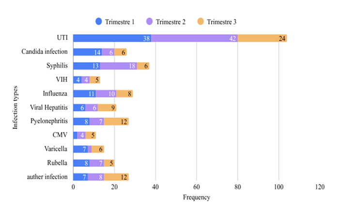
The examination of infection types categorized by pregnancy trimester indicates that urinary tract infections, syphilis, and influenza occur more frequently in the second trimester compared to the first. The possible risk of infection is substantially reduced throughout the third trimester. Therefore, it can be seen that the first two trimesters are characterized by the highest prevalence of infections, followed by the third trimester (Figure 2).
The relationship between infection type and pregnancy trimester was examined using a Pearson chi-square test. The statistical analysis revealed that there is no significant relationship between the two variables (p = 0.360). The results indicate that infection type did not vary significantly between trimesters.
The CBEU is the major test conducted to isolate the uropathogen that causes urinary tract infections in pregnant women. Of the 320 CBEU performed, 110 were determined to be positive, with a bacterial count greater than 10⁵. During this investigation, the prevalence of UTI among 320 pregnant women was determined to be 34.4%, with 110 cases noted (95% CI, 29.4% to 39.8%). In our study, we performed a microbiological investigation of 110 urine samples, which revealed the presence of 110 distinct types of microorganisms, including 82 (74.5%) Gram-negative bacteria, 23 (20.9%) Gram-positive bacteria, and 5 (4.5%) yeast samples from the Candida genus. Escherichia coli formed the most abundant isolate of Gram-negative bacteria n = 51; 46.4%, while Enterococcus spp. was among the most often identified Gram-positive bacteria (n = 15; 13.6%) (Table 1).
The resistance profile of the 82 Gram-negative bacteria reveals high resistance to several antibiotics. Ampicillin (AX 25) has the highest overall resistance rate 93.9%, with complete resistance observed in Escherichia coli, Enterobacter cloacae, and Klebsiella pneumoniae. Relatively high resistance was detected to tetracycline 25.6%, Ceftriaxone 22.0%, and Ciprofloxacin 17.1%, and moderate resistance to Imipenem 14.6% and fosfomycin 13.4%. In contrast, resistance rates were low for chloramphenicol 1.2%, gentamicin 2.4%, and ceftazidime 8.5% (Table 2).
The results of susceptibility testing indicate that Gram-positive bacteria showed complete sensitivity to Vancomycin, except for one strain of Staphylococcus lentus, which was resistant to all the antimicrobial agents tested. Staphylococcus aureus strains were 100% sensitive to gentamicin, resistant 100% to ceftriaxone and penicillin, and moderately sensitive to tetracycline and erythromycin 50%. Enterococcus spp. Strains 93.3% were sensitive to levofloxacin and amoxicillin, and only 13.3% were sensitive to imipenem and gentamicin. Staphylococcus xylosus strains were highly resistant 100% to penicillin (Table 3).
Table 1: The frequency of uropathogens isolated from pregnant women
|
Pathogens |
No. Of isolate |
Percentage (%) |
|
Escherichia coli |
51 |
46.4 |
|
Klebsiella pneumoniae |
20 |
18.2 |
|
Pseudomonas aeruginosa |
3 |
2.7 |
|
Proteus mirabilis |
4 |
3.6 |
|
Candida albicans |
5 |
4.5 |
|
Enterococcus spp. |
15 |
13.6 |
|
Enterobacter cloacae |
4 |
3.6 |
|
Staphylococcus aureus |
5 |
4.5 |
|
Staphylococcus lentus |
1 |
0.9 |
|
Staphylococcus xylosus |
2 |
1.8 |
|
Total |
110 |
100 |
Table 2: Percent of common Gram-negative urinary pathogens that are resistant to antimicrobial agents
|
Bacterial isolates n (%) |
||||||
|
Antimicrobial tested |
Escherichia coli (n=51) |
Proteus mirabilis (n=4) |
Enterobacter cloacae (n=4) |
Pseudomonas aeruginosa (n=3) |
Klebsiella pneumoniae (n=20) |
Total (n=82) |
|
AX 25 |
51 (100.0) |
2 (50.0) |
4 (100.0) |
0 (0.0) |
20 (100.0) |
77 (93.9) |
|
TE 30 |
14 (27.5) |
2 (50.0) |
0 (0.0) |
2 (66.7) |
3 (15.0) |
21 (25.6) |
|
CRO 30 |
9 (17.6) |
1 (25.0) |
2 (50.0) |
0 (0.0) |
6 (30.0) |
18 (22.0) |
|
CIP 5 |
6 (11.8) |
1 (25.0) |
2 (50.0) |
1 (33.3) |
4 (20.0) |
14 (17.1) |
|
IMP 10 |
6 (11.8) |
0 (0.0) |
2 (50.0) |
0 (0.0) |
4 (20.0) |
12 (14.6) |
|
FF 50 |
3 (5.9) |
2 (50.0) |
0 (0.0) |
0 (0.0) |
6 (30.0) |
11 (13.4) |
|
CAZ 30 |
0 (0.0) |
0 (0.0) |
2 (50.0) |
3 (100.0) |
2 (10.0) |
7 (8.5) |
|
FOX 30 |
0 (0.0) |
0 (0.0) |
4 (100.0) |
0 (0.0) |
0 (0.0) |
4 (4.9) |
|
CN 10 |
0 (0.0) |
0 (0.0) |
0 (0.0) |
0 (0.0) |
2 (10.0) |
2 (2.4) |
|
C 30 |
0 (0.0) |
0 (0.0) |
0 (0.0) |
0 (0.0) |
1 (5.0) |
1 (1.2) |
Data are presented as n (%).
*CAZ: Ceftazidime, AX: Amoxicillin, TE: Tetracycline, C: Chloramphenicol, CIP: Ciprofloxacin, IMP: Imipenem, FOX: Cefoxitin, FF: Fosfomycin, CN: Gentamicin, CRO: Ceftriaxone.
Table.3: Resistance Profile of Gram-Positive Bacteria to Antimicrobial Agents
|
Bacterial isolates |
Antimicrobial tested |
||||||||
|
CN 10 |
CRO 30 |
TE 30 |
ERY 15 |
P 10 |
IMP 10 |
VAN 30 |
LEV 5 |
AX 25 |
|
|
Staphylococcus aureus (n=5) |
0 (0.0) |
5 (100.0) |
2 (40.0) |
3 (60.0) |
5 (100.0) |
NA |
0 (0.0) |
NA |
NA |
|
Staphylococcus lentus (n=1) |
1 (100.0) |
1 (100.0) |
1 (100.0) |
1 (100.0) |
1 (100.0) |
NA |
1 (100.0) |
NA |
NA |
|
Staphylococcus xylosus (n= 2) |
1 (50.0) |
1 (50.0) |
1 (50.0) |
1 (50.0) |
2 (100.0) |
NA |
0 (0.0) |
NA |
NA |
|
Enterococcus spp. (n= 15) |
13 (86.7) |
NA |
NA |
8 (53.3) |
NA |
13 (86.7) |
0 (0.0) |
1 (6.7) |
1 (6.7) |
|
Total (n=23) |
15 (65.2)
|
7/8 (87.5)
|
4/8 (50.0)
|
13 (56.5)
|
8/8 (100.0)
|
13/15 (86.7)
|
1 (4.3)
|
1/15 (6.7)
|
1/15 (6.7)
|
Data are presented as n (%)
NA: Not tested, *CN: Gentamycin, CRO: Ceftriaxone, TE: Tetracycline, ERY: Erythromycin, P: Penicillin, IMP; Imipenem, VAN: Vancomycin, LEV: Levofloxacin, AX: Amoxicillin.
Figure 1:
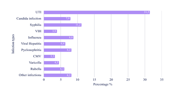
Figure 2:
DISCUSSION :
Research indicates that women experience increased vulnerability to infections during pregnancy, with metabolic, hematological, and immunological changes contributing to this susceptibility [2-11]. A study analyzing 330 records found that women aged 18 to 28 had the highest infection rates, with 157 patients reporting infections. This trend is supported by Laari et al. [11], highlighting greater prevalence in younger women 15 to 25 years due to hormonal fluctuations and less stable immunity. Conversely, the 39 to 48 age group is less affected, likely because of decreased fertility and enhanced monitoring in later pregnancies, as consistent with findings by Hochler et al. [12] that link age with obstetric complications. UTIs are prevalent medical issues affecting the urethra, bladder, and kidneys, particularly common during pregnancy, second only to anemia [13]. A retrospective analysis found a 31.5% occurrence rate among pregnant women, who are more vulnerable due to elevated amino acids, vitamins, and nutrients in their urine that support infections [14]. Additionally, increased plasma volume reduces urine concentration, and about 70% of pregnant women experience glycosuria, further encouraging bacterial growth in urine. In the second study of pregnant women, cytobacteriological analysis revealed that UTIs are prevalent in 34.4% of cases (95% CI, 29.4% to 39.8%) (110/320), aligning with global rates ranging from 13% to 33% [15]. Comparatively, a study by Girish Patill in Central India reported a prevalence of 26.75%, while other studies reported rates from 5% to 47.5% [16-17-18], variations in prevalence are influenced by geographical, environmental, and social factors [19]. Contributing factors to UTIs in pregnant women include gestational age, education level, and underlying risk factors, such as preeclampsia and HIV/AIDS. The study found that Gram-negative bacteria, especially E. coli, are the most common pathogens in (UTIs), accounting for 46.43% of isolates. E. coli prevalence is consistent globally, with 39.7% in South America and 66.01% in China [20-21].
This predominance is linked to the ascending pathophysiology of UTIs and enterobacteria colonization in the perineum [22-23] . Additionally, Enterococcus was isolated in 13.6% of cases as the leading Gram-positive bacteria, and Candida yeasts contributed 4.5%. Overall, Gram-positive bacteria account for 20.9% of UTIs in pregnant women, underscoring the importance of Enterococcus and Candida in the infection landscape, despite their lower pathogen prevalence. Following the genus Staphylococcus, the distribution of species includes Staphylococcus aureus 4.5%, Staphylococcus xylosus 1.8%, and Staphylococcus lentus 0.9%, aligning with Assouma et al. [24] finding that S. aureus represents around 30% of urinary isolates. Coagulase-negative bacteria, likely including S. xylosus, make up the remaining percentage, while Fowoyo et al. [25] identified S. sciuri and S. lentus in 0.8% of urinary tract infections. In contrast to reports identifying S. aureus as the predominant urinary pathogen among Gram-positive cocci, a study by Manjula [14] on UTIs in pregnant Indian women found no uropathogens of the S. aureus species and noted that less than 1% of identified uropathogens were Enterococcus species. Overall, Gram-positive cocci have a minor role in urinary tract infections, but changes in women's genital tracts during pregnancy can heighten the risk of colonization by these bacteria [26]. Treating bacterial infections requires antibiotics, but their effectiveness relies on bacterial sensitivity. In our study, we isolated Gram-positive and Gram-negative bacteria, noting significant resistance to various antibiotic classes. Notably, most Gram-negative isolates showed 100% resistance to amoxicillin, corroborating findings from a previous study in Uganda [15]. E. coli was the primary Gram-negative bacterium identified, exhibiting complete resistance to amoxicillin 100% largely due to the production of TEM-1 β-lactamases, common in urinary tract infections. This resistance is exacerbated by the extensive use of penicillin [27]. Notably, over 90% of E. coli strains from uncomplicated urinary tract infections globally resist amoxicillin. Additionally, 27.45% of E. coli isolates showed resistance to tetracycline linked to tet (A) and tet (B) genes on plasmids. All E. coli species tested were susceptible to several antibiotics, including ceftazidime and Gentamycin. Among Gram-positive isolates, Enterococcus spp. from pregnant women displayed high resistance to Gentamicin and Imipenem 86.7%, but sensitivity to vancomycin 100%, echoing findings from studies in Saudi Arabia [28]. Research on E. faecium in India [29] reported similar resistance patterns, highlighting emerging trends in multidrug resistance. A high resistance of 80% to penicillin and ceftriaxone was observed in S. aureus, aligning with findings in Benin [25-30]. Coagulase-positive staphylococci showed 100% sensitivity to vancomycin and gentamicin, contrasting with Algerian studies that reported high resistance [31]. A Mauritania study [32] found over 90% sensitivity to vancomycin, while Assouma et al. [24] indicated a 52.6% resistance in urinary tract isolates, possibly due to antibiotic exposure. The Staphylococcus lentus strain exhibited high resistance to all antibiotics tested. The findings demonstrate a significant prevalence of antibiotic resistance in Gram-negative bacterial strains, consistent with previous research [33-34]. These findings indicate a significant degree of antibiotic resistance among pregnant women, raising concerns due to its implications for maternal and fetal health, and it is linked to the escalating misuse of antibiotics.
CONCLUSION :
In In summary, our research indicates that urinary tract infections are among the most prevalent illnesses seen during pregnancy. This prompted us to undertake targeted research on this infection to isolate and identify the implicated bacteria and examine their drug resistance. It is crucial to identify the resistance of particular bacteria to antibiotics, as this occurrence poses a substantial public health concern. The emergence of antibiotic-resistant bacteria significantly diminishes treatment efficacy, complicating infection management. This, in turn, can lead to worsening health consequences for patients. Our ultimate goal is to improve therapy options and contribute to better medical care for afflicted individuals.
BIBLIOGRAPHY:
1. Adler H, Lambert JS. Clinical focus: infections in pregnancy. Vol. 42, Hospital practice (1995) 2014. p. 108–24.
2. Kourtis AP, Read JS, Jamieson DJ. Pregnancy and Infection. Obstetric Anesthesia Digest. 2015;35(2):67–8.
3. Richardson AN, Pollak EA, Williams D, Smith MA. Intrauterine Infection. In: Comprehensive Toxicology, Second Edition. Elsevier Inc.; 2010.
4. Yan L, Jin Y, Hang H, Yan B. The association between urinary tract infection during pregnancy and preeclampsia: A meta-analysis. Medicine (United States). Lippincott Williams and Wilkins; 2018;97(36).
5. Balachandran L, Jacob L, Al Awadhi R, Yahya LO, Catroon KM, Soundararajan LP, et al. Urinary Tract Infection in Pregnancy and Its Effects on Maternal and Perinatal Outcome: A Retrospective Study. Cureus. Springer Science and Business Media LLC; 2022; 14(1).
6. Kerure RD, Biradar A V., Lakshetty S, Biradar S. A study of urinary tract infection in pregnancy and its effect on maternal and perinatal outcome. Int J Reprod Contracept Obstet Gynecol. Medip Academy; 2024;13(2):284–9.
7. Ky Loh, N Sivalingam. URINARY TRACT INFECTIONS IN PREGNANCY. Malaysian Family Physician. 2007;2(2):1985–2274.
8. René Caquet. 250 examens de laboratoire Prescription et interprétation. 10e edition. Elsevier Masson SAS. 2008.
9. Bauer AW, Kirby WM, Sherris JC, Turck M. Test de sensibilité aux antibiotiques par une méthode standardisée à disque unique. Am J Clin Pathol. 1966;45:493–6.
10. Melvin P. James S. April M. Shelley Campeau SKMFHGRM. Performance standards for antimicrobial susceptibility testing. Clinical and Laboratory Standards Institute. 29th ed. 2020.
11. Laari JL, Anab M, Jabong DP, Abdulai K, Alhassan AR. Maternal Age and Stage of Pregnancy as Determinants of UTI in Pregnancy: A Case of Tamale, Ghana. Infect Dis Obstet Gynecol. Hindawi Limited; 2022;2022.
12. Hochler H, Lipschuetz M, Suissa-Cohen Y, Weiss A, Sela HY, Yagel S, et al. The Impact of Advanced Maternal Age on Pregnancy Outcomes: A Retrospective Multicenter Study. J Clin Med. Multidisciplinary Digital Publishing Institute (MDPI). 2023;12(17).
13. Farzaneh F, Mokhtari M, Kalemati E. Comparison of the Frequency Asymptomatic Bacteriuria in Patients with Preterm Labor and Term. Zahedan Journal of Research in Medical Sciences. Brieflands. 2018;20(6).
14. Manjula N. G., Girish C. Math., Shripad A. Patil, Subhashchandra M. Gaddad, Channappa T. Shivannavar. Incidence of Urinary Tract Infections and Its Aetiological Agents among Pregnant Women in Karnataka Region. Adv Microbiol. Scientific Research Publishing. 2013;03(06):473–8.
15. Johnson B, Stephen BM, Joseph N, Asiphas O, Musa K, Taseera K. Prevalence and bacteriology of culture-positive urinary tract infection among pregnant women with suspected urinary tract infection at Mbarara regional referral hospital, South-Western Uganda. BMC Pregnancy Childbirth. BioMed Central Ltd. 2021;21(1).
16. Patil G, Patil D, Patil A, Shrikhande S. Microbiological Profile and Antimicrobial Susceptibility Pattern of Uropathogens Isolated from Pregnant Women Attending a Tertiary Care Hospital in Central India. Cureus. Springer Science and Business Media LLC. 2024.
17. Ilusanya O, Ejembi J, Augustine U. Incidence of urinary tract infection (UTI) among Pregnant women in Ibadan, South-Western Nigeria Article in AFRICAN. J Biotechnol. 2009.
18. Barnawi Y, Alghamdi A, Ibrahim A, Al-Anazi L, Alhumaida G, Alotaibi R, et al. Prevalence of urinary tract infections in pregnant women and antimicrobial resistance patterns in women in Riyadh, Saudi Arabia: a retrospective study. BMC Infect Dis. BioMed Central Ltd. 2024;24(1).
19. Abu Aleinein I, Salem Sokhn E. Knowledge and prevalence of urinary tract infection among pregnant women in Lebanon. Heliyon. Elsevier Ltd. 2024;10(17).
20. Medina M, Castillo-Pino E. An introduction to the epidemiology and burden of urinary tract infections. Vol. 11, Therapeutic Advances in Urology SAGE Publications Inc. 2019.
21. He K, Hu Y, Shi JC, Zhu YQ, Mao XM. Prevalence, risk factors and microorganisms of urinary tract infections in patients with type 2 diabetes mellitus: A retrospective study in China. Ther Clin Risk Manag. Dove Medical Press Ltd. 2018;14:403–8.
22. Wiles TJ, Kulesus RR, Mulvey MA. Origins and virulence mechanisms of uropathogenic Escherichia coli. Experimental and Molecular Pathology 2008. 11–9.
23. Matthew A. Mulvey. Adhesion and entry of uropathogenic Escherichia coli. 2002;257–71.
24. Assouma FF, Sina H, Dossou AD, Socohou A, Hounsou MC, Avogbe PH, et al. Antibiotic Resistance Profiling of Pathogenic Staphylococcus Species from Urinary Tract Infection Patients in Benin. Biomed Res Int. Hindawi Limited. 2023.
25. Fowoyo PT, Ogunbanwo ST. Antimicrobial resistance in coagulase-negative staphylococci from Nigerian traditional fermented foods. Ann Clin Microbiol Antimicrob. BioMed Central Ltd. 2017;16(1).
26. Ekuma PU, Ibiam FA, Ekuma MI, Iroha CS, Peter IU, Iroha IR. Evaluating the Bacteria Profile and Drug Susceptibility Patterns of Urinary Tract Infectious Pathogens in Pregnant Women in Abakaliki Metropolis, Nigeria. International Journal of Pathogen Research. Sciencedomain International. 2023;12(5):52–62.
27. Bush K, Bradford PA. Epidemiology of β-lactamase-producing pathogens. Vol. 33, Clinical Microbiology Reviews American Society for Microbiology; 2020.
28. Bazaid AS, Saeed A, Alrashidi A, Alrashidi A, Alshaghdali K, Hammam SA, et al. antimicrobial surveillance for bacterial uropathogens in Ha’il, Saudi Arabia: A five-year multicenter retrospective study. Infect Drug Resist. Dove Medical Press Ltd. 2021;14:1455–65.
29. Hota S, Patil SR, Mane PM. Antimicrobial Resistance Profile of Enterococcal Isolates From Clinical Specimens at a Tertiary Care Hospital in Western Maharashtra, India. Cureus. Springer Science and Business Media LLC. 2024.
30. Dougnon V, Assogba P, Mohammed J, Agbankpe J, Deguenon E, Fabiyi K, et al. Urinary Tract Infections in Benin: Exploring the Virulence Factors and Antibiotic Resistance and Virulence Genes among Bacterial Isolates. International Journal of Pathogen Research. Sciencedomain International. 2021;28–36.
31. Elhassan BENYAGOUB, Elhoucin BENYAGOUB, Habiba BERBAOUI, Chafia RAHMANI, Larbi BENYOUSSEF. Identification and study of the emergence of antibiotic resistance of microorganisms responsible for urinary tract infections in Bechar (Algeria). 2013;5.
32. Hailaji NSM, Ould Salem ML, Ghaber SM. La sensibilité aux antibiotiques des bactéries uropathogènes dans la ville de Nouakchott – Mauritanie. Progres en Urologie. Elsevier Masson SAS. 2016;26(6):346–52.
33. Garaga GSV, Madda DP, Maninithya K, Dayan A. Antibiotic Resistance Patterns in Uropathogens Isolated from Pregnant Women: A Tertiary Hospital Study. J Pharm Bioallied Sci. Ovid Technologies (Wolters Kluwer Health). 2025;17(Suppl 4): S3039–41.
34. Gökçe İ, Çiçek N, Güven S, Altuntaş Ü, Bıyıklı N, Yıldız N, et al. Changes in bacterial resistance patterns of pediatric urinary tract infections and rationale for empirical antibiotic therapy. Balkan Med J. Galenos Publishing House. 2017;34(5):432–5.