Trends in Bioanalytical Method Development for Antiretroviral Drugs: A Review
- Abhijeet Jondhale , Swami Vivekanand University Sagar Madhya Pradesh, India 470228.
- Rani Gaikwad , Arts, Commerce and Science College, Kolhar, Ahmednagar, Maharashtra, India 413710.
- Sunildatta Gore , Department of Pharma. Chemistry, Poona College of Pharmacy, Erandwane, Pune-38.
- Atul Gunjal , Dr. Ithape Institute of Pharmacy, Sangamner, Ahmednagar, Maharashtra, India 422605,
- R. Sudhakar , Swami Vivekanand University Sagar Madhya Pradesh, India 470228.
- Krishna Pathak , Swami Vivekanand University Sagar Madhya Pradesh, India 470228.
Article Information:
Abstract:
Pharmaceutical analysis plays a vital role in ensuring the quality of drugs and pharmaceutical formulations. It serves as a cornerstone for both quality assurance and quality control processes. With the exponential growth of pharmaceutical industry worldwide, there is an increasing demand for novel analytical techniques to meet its evolving needs. As a result, the development of new analytical methods has become a fundamental aspect of pharmaceutical analysis. Throughout history, there has been a persistent pursuit of safe and effective treatments for viral infections. In today's context, with the emergence of new viruses, the development of drugs to combat these infections has become equally imperative. Prior to their market launch, these drugs must undergo a rigorous validation process to ensure their efficacy and safety. This review article provides a critical evaluation of published analytical methods and diverse approaches for investigating antiretroviral drugs in the year 2011-2021. The findings of these studies highlight the widespread use of HPLC, Advanced platforms like LC-MS/MS and UPLC, for the analysis of anti-HIV drugs.
Keywords:
Article :
INTRODUCTION:
Antiretroviral drugs are a key component of HIV treatment, and their development has seen significant advancements. Currently, more than 35-40 antiretroviral drugs are available, including various fixed-dose combinations. These drugs work by targeting different stages of HIV life cycle. Arts & Hazuda (2012), . The antiretroviral drug development has been marked by the introduction of novel formulations and new drug classes to address issues like drug toxicities and resistance Saag et al (2020). The early regimens of antiretroviral therapy were complex, requiring multiple drugs to be taken at different intervals throughout the day, leading to challenges in long-term adherence. However, advancements in drug development have led to the simplification of treatment regimens, with many patients now being able to control their HIV with a Single pill once a day. Bioanalytical methods are vital for measuring antiretroviral drug levels in biological samples, supporting drug monitoring, adherence checks, and treatment evaluation Charbe et al (2019),Mohammad et al (2016). HPLC is widely used to quantify antiretroviral drugs in biological matrices for therapeutic monitoring. Method development and validation are crucial to ensure accurate, reliable measurements, supporting better patient care and treatment outcomes Deshpande et al (2019).
This review highlights key bioanalytical methods, including HPLC-UV, LC-MS/MS, and UPLC, for the estimation of anti-HIV drugs. These techniques are essential for pharmacokinetic evaluation in HIV treatment.
The diagram below FDA approved anti-HIV drugs over the years as depicted in Figure 1.
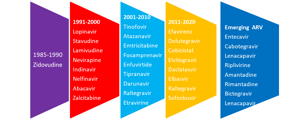
Fig.1 Approved Antiretroviral drugs
MATERIALS AND METHODS:
2.Overview of Antiretroviral Drugs
Antiretroviral drugs represent a cornerstone in the treatment of AIDS. These medications, classified based on their mechanism of action, play a pivotal role in suppressing viral replication. Understanding the classification and mechanisms of action of these drugs is essential for optimizing treatment strategies effectively Pau and George (2014).
2.1 Classification of Antiretroviral Drugs
Antiretroviral drugs are categorized into several classes, each targeting specific steps in the HIV life cycle.
a) Nucleoside/Nucleotide Reverse Transcriptase Inhibitors (NRTIs): NRTIs mimic natural nucleosides and, once incorporated into viral DNA, block further synthesis, halting HIV replication.
b) Non-Nucleoside Reverse Transcriptase Inhibitors (NNRTIs): NNRTIs bind directly to the reverse transcriptase enzyme, inhibiting its function and preventing the conversion of viral RNA into DNA.
c) Protease Inhibitors (PIs): PIs block the activity of the HIV protease enzyme. PIs prevent the formation of infectious viral particles.
d) Integrase Strand Transfer Inhibitors (INSTIs): INSTIs block the integration of viral DNA into the host genome by inhibiting the HIV integrase enzyme and prevent the formation of a persistent viral reservoir replication cycle.
e) Entry Inhibitors: Entry inhibitors interfere with viral entry into host cells by targeting either the viral envelope glycoprotein which is the co-receptor for HIV entry Holec et al (2017).
2.2 Mechanism of Antiretroviral Drugs
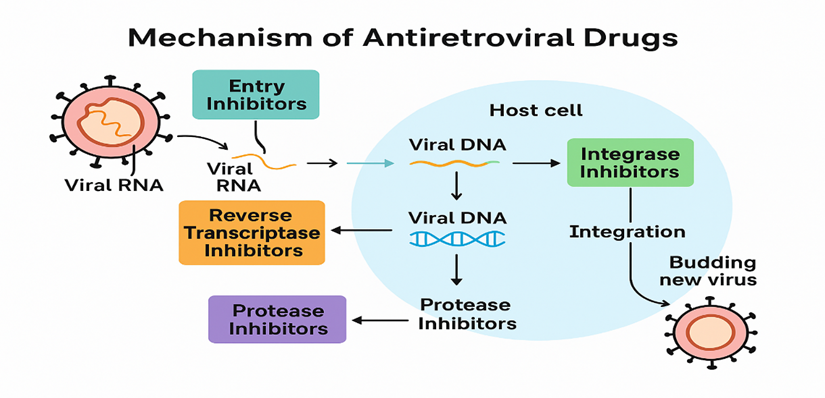
Fig.2 Mechanism of ARV Drugs
RESULT:
3.Bioanalytical Techniques for Estimation of Anti-HIV
3.1 Chromatographic methods
Ultra-rapid chromatographic techniques like HPLC and UPLC are widely used for analyzing antiviral drugs, enabling simultaneous quantification with high sensitivity and specificity. Mass spectrometry (MS) and tandem mass spectrometry (MS/MS) are the primary detectors used, offering excellent sensitivity and selectivity. Among these, LC-MS and LC-MS/MS have become the leading methods for determining ARV drugs Meng (2013). Fluorescence detection (FLD) offers a selective, robust, and cost-effective alternative to MS/MS in chromatographic analysis. Fast and stability-indicating HPLC methods are essential for analyzing antiviral drugs and metabolites. Though liquid-liquid and solid-phase extraction work well for plasma and urine, complex matrices like animal tissues require specialized preparation. A stationary phase in chromatographic systems is vital for the separation process, as the elution or retention of different substances depends on stationary phase properties. Column selection is important for sensitive and rapid analytical results Zuvela (2019).
The type of column packaging materials greatly impacts separation efficiency. Current trends focus on developing durable, high-performance, and environment friendly columns compatible with a wide range of mobile phases. With growing environmental concerns, green analytical chemistry is gaining prominence. Hassana et al. (2020) proposed a micellar HPLC-UV method for simultaneous estimation of Sofosbuvir and Daclatasvir in plasma and urine. HPTLC, an advanced form of TLC, offers several benefits for compound analysis Tuzimski and Sherma (2016). Samia M.et al.(2018) developed and validated a simple, efficient, and cost-effective HPTLC method for the simultaneous determination of Sofosbuvir and Ledipasvir in rabbit plasma. This method provided a straightforward, rapid, and economical alternative compared to reported LC-MS/MS methods.
3.2 Spectroscopic methods for Anti-HIV drugs
Spectrometric methods, based on light absorption or emission, such as UV, VIS, IR, fluorescence, and Raman spectroscopy, are widely used in pharmaceutical research for drug analysis and quality assessment. These techniques are especially valuable in developing regions due to their simplicity, cost-effectiveness, minimal sample requirements and reliable structural insights Mishra et al (2020). In HIV therapy, ARV combinations are vital, but genetic differences can impact drug bioavailability. Monitoring levels using HPLC with UV, MS detection is necessary for optimizing treatment. Tuzimski and Petruczynik (2020). Mansour (2017) introduced a spectrometric method to determine the concentration of Sofosbuvir and Ledipasvir in binary mixtures by identifying the intersection wavelength, both drugs were simultaneously quantified, with specific linear ranges and limits of detection (LODs).
Zaman et al. (2017) establish a simple spectrophotometric Synchronous fluorescence spectroscopy (SFS) method for determining sofosbuvir in vitro dissolution media. SFS offers a simple, selective, and sharp method for the simultaneous quantification of multiple analytes. Fourier transform infrared (FTIR) spectroscopy offers a non-destructive, solvent-free method for directly analyzing Daclatasvir dihydrochloride and Sofosbuvir in tablet formulations Mohamed A.et al., (2018).
Table No.1. Chromatographic methods for determination of anti-HIV drugs
|
Drugs |
Method |
Matrix |
Stationary Phase (Column) |
Mobile Phase |
Flow rate |
Ref |
|
3TC/DTG |
HPTLC |
Tablet |
Hypurity Advance (4.6x 50mm,5µm) |
0.1% formic acid buffer–acetonitrile (20:80%v/v) |
0.25ml/min |
(Venkatesh and Daggumati 2012) |
|
3TC/TDF |
RP-HPLC |
Tablet |
Phenomenax Luna C18 (150mm x 4.6 mm,5µm) |
Acetonitrile: Methanol: Water (30:50:20v/v) |
1.0ml/min |
(Karunakaran et al 2012) |
|
DRV |
HPLC |
Blood mononuclear cells (PBMCs) |
YMC-Pack Pro C18 (250×4.6mm,5µm) |
Potassium phosphate buffer: Acetonitrile (57:43v/v) |
1.0mL/min |
(Nagano et al 2013) |
|
ZDV |
RP-HPLC |
Human Plasma |
Phenomenex C18 (250×4.6 mm, 5 μm |
methanol and 0.1% orthophosphoric acid (40:60 %v/v), |
1.0mL/min |
Nalanda R. Baby (2016) |
|
EFV |
HPLC |
Human Plasma |
BDS hypersil C18 (15cm, 4.6mm x 5μm) |
Acetonitrile: 50mM sodium dihydrogen orthophosphate (pH 5.5,48:52v/v) |
0.8mL/min |
(Deepakumari et al 2017) |
|
ABC/3TC |
HPLC |
Tablet |
Symmetry Premsil C18 (250mm × 4.6mm,5μm) |
Methanol: Water (0.05% OPA, (pH 3,83:17v/v) |
1.0ml/min |
(Sufiyan Ahmad et al 2018) |
|
FTC/TDF |
RP-HPLC |
Tablet |
Inspire C18 (150×4.6mm 5.0μm) |
OPA Buffer (pH 2.5 with NaOH): Methanol (30:70v/v) |
1.0ml/min |
(Sattar and Achanta 2018) |
|
3TC/ABC/DTG |
RP-UPLC |
Tablet |
Zodiac sil RP C18 (4.6 mm × 250 mm, 3.0μm) |
Phosphate buffer: Methanol (30:70%v/v) |
0.25ml/min |
(Dubey and Duggirala 2018) |
|
PAR/RTV/OMB |
HPLC |
Tablet |
RP-C18 Kinetix (150mm×4.6mm,5μm) |
MP-A: 0.15 M SLS, 0.01 M of sodium dihydrogen phosphate dihydrate (pH 6.2) MP-B: Ethanol |
1ml/min. |
(Ibrahim et al 2019) |
|
HPTLC |
Aluminum silica gel 60F254 pre-coated plates (20 ×20 cm) |
Methylene chloride: Methanol: Ethyl acetate: (5:1:3:1v/v/v/v) Ammonia (25%)
|
|
|||
|
DTG |
RP-HPLC |
Tablet |
Kromasil C8(150 × 4.6 mm 5μm) |
MP-A: 0.1% of trifluoroacetic acid MP-B: Methanol (Gradient elution) |
1.0mL/min |
(Venkatnarayana and Jyothi 2020) |
|
LPV |
HPLC‐UV |
Rat Plasma |
Phenomenex Gemini (C18, 150 mm × 2.0 mm, 5 μm) |
water and acetonitrile |
0.3 mL/min |
Chaolong Qin (2020) |
Table No. 2. UPLC-MS/MS methods for determination of anti-HIV drugs
|
Analyte |
Method |
Matrix |
Stationary phase (column) |
Mobile phase |
Flow rate |
Ref |
|
ATV/DRV/RTV |
UPLC-MS/MS |
Human Plasma |
Waters Acquity UPLC C18 (50 × 2.1mm, 1.7μm) |
10mM Ammonium Formate: acetonitrile (50:50 v/v) |
0.300mL/min |
(Mishra and Shrivastav 2014) |
|
3TC/ZDV/LPV/RTV |
UPLC-MS/MS |
Breast milk |
ACQUITY UPLC1BEH C18 (2.1 ×50mm,1.7μm) |
MP-A:0.1% formic acid in water MP-B: ACN (90:10% v/v) |
0.50mL/min |
(Ramirez et al 2018) |
|
DAC/ELB/GZR/ LDV/SIM/SOF/VEL |
UPLC-MS/MS |
Human Plasma |
Acquity UPLC BEH C18 (2.1 x 50mm,1.7μm) |
10mM ammonium formate with MP-A: 0.005% formic acid in water (pH 4.5) MP-B: 0.005 % formic acid in Methanol MP-C: MP B 0.005 % formic acid in a mixture of methanol and acetonitrile (10:90) |
0.4ml/min. |
(Seyen et al 2018) |
|
DTG/EVG/RAL/NVP/ETV |
UPLC-MS/MS |
Human Plasma |
Acquity UPLC BEH C18(2.1 x 50mm1,7μm) |
MP-A:0.1% formic acid in water MP-B:0.1% formic acid in acetonitrile (Gradient elution) |
0.475mL/min |
(Pauline et al 2018) |
|
ABC/ATV/BIC/DTG/ EFV/EVG/FTC/ 3TC/NVP/RAL/RPV/RTV/TFV/ETV |
UPLC-MS/MS |
Human Plasma |
Acclaim TMRSLC 120 C18 (2.1 × 100 mm, 2.2μm) |
0.1% (v/v) formic acid in water: acetonitrile |
0.45mL/min |
(Gouget et al 2020) |
Table No. 3. LC-MS/MS methods for determination of anti-HIV drugs
|
Analyte |
Matrix |
Stationary phase (column) |
Mobile phase |
Flow rate |
Ref |
|
TFV/FTC/3TC |
Human Plasma |
ACE 5 CN column (150 mm × 4.6 mm, 5 μm) |
0.5% formic acid in water and acetonitrile (55:45, v/v). |
1 ml/min |
Manish Yadav et al (2010) |
|
ZDV/ABC/FTC/ 3TC/TFN/RBV
|
Human Plasma |
Synergy Polar RP C18 column (150mm×2.0mm ID,4μm) |
0.1% (v/v) formic acid in water and 0.1% (v/v) formic acid in methanol |
300μL/min |
(W. Kromdijk et al (2013) |
|
LPV/RTV/TFV |
Human Plasma |
Synergi column (100 × 2.0 mm; 4-μm) |
buffers A (water with 0.1% HAc) and B (acetonitrile with 0.1% HAc) |
0.35 ml/min |
Josefin Koehn (2014) |
|
Maraviroc |
Human Plasma |
Waters BEH C8, 50 × 2.1 mm UPLC column, 1.7 μm |
Mobile phase A: water with 0.1% formic acid Mobile phase B: 0.1% formic acid |
1 ml/min |
Emory J. F. (2014) |
|
TFV |
HPLC-MS/MS Human Plasma |
C18 (Phenomenex Kinetex TM 30 mm × 2.1 mm, 2.6 µm) |
10 mmol/L ammonium acetate in water and acetonitrile/methanol (50:50, v/v) |
- |
Mulubwa et al (2015) |
|
EFV |
Dried breast milk |
Fortis TM C18 column: 3 mm, 100 mm×2.1 mm |
1 mM ammonium acetate in water/acetonitrile |
400 mL/min |
Adeniyi Olagunju et al (2015) |
|
BIC |
Human Plasma |
Kinetex EVO C18 column, 50 × 3.0 mm, 5 µm |
80:20 acetonitrile/water with 0.1% formic acid |
0.250 mL/min |
Pavan Kumar Prathipati et al (2018) |
|
EFV/3TC/TDF/FTC |
Dried blood and dried breast milk spots |
Synergi polar column (80A, 150 ×2.0 mm, 4μm |
0.1% formic acid in water and 0.1% formic acid in acetonitrile |
400 μL/min |
Catriona Waitt et al (2017) |
|
TFV/EFV |
Mouse vaginal tissue, Vaginal lavage and Blood plasma |
RP C18 column (3 m, 100 × 2.1 mm) |
0.1% (v/v) formic acid in water and 0.1% (v/v) formic acid in acetonitrile |
0.35 mL min−1 |
Luisa Barreiros et al (2017) |
|
βLAP |
Human Plasma |
Phenomenex Kinetex C18(2.1×50mm, 1.7μm) |
MP-A:0.1% formic acid in water MP-B:0.1% formic acid in acetonitrile (Gradient elution) |
0.5mL/min. |
(Putnam et al 2020) |
CONCLUSION :
This review summarizes key analytical methods from 2011 to 2020 for detecting antiviral agents in biological, environmental and pharmaceutical samples. Techniques like, spectrophotometry, HPTLC, HPLC, UPLC, LC-MS, LC-MS/MS are widely used, especially in serum and plasma analysis. HPLC, particularly with reverse-phase C18/C8 columns and acidic mobile phases, remains the preferred method due to its reliability, cost-effectiveness, and broad accessibility. UV detection often lacks the sensitivity needed for antiviral analysis in biological samples. LC-MS/MS is the gold standard for pharmaceutical analysis in complex matrices due to its high sensitivity and specificity. The rising use of antivirals, developing robust, selective and sensitive analytical/Bioanalytical methods is crucial.
ACKNOWLEDGEMENT: Not applicable
SOURCE OF FUNDING: Not applicable
CONFLICT OF INTEREST: None.
ABBREVIATIONS:
|
OPA |
Ortho Phosphoric acid |
|
SOF |
Sofosbuvir |
|
TFV |
Tenofovir |
|
DRV |
Darunavir |
LDV |
Ledipasvir |
EFV |
Efavirenz |
||
|
DTG |
Dolutegravir |
3TC |
Lamivudine |
TDF |
Tenofovir Disoproxil Fumarate |
||
|
ZDV |
Zidovudine |
RTV |
Ritonavir |
TAF |
Tenofovir Alafenamide |
||
|
ABC |
Abacavir |
FTC |
Emtricitabine |
NVP |
Nevirapine |
||
|
DAC |
Daclatasvir |
βLAP |
β-lapachone |
BIC |
Bictegravir |
||
|
COBI |
Cobicistat |
EVG |
Elvitegravir |
RPV |
Rilpivirine |
||
|
LPV |
Lopinavir |
ELB |
Elbasvir |
GZR |
Grazoprevir |
||
|
SIM |
Simeprevir |
VEL |
Velpatasvir |
PAR |
Paritaprevir |
||
|
OMB |
Ombitasvir |
RAL |
Raltegravir |
ETV |
Entecavir |
||
|
ATV |
Atazanavir |
RBV |
Ribavirin |
EVG |
Elvitegravir |
||
|
|
|
|
|
|
|
|
|
BIBLIOGRAPHY:
1. Arts E.J., Hazuda D.J.,2012. HIV-1 antiretroviral drug therapy. Cold Spring Harb Perspect Med.;2(4):1-24. https://doi.org/10.1101/cshperspect.a007161.
2. Saag M.S., Gandhi R.T., Hoy J.F., Landovitz R.J., Thompson M.A., Sax P.E., Smith D.M., Benson C.A., Buchbinder S.P., Rio C.D., Eron J.J.,2020. Antiretroviral Drugs for Treatment and Prevention of HIV Infection in Adults 2020 Recommendations of the International Antiviral Society-USAPanel. Clin. Rev.Educ.;324(16):1651–1669 https://doi.org/10.1001/jama.2020.17025.
3. Charbe N.B., Zacconi FC, Amnerkar N., Ramesh B., Tambuwala M.M., Clementi E.,2018. Bio-analytical Assay Methods used in Therapeutic Drug Monitoring of Antiretroviral Drugs-A Review. Curr. Drug ther.; 14(1):16–57. https://doi.org 10.2174/1574885514666181217125550.
4. Mohammad M.M., Beqqali A.E., Rehim M.A.,2016. Bioanalytical method development and validation:critical concepts and strategies. J. Chromatogr. B;1-38, http://dx.doi.org/10.1016/j.jchromb.2016.09.028.
5. Deshpande M.M., Kasture V.S., Mohan M., Chavan M.J.,2019. Bioanalytical Method Development and Validation: A Review. Recent Adv. Anal. Chem.; 1-19, http://dx.doi.org/10.5772/intechopen.81620.
6. Pau A.K., George J.M., 2014. Antiretroviral Therapy Current Drugs, Am. J. Infect. Dis. ;28:371–402. http://dx.doi.org/10.1016/j.idc.2014.06.001.
7. Holec A.D., Mandalb S., Prathipatib P.K., Destacheb C.J.,2017. Nucleotide Reverse Transcriptase Inhibitors: A Thorough Review, Present Status and Future Perspective as HIV Therapeutics. Curr. HIV.Res.;15:411-421. https://doi.org/10.2174/1570162X15666171120110145.
8. Ammaranond P., Sanguansittianan S.,2012. Mechanism of HIV antiretroviral drugs progress toward drug resistance.Fundam Clin Pharmacol.; 26(1):146–161. https://doi.org/10.1016/B978-0-12-814515-9.00126-0.
9. Laskey S.B., Siliciano R.F.,2014. A mechanistic theory to explain the efficacy of antiretroviral therapy. Nat. Rev. Microbiol.;12(11):772–780. https://doi.org/10.1038/nrmicro3351.
10. Meng M., Wang L., Voelker T., Reuschel S., Van Horne K.C., Bennett P., 2013. A systematic approach for developing a robust LC-MS/MS method for bioanalysis. Bioanalysis.;5(1):91–115. https://doi.org/ 10.4155/BIO.12.295.
11. Zuvela P., Skoczylas M., Liu J.J, Baczek T., Kaliszan R., Wong M.W., Buszewski B.,2019. Column Characterization and Selection Systems in Reversed-Phase High-Performance Liquid Chromatography. Chem Rev;119(6):3674–3729. https://doi.org/10.1021/acs.chemrev.8b00246.
12. Hassan W.S., Elmasry M.S., Shalaby A., Sayed H.M., Zidan D.W.,2020. Micellar high performance liquid chromatographic method for separation and validation of two anti-hepatitis C- virus drugs in pure form, human plasma and human urine. Ann Pharm Fr.;78(3):217–29. https://doi.org/10.1016/j.pharma.2020.01.007.
13. Tuzimski T., Sherma J..2015.High-Performance Liquid Chromatography versus Other Modern Analytical Methods for Determination of Pesticides.;491–530.
14. Samia M., Gizawy E., Salwa R., Shaboury E., Noha N. A., Abo-Zeid M.N.,2018. New, simple and sensitive HPTLC method for simultaneous determination of anti-hepatitis C Sofosbuvir and Ledipasvir in rabbit plasma,J.Chromatogr. B; 1092:432–439. https://doi.org/10.1016/j.jchromb.2018.06.033.
15. Mishra R.K.,Chaubey N., Patel J.R.,Mishra S., Singh R.,2020. A Review Of Analytical Techniques For Determination of Anti-Hiv Drugs, Int J App Pharm,;12(6):41-50, http://dx.doi.org/10.22159/ijap.2020v12i6.39040.
16. Tuzimski T., Petruczynik A.,2020. Review of Chromatographic Methods Coupled with Modern Detection Techniques Applied in the Therapeutic Drugs Monitoring (TDM). Molecules.;25(17):2-17. http://dx.doi.org/10.3390/molecules25174026.
17. Mansour F.R.,2018. A new innovative spectrophotometric method for the simultaneous determination of sofosbuvir and ledipasvir, Spectrochim. Acta A Mol. Biomol. Spectrosc. 188:626–632. http://dx.doi.org/ 10.1016/j.saa.2017.07.066.
18. Zaman B., Hassan W., Khan A., Noreen H.,2017.Development and validation of spectrophotometric method for assay determination and in vitro dissolution studies of sofosbuvir tablets. J Chem Soc Pakistan.;39(6):962–969.
19. Abdel-Lateef M.A., Omar M.A., Ali R., Derayea S.M.,2019. Xanthene based spectroscopic probe for the analysis of HCV antiviral, daclatasvir dihydrochloride, through feasible complexation reaction. Microchem J.;145:672–675. https://doi.org/10.1016/j.microc.2018.11.038.
20. Venkatesh P., Daggumati M.,2012. Development and validation of a normal-phase HPTLC method for the simultaneous analysis of Lamivudine and Zidovudine in fixed-dose combination tablets. J Pharm Anal.;2(2):152–155. https://doi.org/10.1016/j.jpha.2011.11.002.
21. Karunakaran A., Kamarajan K., Thangarasu V.,2012. A validated RP-HPLC method for simulataneous estimation of Lamivudine and Tenofovir disoproxil fumarate in pure and in tablet dosage form. Eurasian J Anal Chem.;7(2):56–66.
22. Nagano D., Araki T., 1akamura T., Yamamoto K.,2013. Determination of Intracellular Darunavir by Liquid Chromatography Coupled with Fluorescence Detection. J. Chromatogr. Sci. 52:1021–1025. https://doi.org/10.1093/chromsci/bmt147.
23. Baby, N. R., Rao, S. A., Naidu, P. S., Surla, D., & Bollab, N. 2016. Simple and rapid method development and validation of RP-HPLC method for the determination of zidovudine in human plasma. Journal of Pharmacy Research, 10(4), 160-166.
24. Deepakumari H.N., Mallegowda S.M., Prashanth M.K., Revanasiddappa H.D.,2017. Bio Analytical Method Development Of an Anti-HIV-1 Drug Efavirenz Using High Performance Liquid Chromatography In Human Plasma. Materials Today;4: 11939–11946.
25. Sufiyan A.R., Patil L., Mohammed R.U., Mohammad I., Rashid A.,2018.Analytical Method Development and Validation for the Simultaneous Estimation of Abacavir and Lamivudine by Reversed phase High performance Liquid Chromatography in Bulk and Tablet Dosage Forms. Pharmacogn. Res.;10(1):92-97. https://doi.org/10.4103/pr.pr_96_17.
26. Sattar A., Achanta S.,2018.Analytical Method Development and Validation for the Determination of Emtricitabine and Tenofovir Disoproxil Fumarate Using Reverse Phase HPLC Method in Bulk and Tablet Dosage Form, J. Pharm. Sci. & Res.;10(5):1207-1212.
27. Dubey S.,Duggirala M.,2018. Simultaneous Estimation of Lamivudine, Abacavir and Dolutegravir by Uplc Method.International Journal of Applied Pharmaceutics;10(1):46-52. http://dx.doi.org/10.22159/ijap.2018v10i1.21156.
28. Ibrahim A.E., Saraya R.E., Saleh H., Elhenawee M.,2019. Development and validation of eco-friendly micellar-HPLC and HPTLC-densitometry methods for the simultaneous determination of paritaprevir, ritonavir and ombitasvir in pharmaceutical dosage forms. Heliyon;5:1-13. https://doi.org/10.1016/j.heliyon.2019.e01518.
29. Venkatnarayana M., Siva J. N.,2020. Development of Validation and Stability Indicating Method of Anti-HIV Dolutegravir Drug and its Related Impurities by Using RP-HPLC. J. Chromatogr. Sep. Tech.;11(1):1-7. https://dx.doi.org/10.35248/2157-7064.20.11.426.
30. Qin, C., Feng, W., Chu, Y., Lee, J. B., Berton, M., Bettonte, S., ... & Gershkovich, P. 2020. Development and validation of a cost‐effective and sensitive bioanalytical HPLC‐UV method for determination of lopinavir in rat and human plasma. Biomedical Chromatography, 34(11), e4934.
31. Mishra T., Shrivastav P.S.,2014. Validation of Simultaneous Quantitative Method of HIV Protease Inhibitors Atazanavir, Darunavir and Ritonavir in Human Plasma by UPLC-MS/MS. The ScientificWorld Journal;1-12. http://dx.doi.org/10.1155/2014/482693.
32. RamõÂrez.A., ElõÂas S.S., Giselle L.F.,NoemõÂ P.C., Rosa Georgina R.D., Ricardo F.D., Mauricio D.C., Margarita L.M., Zayra F.G., Jessica H.P.,2018. Simultaneous quantification of four antiretroviral drugs in breast milk samples from HIV-positive women by an ultra-high performance liquid chromatography tandem mass spectrometry (UPLC-MS/MS) method. Plos One;13(1):1-15. https://doi.org/10.1371/journal.pone.0191236.
33. Seyen M.V., Marga J.A. Teulen D.G., Nielka P. van Erp, Burger D.M.,2019. Quantification of second generation direct-acting antivirals daclatasvir, elbasvir, grazoprevir, ledipasvir, simeprevir, sofosbuvir and velpatasvir in human plasma by UPLC-MS/MS.Journal of Chromatography B; https://doi.org/10.1016/j.jchromb.2019.01.024.
34. Pauline D.J.,Bollen, Marga J.A., eulen D.G., Schalkwijk S., Nielka P., Erp V.,David M., Burger,2018. Development and validation of an UPLC-MS/MS bioanalytical method for simultaneous quantification of the antiretroviral drugs dolutegravir, elvitegravir, raltegravir, nevirapine and etravirine in human plasma. J. Chromatogr. B.1-29. https://doi.org/10.1016/j.jchromb.2018.12.008.
35. Gougeta H.,Noéb G., Aurélie B.T., FurlanbV.,2019.UPLC–MS/MS method for the simultaneous quantification ofbictegravir and 13 others antiretroviral drugs plus cobicistat andritonavir boosters in human plasma. J Pharm Biomed Anal. ;181:1-8. https://doi.org/10.1016/j.jpba.2019.113057.
36. Yadav, M., Singhal, P., Goswami, S., Pande, U. C., Sanyal, M., & Shrivastav, P. S. 2010. Selective determination of antiretroviral agents tenofovir, emtricitabine, and lamivudine in human plasma by a LC-MS-MS method for a bioequivalence study in healthy Indian subjects. J Chromatogr Sci, 48(9), 704-713.
37. W. Kromdijk, S.A. Pereira, H. Rosing, J.W. Mulder, J.H. Beijnen, A.D.R. Huitema,2013. Development and validation of an assay for the simultaneous determination of zidovudine, abacavir, emtricitabine, lamivudine, tenofovir and ribavirin in human plasma using liquid chromatography–tandem mass spectrometry.Journal of Chromatography B.919–920,43-51. https://doi.org/10.1016/j.jchromb.2013.01.005.
38. Koehn, J. and Ho, R.J., 2014. Novel liquid chromatography-tandem mass spectrometry method for simultaneous detection of anti-HIV drugs lopinavir, ritonavir, and tenofovir in plasma. Antimicrob. Agents Chemother, 58(5).2675-2680.
39. Emory, J. F., Seserko, L. A., & Marzinke, M. A. 2014. Development and bioanalytical validation of a liquid chromatographic-tandem mass spectrometric (LC-MS/MS) method for the quantification of the CCR5 antagonist maraviroc in human plasma. Clinica Chimica Acta, 431, 198-205.
40. Mulubwa, M., Rheeders, M., Du Plessis, L., Grobler, A., & Viljoen, M. 2015. Development and validation of high performance liquid chromatography tandem mass spectrometry (HPLC-MS/MS) method for determination of tenofovir in small volumes of human plasma. J. Chromatogr. Sep. Tech., 6(7), 1.
41. Olagunju, A., Bolaji, O. O., Amara, A., Waitt, C., Else, L., Soyinka, J., ... & Khoo, S. 2015. Development, validation and clinical application of a novel method for the quantification of efavirenz in dried breast milk spots using LC-MS/MS. J Antimicrob Chemother 2015; 70: 555 –561 doi: https://doi.org/10.1093/jac/dku420.
42. Waitt, C., Penchala, S. D., Olagunju, A., Amara, A., Else, L., Lamorde, M., & Khoo, S. 2017. Development, validation and clinical application of a method for the simultaneous quantification of lamivudine, emtricitabine and tenofovir in dried blood and dried breast milk spots using LC–MS/MS. Journal of Chromatography B, 1060, 300-307. http://dx.doi.org/10.1016/j.jchromb.2017.06.012.
43. Barreiros, L., Cunha-Reis, C., Silva, E. M., Carvalho, J. R., das Neves, J., Sarmento, B., & Segundo, M. A. 2017. Development and validation of a liquid chromatography-MS/MS method for simultaneous quantification of tenofovir and efavirenz in biological tissues and fluids. J. Pharm. Biomed. Anal., 136. 120-125.
44. Prathipati, P. K., Mandal, S., & Destache, C. J. 2019. Development and validation of LC–MS/MS method for quantification of bictegravir in human plasma and its application to an intracellular uptake study. Biomedical Chromatography, 33(2), e43qin79.
45. Putnam W.C., Kallem R.R., Subramaniyan I., Beg M.S., Edpuganti V.,2020. Bioanalytical method development and validation of a liquid chromatography-tandem mass spectrometry method for determination of -lapachone in human plasma. J. Pharm. Biomed. Anal. 1-33. https://doi.org/10.1016/j.jpba.2020.113466.