“EFFECT OF PROPOFOL PRIMING ON INDUCTION DOSE REQUIREMENT AND HAEMODYNAMIC RESPONSE: A RANDOMIZED CONTROLLED TRIAL”
- Jyoti Kunal Nikale , Senior Consultant Dept. of Anaesthesiology, Ashoka Medicover Hospital. Nashik
Article Information:
Abstract:
Due to its quick onset, smooth recovery, and good intubating conditions, propofol is often used to start general anesthesia. But it does cause cardiovascular depression that depends on the dose. The priming principle, which involves giving a sub-anesthetic dose before the full induction dose, may lower the total amount of propofol needed and make hemodynamic instability less severe. Objective: To assess the impact of propofol priming on the total induction dose requirement and the hemodynamic response during intubation. Materials and Methods: This prospective, randomized, controlled study involved 100 ASA physical status I and II patients aged 18–55 years undergoing elective surgeries under general anesthesia. Patients were randomly assigned to two groups (n=50 each). Group I (the control group) got 2 mg/kg of propofol through an IV until they lost their eyelash reflex. Group II (priming group) got 20% of the calculated dose as a priming dose 30 seconds before the rest of the dose was given, which was adjusted until the eyelash reflex was lost. The total induction dose, heart rate, systolic blood pressure (SBP), diastolic blood pressure (DBP), mean arterial pressure (MAP), and any complications were all noted. We used Student's unpaired t-test and Chi-square test to do the statistical analysis. Results: The initial demographic variables were similar across groups. The average induction dose in the priming group (72.28 ± 15.36 mg) was much lower than in the control group (96 ± 13.77 mg), which is a 28.05% drop (p < 0.001). Changes in heart rate were similar, except for 5 minutes after induction, when the priming group had better maintenance (p = 0.046). At one minute after induction, right after intubation, and five minutes after induction, the priming group had much better SBP, DBP, and MAP (p < 0.001 at most time points). The control group had a higher rate of hypotension and apnea, but this was not statistically significant. Fasciculations after suxamethonium were noted in 12% of patients in the priming group. Conclusion: Using the priming principle greatly lowers the amount of propofol needed to start an induction and makes the patient's blood pressure more stable during intubation. This easy and cheap plan might work best for people who are worried about their heart health.
Keywords:
Article :
INTRODUCTION:
The introduction of intravenous anesthetics was a major step forward in the history of anesthesia. Before they were used, general anesthesia was mostly done with inhaled gases or volatile agents, which was often uncomfortable and not well tolerated by patients [1].
The creation of intravenous induction agents not only made patients more comfortable, but it also made starting anesthesia smoother and more controlled.
Intravenous induction agents with rapid onset are usually very strong and can cause big changes in blood flow. Of these drugs, propofol is one of the most commonly used and preferred for starting general anesthesia. Propofol has many benefits over traditional induction agents. These include a quick onset of hypnosis, antiemetic properties, a faster recovery profile, effective suppression of pharyngeal, laryngeal, and tracheal reflexes, and good intubating conditions with enough depth of anesthesia.
Even with these benefits, propofol has a major drawback: it can cause cardiovascular depression that depends on the dose. Rapid induction with propofol typically leads to a considerable decrease in systemic arterial blood pressure. Research indicates that the administration of propofol at a dosage of 2 mg/kg may lead to a decrease of approximately 26–28% in systolic blood pressure and 18–20% in diastolic blood pressure [2,3]. These kinds of changes in blood flow can be important for patients, especially those with limited cardiovascular reserve.
To lessen these negative effects, researchers have looked into a number of ways to lower the amount of propofol needed to start an induction. These include giving nitrous oxide [4–6], opioids [7,8], barbiturates like thiopentone [9], benzodiazepines like midazolam [10], clonidine [11], and local anaesthetics [12], magnesium sulphate [11], and using the "priming principle" [13,14]. Even though these methods may lower the total induction dose, they are often linked to more side effects, like too much sedation, breathing problems, or more problems with blood flow.
The "priming principle" entails the administration of a sub-anesthetic dose of a drug preceding the full induction dose. This idea has been used a lot when using non-depolarizing neuromuscular blocking agents. In this context, priming has been demonstrated to decrease the onset time of neuromuscular blockade, enhance intubating conditions, and lower the overall dosage requirement of the muscle relaxant [15,16]. The priming technique has other benefits, such as lowering the number and severity of dose-related side effects and, for neuromuscular blockers, a smaller total dose than the usual intubating dose. If the priming principle can also lower the induction dose of propofol, it may have two important benefits for patients. First, it was possible to lessen the hemodynamic problems that come with quick induction. Second, the total cost of anesthesia may be lower, which is especially important because propofol is more expensive than older induction agents.
In this study, a priming dose of propofol equal to 20% of the calculated induction dose (2 mg/kg) was given. After a 30-second break, the rest of the dose was titrated until the eyelash reflex was lost. The purpose of this study was to assess the impact of this priming strategy on the overall propofol requirement and the corresponding hemodynamic responses during the induction of general anesthesia.
MATERIALS AND METHODS:
Individuals classified as ASA physical status III or IV, those declining participation, pregnant or lactating women, patients with allergies to study medications, and individuals with uncontrolled cardiovascular, respiratory, hepatic, or renal conditions were excluded. The Institutional Ethics Committee approved this prospective, randomized, controlled study before it began. All participants provided written informed consent. The study enrolled 100 patients, aged 18–55 years, of either sex, classified as American Society of Anesthesiologists (ASA) physical status I and II, and scheduled for elective surgeries under general anesthesia.
All patients received a thorough pre-anesthetic evaluation, which included a medical history, a physical exam, and an assessment of their airways. Baseline parameters, including heart rate and blood pressure, were documented as the mean of two measurements obtained at least 10 minutes apart, 30 minutes before surgery. Routine investigations encompassed hemoglobin assessment and urinalysis; supplementary tests, including blood glucose measurement, electrocardiography, and chest radiography, were conducted as clinically warranted.
Using a random number table and a sealed envelope, participants were randomly put into two equal groups, each with 50 people. Standard monitoring, such as pulse oximetry, non-invasive blood pressure, electrocardiography, and capnography, was put in place. We got intravenous access and started the Ringer's lactate infusion. All patients were given glycopyrrolate 0.004 mg/kg through an IV, midazolam 0.03 mg/kg through an IV 15 minutes before induction, and fentanyl 1 µg/kg through an IV 30 seconds before induction.
In Group I (the control group), propofol 2 mg/kg was given through an IV at a rate of 30 mg every 10 seconds until the eyelash reflex was lost. In Group II (the priming group), 20% of the calculated induction dose of propofol (2 mg/kg) was given as a priming dose. After 30 seconds, the rest of the dose was given at the same rate until the eyelash reflex was lost. After induction, neuromuscular blockade was attained with 1 mg/kg of suxamethonium administered intravenously, and endotracheal intubation was executed. Anaesthesia was sustained with a mixture of oxygen and nitrous oxide (50:50), supplemented by vecuronium as necessary. There was no surgical stimulus during the first five minutes after induction.
The main variable that was measured was the total amount of propofol needed for induction, which included the priming dose in Group II. Secondary outcomes encompassed heart rate, systolic blood pressure, diastolic blood pressure, and mean arterial pressure measured at the following intervals: pre-induction, one minute post-induction, immediately post-intubation, and five minutes post-induction. Complications, including hypotension (systolic blood pressure <90 mmHg), apnea exceeding 30 seconds, vomiting, involuntary movements, laryngospasm, coughing, and fasciculations, were recorded.
The sample size was calculated by assuming an 80% study power and a significance level (α) of 0.05, which meant that there would be 50 patients in each group. Continuous variables were represented as mean ± standard deviation and analyzed using Student’s unpaired t-test. The Chi-square test was used to look at categorical variables. A p-value of less than 0.05 was deemed statistically significant.
RESULT:
One hundred ASA I and II patients aged 18 to 55 who were having elective surgeries under general anesthesia were randomly assigned to two groups (n = 50 each). Group I (control) received propofol at a dosage of 2 mg/kg for induction, while Group II (priming group) received 20% of the calculated dose as a priming dose, subsequently followed by titration of the remaining dosage until the loss of the eyelash reflex occurred.
Demographic Characteristics: The demographic variables were similar in both groups. The average age in Group I was 29.60 ± 6.51 years, and the average age in Group II was 29.68 ± 7.34 years (p = 0.954). The groups had a similar number of males and females (Group I: 13 males, 37 females; Group II: 15 males, 35 females; p = 0.82). The average body weight for Group I was 50.66 ± 7.44 kg and for Group II it was 52.74 ± 9.81 kg (p = 0.235). These differences were not statistically significant.(Table 1)
Table 1: Baseline Characteristics of Study Participants and mean induction dose used among study subjects
|
Variable |
Group I (n=50) |
Group II (n=50) |
p-value |
|
Age (years) |
29.60 ± 6.51 |
29.68 ± 7.34 |
0.954 |
|
Male/Female |
13 / 37 |
15 / 35 |
0.82 |
|
Weight (kg) |
50.66 ± 7.44 |
52.74 ± 9.81 |
0.235 |
|
Mean induction dose (mg) |
96 ± 13.77 |
72.28 ± 15.36 |
<0.001* |
Induction Dose Requirement: The average dose of propofol used to start the control group (96 ± 13.77 mg) was much higher than the dose used to start the priming group (72.28 ± 15.36 mg) (p < 0.001). This means that the total amount of induction dose needed is 28.05% less when the priming principle is used.(Table 1)
Heart Rate: The baseline heart rate was similar between the two groups (84.66 ± 10.41 vs 87.08 ± 11.48 beats/min; p = 0.272). One minute after induction and right after intubation, the heart rate went up in both groups, but the difference between the two groups was not statistically significant (p = 0.496 and p = 0.311, respectively). Five minutes after induction, though, Group II's heart rate was much better maintained (88.00 ± 11.71 beats/min) than Group I's (83.58 ± 10.13 beats/min) (p = 0.046).(Table 2)
Table 2: Heart Rate
|
Time Point |
Group I |
Group II |
p-value |
|
Pre-induction |
84.66 ± 10.41 |
87.08 ± 11.48 |
0.272 |
|
1 min after induction |
91.78 ± 10.90 |
93.42 ± 11.64 |
0.496 |
|
Immediately after intubation |
97.68 ± 10.44 |
100.24 ± 11.37 |
0.311 |
|
5 min after induction |
83.58 ± 10.13 |
88.00 ± 11.71 |
0.046* |
Systolic Blood Pressure: The baseline systolic blood pressure (SBP) was marginally elevated in Group II (116.88 ± 7.50 mmHg) relative to Group I (113.44 ± 8.16 mmHg) (p = 0.031). One minute after induction, SBP dropped a lot in Group I (101.84 ± 6.60 mmHg) compared to Group II (112.76 ± 7.50 mmHg) (p < 0.001). Right after intubation, SBP went up in both groups, but it stayed much higher in Group II (123.72 ± 9.07 mmHg) than in Group I (116.40 ± 9.80 mmHg) (p < 0.001). Five minutes after induction, SBP was still much higher in Group II (116.48 ± 7.93 mmHg) than in Group I (103.04 ± 6.36 mmHg) (p < 0.001).(Table 3)
Table 3: Systolic Blood Pressure
|
Time Point |
Group I |
Group II |
p-value |
|
Pre-induction |
113.44 ± 8.16 |
116.88 ± 7.50 |
0.031* |
|
1 min after induction |
101.84 ± 6.60 |
112.76 ± 7.50 |
<0.001* |
|
Immediately after intubation |
116.40 ± 9.80 |
123.72 ± 9.07 |
<0.001* |
|
5 min after induction |
103.04 ± 6.36 |
116.48 ± 7.93 |
<0.001* |
Diastolic Blood Pressure: The baseline diastolic blood pressure (DBP) was similar across groups (p = 0.059). One minute after induction, DBP dropped a lot in Group I (67.60 ± 5.03 mmHg) compared to Group II (76.48 ± 8.10 mmHg) (p < 0.001). After intubation, Group II had a much higher DBP (83.96 ± 9.29 mmHg) than Group I (78.60 ± 7.25 mmHg) (p = 0.008). Five minutes after induction, DBP was still much higher in Group II (78.12 ± 7.24 mmHg) than in Group I (68.64 ± 4.30 mmHg) (p < 0.001).(Table 4)
Table 4: Diastolic Blood Pressure
|
Time Point |
Group I |
Group II |
p-value |
|
Pre-induction |
75.64 ± 6.15 |
78.20 ± 7.19 |
0.059 |
|
1 min after induction |
67.60 ± 5.03 |
76.48 ± 8.10 |
<0.001* |
|
Immediately after intubation |
78.60 ± 7.25 |
83.96 ± 9.29 |
0.008* |
|
5 min after induction |
68.64 ± 4.30 |
78.12 ± 7.24 |
<0.001* |
Mean Arterial Pressure: At all measured time points, the priming group had a much better mean arterial pressure (MAP). One minute after induction, the mean arterial pressure (MAP) was 79.02 ± 4.96 mmHg in Group I and 88.56 ± 7.33 mmHg in Group II (p < 0.001). Right after intubation, the MAP was 91.19 ± 7.66 mmHg in Group I and 97.19 ± 8.53 mmHg in Group II (p < 0.001). Five minutes post-induction, MAP was significantly elevated in Group II (91.16 ± 6.70 mmHg) relative to Group I (80.19 ± 4.38 mmHg) (p < 0.001). These results show that the priming group had less variation in their blood flow.(Table 5)
Table 5: Mean Arterial Pressure (mmHg)
|
Time Point |
Group I |
Group II |
p-value |
|
Pre-induction |
88.23 ± 6.28 |
91.09 ± 6.62 |
0.030* |
|
1 min after induction |
79.02 ± 4.96 |
88.56 ± 7.33 |
<0.001* |
|
Immediately after intubation |
91.19 ± 7.66 |
97.19 ± 8.53 |
<0.001* |
|
5 min after induction |
80.19 ± 4.38 |
91.16 ± 6.70 |
<0.001* |
Complications: The overall rate of complications was 20% in Group I and 18% in Group II. Four percent of patients in Group I and two percent of patients in Group II felt pain when they got the shot. Eight percent of patients in Group I had low blood pressure (SBP <90 mmHg), while only two percent of patients in Group II had low blood pressure. Clinically significant apnea (>30 seconds) was observed in 8% of patients in Group I and 2% in Group II. However, fasciculations after suxamethonium were seen in 12% (6/50) of patients in Group II, but none were seen in Group I.(Figure 1)
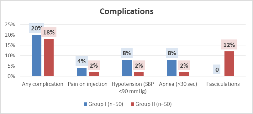
Figure 1: Incidence of Complications
DISCUSSION :
Induction of anesthesia is a very important part of giving general anesthesia because it is when changes in blood flow and drug-related side effects are most likely to happen. Before intravenous anesthetics were available, induction had to be done with gases or vapors that patients often found unpleasant and hard to handle. The introduction of thiopentone in 1934 represented a significant advancement in intravenous anaesthesia, and the subsequent development of newer agents further enhanced its clinical applicability [2].
Propofol (2,6-di-isopropyl phenol) has become one of the most popular modern intravenous induction agents because it works quickly, lasts a short time, stops nausea, has a smooth recovery profile, and does a better job of suppressing airway reflexes during intubation [3,18,19]. However, propofol is known to cause cardiovascular depression that depends on the dose. This is shown by lower systemic vascular resistance and arterial blood pressure [20,21]. Consequently, strategies designed to diminish the induction dose of propofol without sacrificing anaesthetic depth are of clinical significance.
Different methods have been tried to lower the amount of propofol needed to start anesthesia, such as giving nitrous oxide [4–6], opioids [7,8], barbiturates [9], benzodiazepines [10], clonidine, magnesium sulfate [11], and other drugs at the same time. These methods may lower the amount of medicine needed, but they can also cause problems like too much sedation, breathing problems, or more unstable blood flow.
The "priming principle," commonly utilized with non-depolarizing neuromuscular blockers to decrease onset time and enhance intubation conditions [15,16], has been examined as a method to lower the induction dose of propofol. Maroof et al. [13] were the first to use this idea with propofol on a small group of people and showed that it significantly lowered the amount of the drug needed.
The current study aimed to assess the efficacy of the priming principle—administering 20% of the calculated induction dose of propofol 30 seconds prior to the remaining dose—in reducing the total propofol requirement and mitigating hemodynamic changes during induction and intubation.
Requirement of Induction Dose: The implementation of the priming principle in this study led to a 28.05% decrease in the mean induction dose (96 ± 13.77 mg vs 72.28 ± 15.36 mg; p < 0.001). This reduction is more than what Maroof et al. [13] found, which was a 21.4% drop, but less than what Naphade et al. [14] found, which was a 35% drop.
The extent of reduction noted in our study may be ascribed to concurrent premedication with midazolam (0.03 mg/kg), which has demonstrated efficacy in diminishing propofol requirements [10]. Benzodiazepine pre-treatment augments GABAergic transmission, consequently reducing the hypnotic threshold and amplifying the effects of propofol.
Receptor theory can explain why the dose needed to get the same effect is lower at the molecular level. Miller [2] describes classical ligand–receptor interaction models that say giving a sub-hypnotic "priming" dose may make it more likely that receptors will be activated by partially filling GABAA receptor complexes. The subsequent administration of the remaining dose necessitates a reduced quantity of the drug to attain the equivalent hypnotic endpoint. Propofol enhances GABAA receptor-mediated chloride conductance by augmenting agonist affinity and extending channel opening [25]. Sub-hypnotic doses also have an anxiolytic effect, lowering sympathetic drive before induction [25,26]. Anderson et al. [25] and Djaiani et al. [26] have reported analogous findings, illustrating diminished propofol requirements when sub-hypnotic doses were given prior to induction.
Effects on blood flow : The current study exhibited enhanced hemodynamic stability in the priming group. Heart rate changes were similar right after induction and intubation, but five minutes later, the priming group had a much better heart rate (p = 0.046). Propofol is known to cause a biphasic response in the heart and blood vessels. After administration, systemic vascular resistance drops right away, which causes reflex sympathetic activation and a temporary increase in heart rate. After that, baroreceptor resetting may happen, which would lower the heart rate and arterial pressure [27]. In our study, the more significant decrease in heart rate noted in the control group at five minutes likely indicates the larger total dose administered.
At one minute after induction, right after intubation, and five minutes after induction, the control group had much lower systolic, diastolic, and mean arterial pressures (p < 0.001 at most times). These results corroborate the dose-dependent characteristics of propofol-induced hypotension as delineated by Major et al. [23] and Pauline et al. [20], who illustrated progressively more significant reductions in arterial pressure with increased induction doses.
Caleys et al. [22] ascribed propofol-induced hypotension chiefly to afterload reduction without a compensatory increase in cardiac output, whereas Pensado et al. [21] noted diminished systemic vascular resistance without a substantial decrease in cardiac output. Fairfeild et al. [27] also found that systolic, diastolic, and mean arterial pressures all went down after propofol induction.
Our results are consistent with these studies and show that priming can effectively reduce dose fluctuations in the blood flow. The smaller difference from baseline haemodynamic parameters in the priming group shows that cardiovascular stability has improved.
Complications: There were slightly more complications in the control group (20%) than in the priming group (18%), but this difference was not statistically significant. The control group experienced a higher incidence of hypotension and clinically significant apnea, aligning with the dose-dependent cardiorespiratory depressant effects of propofol [23]. The occurrence of pain upon injection was minimal (3%), in contrast to previously documented rates of 28–90% [28]. This probably means that the injection went through a big antecubital vein and that fentanyl was given before, which has been shown to lessen the pain of propofol injection [29].
Interestingly, 12% of the patients in the priming group had fasciculations after taking suxamethonium, but none of the patients in the control group did. The reported occurrence of suxamethonium-induced fasciculations varies significantly, ranging from 1.5% to 89%, with a higher prevalence observed in females [30]. The lack of fasciculations in the control group may be due to a greater depth of anesthesia achieved with a higher bolus dose of propofol. It has been recorded that the frequency of fasciculations fluctuates with the depth of anaesthesia during the administration of neuromuscular blockers [31,32]. The relatively lighter depth in the priming group, attributable to a reduced total propofol dose, may elucidate the observed increased incidence.
The current study illustrates that the implementation of the priming principle in propofol induction leads to a substantial decrease in the total induction dose and enhanced hemodynamic stability, without a clinically significant rise in adverse effects. These results bolster the notion that dose-dependent cardiovascular depression linked to propofol can be mitigated via strategic dose fractionation.
CONCLUSION :
The present randomized prospective study demonstrates that application of the priming principle to propofol induction significantly reduces the total induction dose requirement. Administration of 20% of the calculated dose as a priming dose 30 seconds prior to the remaining dose resulted in a mean dose reduction of 28.05%, with an average effective induction dose of approximately 1.36 mg/kg.
This reduction in propofol requirement was associated with improved peri-intubation haemodynamic stability. Patients in the priming group exhibited significantly lesser reductions in systolic, diastolic, and mean arterial pressures following induction and intubation compared to the control group. The haemodynamic fluctuations observed were consistent with the dose-dependent cardiovascular depressant effects of propofol. The overall incidence of complications such as hypotension and apnea was lower in the priming group, although the difference did not reach statistical significance.
However, an increased incidence of suxamethonium-induced fasciculations (12%) was observed in the priming group, likely attributable to relatively lighter depth of anaesthesia at the time of neuromuscular blocker administration due to reduced propofol dosing.
In summary, the priming principle represents a simple, cost-effective, and clinically practical strategy to reduce propofol induction dose while maintaining haemodynamic stability. Its use may be particularly advantageous in patients where cardiovascular stability is of concern. Further studies in higher-risk populations and with larger sample sizes may help to better define its safety profile and clinical applicability.
CONFLICT OF INTEREST: NONE TO DECLARE
SOURCES OF FUNDING: NONE
BIBLIOGRAPHY:
1. Douglas RB, Wilkinson DJ. Great moments in the history of anesthesia. In: Healy TEJ, Knight PR, editors. Wylie and Churchill-Davidson’s A Practice of Anesthesia. London: Arnold Publishers; 2003. p. 10-11.
2. Lason MD. History of anesthetic practice. In: Miller RD, editor. Miller’s Anesthesia. Philadelphia: Natasha Andjelkovic; 2005. p. 28-29.
3. Glen JB. Animal studies of the anaesthetic activity of ICI 35 868. Br J Anaesth. 1980;52:731-741.
4. Wu NN, Hwang NC. Inhaling nitrous oxide reduces the induction dose requirements of propofol. Anesth Analg. 2000;90:1213-1216.
5. Prys-Roberts C, Pavies FR, Calverely RK, Goodman NW. Haemodynamic effects of infusions of di-isopropyl phenol (IC 35 868) during nitrous oxide anaesthesia in man. Br J Anaesth. 1983;55:105.
6. Coates DP, Monk CR, Prys-Roberts C, Turtle M. Haemodynamic effects of infusions of the emulsion formulation of propofol during nitrous oxide anaesthesia in humans. Anesth Analg. 1987;66:64-70.
7. Ben-Shlomo I, Finger J, Bav-AV E, Etchin A, Pere AZ, Tverskoy M. Propofol and fentanyl act additively for induction of anaesthesia. Anaesthesia. 1993;48:111-113.
8. Richards MJ, Skues MA, Jarvis AP, Prys-Roberts C. Total intravenous anaesthesia with propofol and alfentanil: Dose requirements for propofol and the effect of premedication with clonidine. Br J Anaesth. 1990;65:157-163.
9. Naguib M, Sari-Kouzel A. Thiopentone–propofol hypnotic synergism in patients. Br J Anaesth. 1991;67:4-6.
10. Cressy DM, Claydon P, Bhaskaran NC, Reilly CS. Effect of midazolam pretreatment on induction dose requirements of propofol in combination with fentanyl in older adults. Anaesthesia. 2001;56:108-113.
11. Altan A, Turgut N, Yildiz F, Turkmen A, Ustun H. Effects of magnesium sulphate and clonidine on propofol consumption, haemodynamics and postoperative recovery. Br J Anaesth. 2005;94(4):438-441.
12. Ben-Shlomo I, Tverskoy M, Fleyshman G, Cherniavsky G. Hypnotic effect of IV propofol is enhanced by IM administration of either lignocaine or bupivacaine. Br J Anaesth. 1997;78:375-377.
13. Maroof M, Khan RM. ‘Priming principle’ and the induction dose of propofol. Anesth Analg. 1996;82:S301.
14. Naphade RW, Agarwal PI. Effect of priming principle on the induction dose of propofol. ISA GoldCON. 2002;2-3.
15. Donati F. Editorial – The priming saga: where do we stand now? Can J Anaesth. 1988;35(1):1-4.
16. Baumgarten RK, Carter CE, Reynolds WN, Brown JL. Priming with nondepolarizing relaxants for rapid tracheal intubation: A double-blind evaluation. Can J Anaesth. 1988;35(1):5-11.
17. Fahy LT, Van Mourik GA, Utting JE. A comparison of the induction characteristics of thiopentone and propofol (2,6-di-isopropyl phenol). Anaesthesia. 1985;40:939-944.
18. Gold MI, Abraham EC, Herrington C. A controlled investigation with propofol, thiopentone and methohexitone. Can J Anaesth. 1987;34(5):478-483.
19. McKeating K, Bali IM, Dundee JW. The effects of thiopentone and propofol on upper airway integrity. Anaesthesia. 1988;43:638-640.
20. Cullen PM, Turtle M, Prys-Roberts C, Way WL, Dye J. Effect of propofol anaesthesia on baroreceptor reflex activity in humans. Anesth Analg. 1987;66:1115-1120.
21. Pensado A, Molins N, Alvarez J. Haemodynamic effects of propofol during coronary bypass surgery. Br J Anaesth. 1993;71:586-588.
22. Caleys MA, Gepts E, Camu F. Haemodynamic changes during anaesthesia induced and maintained with propofol. Br J Anaesth. 1988;60:3-9.
23. Major R, Veniquet AJW, Waddell JM, Savege TM, Hoffler DE, Aveling W. A study of three doses of ICI 35 868 for induction and maintenance of anaesthesia. Br J Anaesth. 1989;33:267-272.
24. Kopman AF, Khan NA, Neuman GG. Precurarization and priming: A theoretical analysis of safety and timing. Anesth Analg. 2001;93:1253-1256.
25. Anderson BJ, Robb H. A comparison of midazolam co-induction with propofol predosing for induction of anaesthesia. Anaesthesia. 1998;53:1117-1129.
26. Djaiani G, Ribes-Pastor MP. Propofol auto co-induction as an alternative to midazolam co-induction for ambulatory surgery. Anaesthesia. 1998;54:51-85.
27. Fairfield JE, Dritsas A, Beale RJ. Haemodynamic effects of propofol: Induction with 2.5 mg kg⁻¹. Br J Anaesth. 1991;67:618-620.
28. Tan CH, Onsiong MK. Pain on injection of propofol. Anaesthesia. 1998;53:468-478.
29. Chan CS, Wong AKY, Au Yeung YC, Li KC. Clinical experience with propofol emulsion as an intravenous anaesthetic. J Hong Kong Med Assoc. 1987;38(2):93-95.
30. Wong SF, Chung F. Succinylcholine-associated postoperative myalgia. Anaesthesia. 2000;55:144-152.
31. Manani G, Valenti S, Segatto A, Angel A, Meroni M, Giron GP. The influence of thiopentone and alfathesin on succinylcholine-induced fasciculations and myalgia. Can Anaesth Soc J. 1981;28(2):253-257.
32. Kararmaz A, Kaya S, Turhanoglu S, Ozyilmaz MA. Effects of high-dose propofol on succinylcholine-induced fasciculations and myalgia. Acta Anaesthesiol Scand. 2003;47(2):180-184.