Diagnostic Accuracy of Pre-Operative Total Leukocyte Count for Prediction of Acute Appendicitis in Tertiary Care Hospital
- Asad Ishaq , Post Graduate Resident (General Surgery), Surgical Unit-I, Allied Hospital Faisalabad, Pakistan.
- Dilawaiz Mujahid , Assistant Professor, Surgical Unit-I, Allied Hospital Faisalabad, Pakistan.
- Memoona Aslam , Post Graduate Resident, Surgical Unit-I, Allied Hospital Faisalabad, Pakistan.
- Muhammad Hamid Raza , Post Graduate Resident (PGR), Surgical Unit-I, Allied Hospital Faisalabad, Pakistan.
- Aatif Hussnain , Post Graduate Trainee, Surgical Unit-I, Allied Hospital Faisalabad, Pakistan.
- Muhammad Hassan Saeed , Post Graduate Trainee, General Surgery, Surgical Unit-I, Allied Hospital Faisalabad, Pakistan
Article Information:
Abstract:
Objective: To determine the diagnostic accuracy of pre-operative total leukocyte count for prediction of acute appendicitis taking histopathology as gold standard. Study Design: Cross sectional validation study. Place and Duration of Study: Department of Surgery, Allied Hospital, Faisalabad. Study Duration: 13 August 2025 to 13 November 2025. Methodology: A total of 290 cases between 18-65 years of age of either gender, and undergoing appendectomy with suspicious of Acute Appendicitis were enrolled, after informed consent of the patients, complete history and clinical examination of the subjects were done. A 4cc blood in a disposable syringe was taken from the patients and sent to the hospital laboratory for evaluation of TLC count. The TLC was recorded for each patient separately. Each patient suspected clinically for appendicitis undergoing surgery by the consultant (General Surgery) and then the patients were undergoing for appendectomy. Diagnosis accuracy of TLC was compared with histopathology. Results: Of 290 cases, mean age was 37.83+9.80 years, 60.3%(175) were male subjects whereas 39.7%(115) were females, 80.3%(233) cases had positive histopathology for acute appendicitis, and 70.3%(204) had TLC positive.The diagnostic accuracy of TLC for acute appendicitis was calculated as 194 cases of true positive, 10 for false positive, 39 for false negative and 47 had true negative findings, which calculated as 83.3% sensitivity of TLC, 82.5% specificity, 95.1% PPV, 54.7% NPV, overall diagnostic accuracy was calculated as 83.1%, PLR(+) was 4.75 and 0.20 for NLR(-). Conclusion: The diagnostic accuracy of pre-operative total leukocyte count is higher for prediction of acute appendicitis taking histopathology as gold standard.
Keywords:
Article :
INTRODUCTION:
Acute appendicitis is a common condition1 characterized by inflammation of the appendix, primarily affecting individuals experiencing abdominal pain,2 with a notable prevalence among those aged between 10 and 19 years. Studies indicate that the risk of acute appendicitis is higher in males, with a risk ratio of 8.6%, compared to females, who have a risk ratio of 6.7%.3 Moreover, the lifetime risk of developing acute appendicitis is estimated at 7%.4 However, its pattern and lifestyle factors vary worldwide.5 There is no specific cause of appendicitis but in majority of cases it is caused by appendiceal lumen obstruction.6 Typical symptoms may be periumbilical pain, nausea, anorexia and vomiting. About 30% of patients are having atypical symptom.3
Appendix is a part of the digestive system lying in the Right Lower Quadrant (RLQ) of abdomen originates from the postero-medial region of the caecum approximately 2cm below the ileocecal valve. Appendix length ranges from 2-20 cm.7 USG is known as the principal imaging modality for determination of appendicitis with various advantages but having limitations in performance according to settings.8Leukocyte count estimation is one of the most helpful and first line investigations for patients presenting with acute abdomen. It is an easily available, simple and economical laboratory investigation that can be performed in all patients presenting with right lower quadrant abdominal pain mimicking acute appendicitis.9
A previous study revealed the diagnostic accuracy of increased TLC for diagnosing of acute appendicitis was recorded 83.10% sensitivity, 82.14% specificity and 82.94% as accuracy rate taking histopathological as gold standard. The histopathology reports of surgically removed appendices showed that 142 (80.22%) cases actually had acute appendicitis.9 Another study reveal that TLC had a Sensitivity of 90.7%, Specificity of 96.2%, PPV of 95.1%, NPV of 92.6%, and Diagnostic Accuracy of 93.7% to diagnose acute appendicitis taking histopathology as gold standard.10
Objective
The rationale for this study stems that the above studies show significant variability in diagnostic metrics. This discrepancy suggests that diagnostic performance vary based on population characteristics, healthcare settings, and diagnostic protocols. A new study could help reconcile these differences and provide more definitive data.
METHODOLOGY:
This Cross sectional validation study was conducted at Department of Surgery, Allied Hospital, Faisalabad during 13 August 2025 to 13 November 2025. The sample size was calculated by sensitivity and specificity calculator at Confidence level = 95%, anticipated sensitivity = 83.10%⁵, anticipated specificity = 82.14%⁵, prevalence of acute appendicitis = 80.22%⁵, margin of error = 10%, sample size = 290. Sampling technique used in this study was Non-probability: Purposive Sampling. The included cases were between 18-65 years of age of either gender, and undergoing appendectomy with suspicious of Acute Appendicitis (in histopathological examination of the appendix, neutrophils are found infiltrating the mucosa, submucosa, and muscularis propria layers. Additionally, the normal shiny, smooth serosal surface of the appendix is altered, becoming a dull, rough, red membrane) having complaint of pain in right iliac fossa. Whereas patients with a history of diabetes mellitus (BSR>186mg/dl), those who are taking non-steroidal anti-inflammatory or immuno-suppressive medicines within 2 weeks’ prior surgery, and patients with a perforated appendix (based on clinical examination and ultrasound). An informed consent of the patients was obtained from the patients to include their data in the study with the assurance of confidentiality of their record. Complete history and clinical examination of the subjects were done including general physical, local and systemic examination by the researcher himself. A 4cc blood in a disposable syringe was taken from the patients with the help of paramedical staff and sent to the hospital laboratory for evaluation of TLC count. The TLC was recorded for each patient separately. Each patient suspected clinically for appendicitis undergoing surgery by the consultant (General Surgery) and then the patients were undergoing for appendectomy.
Patients undergo appendectomy as per departmental protocol and sample was sent for histopathology and acute appendicitis was labeled as per operational definition. Diagnosis accuracy of TLC was compared with histopathology. All this information was recorded on a pre-designed proforma. The collected data was entered in computer software SPSS software (version 25.0).
Mean and S.D was calculated for quantitative data like age, TLC, frequency and percentage for qualitative variables like gender and AA was presented as frequency and percentage. A 2X2 table was drawn to calculate the sensitivity, specificity, positive predictive value, negative predictive value and diagnostic accuracy of TLC taking histopathology as gold standard. The data was stratified for age, gender and BMI to control the effect modifiers. Post stratification diagnostic accuracy of TLC was calculated. ROC was plotted and likelihood ratio was calculated.
RESULTS:
Baseline demographics of the study participants are 37.83+9.80 years for age(mean), 25.78 ± 3.67 for BMI, 13.22 ± 4.06 for TLC(×10⁹/L), 60.3%(175) were male subjects whereas 39.7%(115) were females, 80.3%(233) cases had positive histopathology for acute appendicitis, and 70.3%(204) had TLC positive. (Table 1).
Table 1. Baseline characteristics of study participants (n=290)
|
Variable |
Result |
|
Age (years), Mean ± SD |
37.83 ± 9.80 |
|
BMI (kg/m²), Mean ± SD |
25.78 ± 3.67 |
|
TLC (×10⁹/L), Mean ± SD |
13.22 ± 4.06 |
|
Male |
175 (60.3%) |
|
Female |
115 (39.7%) |
|
Histopathology positive for acute appendicitis |
233 (80.3%) |
|
Histopathology negative |
57 (19.7%) |
|
TLC positive |
204 (70.3%) |
|
TLC negative |
86 (29.7%) |
The diagnostic accuracy of TLC for acute appendicitis was calculated as 194 cases of true positive, 10 for false positive, 39 for false negative and 47 had true negative findings, which calculated as 83.3% sensitivity of TLC, 82.5% specificity, 95.1% PPV, 54.7% NPV, overall diagnostic accuracy was calculated as 83.1%, PLR (+) was 4.75 and 0.20 for NLR (-). (Table 2).
Table 2. Diagnostic accuracy of TLC for acute appendicitis taking histopathology as gold standard (n=290)
|
TLC result |
Histopathology Positive |
Histopathology Negative |
Total |
|
Positive |
194 (TP) |
10 (FP) |
204 |
|
Negative |
39 (FN) |
47 (TN) |
86 |
|
Total |
233 |
57 |
290 |
|
Diagnostic parameter |
Value |
|
Sensitivity |
83.3% |
|
Specificity |
82.5% |
|
Positive predictive value (PPV) |
95.1% |
|
Negative predictive value (NPV) |
54.7% |
|
Diagnostic accuracy |
83.1% |
|
Positive likelihood ratio (LR+) |
4.75 |
|
Negative likelihood ratio (LR−) |
0.20 |
In the following Table III, we stratified diagnostic accuracy according to age, gender and BMI (Table 3).
Table 3. Stratified diagnostic accuracy of TLC according to age, gender and BMI
|
Stratification |
TP |
FP |
TN |
FN |
Sensitivity % |
Specificity % |
PPV % |
NPV % |
Accuracy % |
|
Age 18–30 years |
55 |
2 |
13 |
9 |
85.9 |
86.7 |
96.5 |
59.1 |
86.1 |
|
Age 31–65 years |
139 |
8 |
34 |
30 |
82.2 |
81.0 |
94.6 |
53.1 |
82.0 |
|
Male |
112 |
5 |
33 |
25 |
81.8 |
86.8 |
95.7 |
56.9 |
82.9 |
|
Female |
82 |
5 |
14 |
14 |
85.4 |
73.7 |
94.3 |
50.0 |
83.5 |
|
BMI 18–25 kg/m² |
83 |
6 |
17 |
17 |
83.0 |
73.9 |
93.3 |
50.0 |
81.3 |
|
BMI 26–30 kg/m² |
91 |
4 |
23 |
16 |
85.0 |
85.2 |
95.8 |
59.0 |
85.1 |
|
BMI >30 kg/m² |
20 |
0 |
7 |
6 |
76.9 |
100.0 |
100.0 |
53.8 |
81.8 |
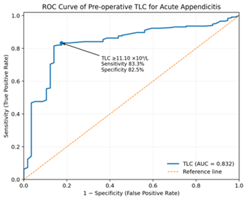
ROC curve analysis of pre-operative TLC for acute appendicitis shwos AUC as 0.832, standard was 0.030, 95% CI for AUC was 0.774–0.890, p-value <0.001, TLC threshold was ≥11.10 ×10⁹/L, sensitivity at this threshold was 83.3% and specificity at this threshold was 82.5% (Table 4).
Table 4. ROC curve analysis of pre-operative TLC for acute appendicitis
|
ROC parameter |
Result |
|
Area under the curve (AUC) |
0.832 |
|
Standard error |
0.030 |
|
95% CI for AUC |
0.774–0.890 |
|
p-value |
<0.001 |
|
TLC threshold |
≥11.10 ×10⁹/L |
|
Sensitivity at threshold |
83.3% |
|
Specificity at threshold |
82.5% |
DISCUSSION:
Acute appendicitis remains one of the most frequently encountered surgical emergencies, yet accurate pre-operative diagnosis continues to be challenging because of variability in clinical presentation and overlap with other causes of acute abdominal pain. Contemporary diagnostic strategies therefore combine clinical assessment with laboratory parameters, scoring systems and imaging modalities rather than relying on any single investigation. ¹ In resource-constrained healthcare settings, total leukocyte count (TLC) remains particularly relevant because it is inexpensive, rapidly available and routinely performed in patients presenting with suspected acute appendicitis.
In the present study, 290 patients with suspected acute appendicitis were evaluated, with a mean age of 37.83 ± 9.80 years. Males constituted 60.3% of the study population, indicating a moderate male predominance. Histopathological examination confirmed acute appendicitis in 233 (80.3%) patients, whereas 57 (19.7%) had negative histopathological findings. The predominance of males is broadly consistent with the established epidemiological pattern of acute appendicitis, which is reported to occur somewhat more frequently among males. ¹ Furthermore, the relatively high proportion of histopathologically confirmed cases reflects the selection of patients who had sufficient clinical suspicion to undergo appendectomy.
The principal finding of the present study was that pre-operative TLC demonstrated good diagnostic performance for acute appendicitis. TLC correctly identified 194 true-positive and 47 true-negative cases, while 10 patients were false-positive and 39 were false-negative. This resulted in a sensitivity of 83.3%, specificity of 82.5%, positive predictive value (PPV) of 95.1%, negative predictive value (NPV) of 54.7%, and overall diagnostic accuracy of 83.1%. These findings indicate that an elevated TLC is strongly associated with histopathologically confirmed appendicitis. The particularly high PPV suggests that, within a clinically selected population undergoing appendectomy, a raised TLC provides useful supportive evidence for the diagnosis. Conversely, the relatively modest NPV indicates that a normal TLC cannot reliably exclude acute appendicitis.
The findings of the present study are comparable with several previously reported studies. Saleem et al. evaluated 180 patients and reported that leukocytosis had a sensitivity of 93.5%, specificity of 64.3%, PPV of 89.6%, NPV of 75.1%, and diagnostic accuracy of 86.7%. ¹¹ Although their sensitivity was higher than the 83.3% observed in the present study, our specificity was considerably higher at 82.5%. Their conclusion that leukocytosis has high sensitivity but should not be considered a sole diagnostic marker supports the interpretation of our findings, particularly considering the relatively low NPV observed in the present study. Similarly, Brohi et al. evaluated TLC, neutrophil count and C-reactive protein among patients with clinically suspected acute appendicitis. They reported a sensitivity of approximately 79.5% for TLC and an overall diagnostic precision of approximately 76%,¹² findings that are reasonably close to the sensitivity observed in our study. The differences between studies may be explained by variation in TLC cut-off values, disease severity, duration of symptoms, patient selection and prevalence of histopathologically confirmed appendicitis. Khan et al. also demonstrated substantial diagnostic value of TLC in acute appendicitis. In their study of 320 patients, TLC showed a sensitivity of 87.5%, specificity of 80%, PPV of 95.9% and NPV of 54.5% for acute appendicitis. ¹³ These results are particularly comparable with the present findings of 83.3% sensitivity, 82.5% specificity, 95.1% PPV and 54.7% NPV. The close similarity in specificity, PPV and NPV between the two studies strengthens the evidence that TLC is useful for supporting a diagnosis of acute appendicitis but has limited ability to exclude the disease when the leukocyte count is not elevated. Khan et al. further demonstrated improved diagnostic performance when TLC was combined with CRP and neutrophil percentage, suggesting that multiple inflammatory markers may provide greater diagnostic value than TLC alone.
In contrast, Bibi et al., in a study conducted in Faisalabad, reported considerably poorer diagnostic performance of WBC count, with sensitivity of 27% and specificity of 43.75%. ¹⁴ The authors concluded that WBC count was not sufficiently reliable as an independent investigation for acute appendicitis. The marked difference from our results illustrates the considerable heterogeneity reported in the literature. Differences in study population, definitions of an elevated leukocyte count, timing of blood sampling, spectrum of disease and selection of patients for surgery may partly account for this discrepancy. Importantly, however, their findings reinforce the clinical principle that appendicitis may occur despite a normal leukocyte count. Maghsoudi et al. evaluated multiple complete blood count-associated parameters in 200 patients, of whom 15% underwent negative appendectomy. They found significantly higher WBC and neutrophil-related parameters among patients with positive appendectomy; however, the neutrophil-to-lymphocyte ratio showed the greatest diagnostic performance, with sensitivity of 83.5% and specificity of 90%. ¹⁵ The authors nevertheless emphasized that neither NLR nor other CBC-related parameters were sufficient as stand-alone pre-operative diagnostic tests. This is consistent with our findings, as the presence of 39 false-negative cases and an NPV of only 54.7% demonstrates that reliance solely on TLC could result in missed cases of histologically proven acute appendicitis.
The present study also demonstrated a positive likelihood ratio (LR+) of 4.75 and negative likelihood ratio (LR−) of 0.20. These values further support the clinical usefulness of TLC. An elevated TLC substantially increases the probability of appendicitis in an appropriately selected patient, while a negative result reduces—but does not eliminate—the probability of disease. This observation is important because diagnostic decisions in acute appendicitis should integrate laboratory findings with symptoms, physical examination, clinical scoring systems and, where appropriate, imaging. Receiver operating characteristic analysis provided additional evidence regarding the discriminatory ability of TLC. The area under the ROC curve was 0.832 (95% CI: 0.774–0.890; p<0.001), indicating good discrimination between histopathologically positive and negative cases. At an optimal TLC threshold of ≥11.10 ×10⁹/L, sensitivity was 83.3% and specificity was 82.5%. Recent evidence also supports the discriminatory ability of leukocyte count in appendiceal inflammation. Ali et al. reported an AUC of 0.84 for WBC count in predicting perforated appendicitis, with sensitivity of 85.0% and specificity of 73.3%. ¹⁶ Although their outcome was perforated rather than overall acute appendicitis and CT was used as the reference standard, their AUC is remarkably similar to the 0.832 obtained in the present study, supporting the overall diagnostic potential of leukocyte count.
The stratified analysis in the present study demonstrated relatively stable diagnostic performance across different demographic categories. Accuracy was 86.1% among patients aged 18–30 years and 82.0% among those aged 31–65 years. Similarly, diagnostic accuracy was 82.9% among males and 83.5% among females. These findings suggest that the diagnostic performance of TLC was broadly maintained across age and gender groups. Some variation was observed in specificity, particularly between males (86.8%) and females (73.7%), although TLC retained high PPV in both groups. Likewise, accuracy across BMI categories ranged from 81.3% to 85.1%. The apparent specificity of 100% among patients with BMI >30 kg/m² should be interpreted cautiously because of the relatively small number of histopathologically negative cases in this subgroup. The clinical relevance of these findings lies in the simplicity and accessibility of TLC. Advanced imaging, particularly computed tomography, provides high diagnostic accuracy but may not always be immediately available and entails additional cost and radiation exposure. Clinical scoring systems and imaging are therefore increasingly used selectively according to the probability of appendicitis. Noori et al. demonstrated that CT was particularly beneficial in patients with equivocal clinical scores, whereas patients with high clinical scores derived comparatively little additional diagnostic benefit from CT.¹⁷ Thus, TLC may contribute effectively to an integrated diagnostic pathway, particularly when interpreted alongside clinical findings and validated scoring systems.
Overall, the present study demonstrates that pre-operative TLC has good diagnostic accuracy for acute appendicitis when histopathology is used as the reference standard. Its high PPV, LR+ of 4.75 and AUC of 0.832 indicate meaningful diagnostic utility, particularly for supporting the diagnosis in patients with compatible clinical findings. Nevertheless, its relatively low NPV and the presence of false-negative cases indicate that a normal TLC should not be used to exclude acute appendicitis. TLC should therefore be regarded as an accessible and valuable adjunct to clinical assessment rather than a replacement for comprehensive evaluation, clinical scoring or appropriate imaging.
Limitations
The present study has certain limitations. It was conducted at a single tertiary-care centre using a non-probability purposive sampling technique, which may limit the generalizability of the findings. Moreover, only patients proceeding to appendectomy were evaluated, potentially producing spectrum and selection bias and contributing to the high prevalence of histopathologically confirmed appendicitis and consequently the high PPV. Other inflammatory biomarkers such as CRP, neutrophil percentage and NLR were not incorporated into the primary diagnostic model. Future prospective multicentre studies should evaluate combinations of TLC with these biomarkers, clinical scoring systems and imaging findings and assess whether integrated diagnostic algorithms can reduce unnecessary appendectomies while avoiding delays in treatment.
CONCLUSION:
The diagnostic accuracy of pre-operative total leukocyte count is higher for prediction of acute appendicitis taking histopathology as gold standard and can be used in settings where advanced level of diagnostic modalities like USG/MRI is not available.
REFERENCES:
Kumar HR. The epidemiology and diagnosis of acute appendicitis: narrative review article. SAR J Med 2026;7(3):75-81. doi:10.36346/sarjm.2026.v07i03.001
2. Doleman B, Fonnes S, Lund JN, Boyd-Carson H, Javanmard-Emamghissi H, Moug S, Hollyman M, Tierney G, Tou S, Williams JP. Appendectomy versus antibiotic treatment for acute appendicitis. Cochrane Database Syst Rev 2024;4(4):CD015038. doi: 10.1002/14651858.CD015038.pub2.
3. Akbulut S, Sahin IT. Comment on the correlation between complete blood count parameters and appendix diameter for the diagnosis of acute appendicitis. Healthcare 2020;8(4):1-10.
4. Peksöz R, Bayar B. The role of complete blood count parameters in diagnosing acute appendicitis and measuring the severity of inflammation. Ulus Travma Acil Cerrahi Derg 2021;27(6):654-61.
5. Kailasia Y, Jain S, Garg A. Epidemiological profile and risk factors of acute appendicitis in India. Bioinformation. 2026 Apr 30;22(4):2376-2379. doi: 10.6026/973206300222376. PMID: 42282377; PMCID: PMC13252261.
6. Doleman B, Fonnes S, Lund JN, Boyd-Carson H, Javanmard-Emamghissi H, Moug S, et al. Appendectomy versus antibiotic treatment for acute appendicitis. Cochrane Database Syst Rev 2024;4:e01503.
7. Rios KD, Salazar CC, Morocho JF, León JL, Guzmán JM. Apendicectomía negativa en pacientes diagnosticados de apendicitis aguda Hospital Homero Castanier Crespo. Conciencia Digital 2023;6(1.2):6-21.
8. Johnson, C.A., Pivalizza, C.M., Chan, H.K. et al. Diagnosis of acute appendicitis at a pediatric emergency department within a general hospital. Emerg Radiol 2026;33:503–511. https://doi.org/10.1007/s10140-026-02442-w
9. Fatima SR, Zaheer F, Moosa FA, Arqam SM, Mussab RM, Choudhry MS, et al. Combined diagnostic accuracy of total leukocyte count, neutrophil count, and ultrasonography for the diagnosis of acute appendicitis. Cureus 2021;13(2):e13086.
10. Afzal DJ, Arshad DH, Raheem DH, Jameel MK, Hameed DF, Shiraz DD. Diagnostic Value of Total and Differential Leukocyte Counts for the Diagnosis of Acute Appendicitis. Esculapio J Services Inst Med Sci 2023;19(01):100-4.
11. Saleem MM, Pervaiz M, Chaudhary IA. Predictive value of leukocytosis for diagnosing acute appendicitis. Pak Armed Forces Med J. 2017;67(Suppl-3):S292-6. The study reported sensitivity 93.5%, specificity 64.3%, PPV 89.6%, NPV 75.1% and accuracy 86.7%.
12. Brohi LB, Mooghal M, Ahmed H, Khan WA, Malik J, Malik TM, et al. Diagnostic precision of C-reactive protein levels, neutrophil count and total leukocyte count in identifying acute appendicitis clinically. J Surg Res. 2021;4(3):441-9. doi:10.26502/jsr.10020153.
13. Khan AA, Manan A, Ahmed I, Mahar T, Usman M, Qadir A. C-reactive protein, leukocyte count and neutrophils: a diagnostic aid in acute appendicitis. J Islamabad Med Dental Coll. 2022;11(4):222-8. doi:10.35787/jimdc.v11i4.741. Their TLC results—87.5% sensitivity, 80% specificity, 95.9% PPV and 54.5% NPV—provide an especially strong comparison with your results.
14. Bibi S, Saqib K, Saleem M, Jadaan A, Ahmad H, Rana H. Role of white blood cell count in diagnostic accuracy of acute appendicitis. J Aziz Fatm Med Den College. 2021;3(2):56-60.
15. Haji Maghsoudi L, Kabir K, Soltanian A, Pak H, Ahmadinejad M, Khas Ahmadi A. A cross-sectional study on evaluation of complete blood count-associated parameters for the diagnosis of acute appendicitis. Health Sci Rep. 2021;4:e326. doi:10.1002/hsr2.326.
16. Ali I, Talpur AA, Kumar S, Khatti SN, Ahmed U. Diagnostic accuracy of white blood cell count, serum bilirubin, and C-reactive protein for predicting perforated appendicitis: a cross-sectional study. J Saidu Med Coll. 2026;16(2). doi:10.52206/jsmc.2026.16.2.1428.
17. Noori IF, Jabbar AS, Noori AF. Clinical scores (Alvarado and AIR scores) versus imaging (ultrasound and CT scan) in the diagnosis of equivocal cases of acute appendicitis: a randomized controlled study. Their study found CT particularly useful in equivocal-score patients and supported selective rather than indiscriminate imaging.