Diagnostic Accuracy of Coronary Computed Tomography Angiography in Diagnosing Coronary Artery Stenosis, Taking Catheter Angiography as Gold Standard
- Azra Mahmood , Training Medical Officer, Department of Radiology, Bacha Khan Medical Complex/ MTI Mardan Medical Complex, Mardan, Pakistan
- Zubair Janan Orakzai , Associate Professor, Department of Radiology, Bacha Khan Medical Complex/MTI, Mardan Medical Complex, Mardan, Pakistan
- Maria Ahmad , Training Medical Officer, Department of Radiology, Bacha Khan Medical Complex/ MTI Mardan Medical Complex, Mardan, Pakistan
- Sumaira Noureen , Assistant Professor, Department of Radiology, Bacha Khan Medical Complex/MTI Mardan Medical Complex, Mardan, Pakistan
- Hajra Shah , Training Medical Officer, Department of Radiology, Bacha Khan Medical Complex/ MTI Mardan Medical Complex, Mardan, Pakistan
- Nadia , Training Medical Officer, Department of Radiology, Bacha Khan Medical Complex/ MTI Mardan Medical Complex, Mardan, Pakistan
Article Information:
Abstract:
Coronary artery disease (CAD) is a significant cause of death and disability throughout the world. Catheter angiography is regarded as the gold standard of the diagnosis of coronary artery stenosis, but is invasive and can carry a risk of the procedure itself. CCTA is a non-invasive imaging technique that may be useful in the detection of coronary artery stenosis and subsequent management. Objective: To determine the diagnostic accuracy of coronary computed tomography angiography in diagnosing coronary artery stenosis, taking catheter angiography as the gold standard. Methods: The study was a cross-sectional validation study which was conducted from 10th May 2025 to 10th August 2025 in the Department of Radiology, Mardan Medical Complex, Mardan. Non-probability consecutive sampling was used including a total of 127 patients with suspected CAD. Patients of both genders aged 30 to 70 years, with suspected coronary artery disease (CAD) for ≤48 hours were recruited. Coronary artery stenosis was considered to be ≥50% narrowing of one or more major coronary arteries. Coronary CTA was performed in all patients, and patients underwent catheter angiography. Catheter angiography was used as the gold standard. Sensitivity, specificity, positive predictive value, negative predictive value and diagnostic accuracy were determined. Results: Out of 127 patients, catheter angiography confirmed coronary artery stenosis in 47 (37.0%) patients, while CCTA detected stenosis in 51 (40.2%) patients. There were 43 true-positive, 72 true-negative, 8 false-positive, and 4 false-negative cases. The sensitivity, specificity, positive predictive value, negative predictive value, and diagnostic accuracy of CCTA were 91.5%, 90.0%, 84.3%, 94.7%, and 90.6%, respectively. The association between CCTA and catheter angiography findings was statistically significant (p<0.001). Conclusion: Coronary computed tomography angiography showed high diagnostic accuracy for detecting coronary artery stenosis. It is a reliable and non-invasive diagnostic tool, especially for excluding significant coronary artery stenosis in patients with suspected coronary artery disease.
Keywords:
Article :
INTRODUCTION:
Coronary artery disease is one of the most common causes of cardiovascular morbidity and mortality in adult population. It is usually caused by narrowing of the coronary arteries (atherosclerosis), which decreases blood flow to the heart, and can cause angina, myocardial ischemia, acute coronary syndrome, myocardial failure or sudden death. Chest pain is the most common presenting symptom; however, many of those who suffer from this disease may have atypical symptoms or a silent disease and may not be diagnosed and treated until much later. Thus, it is very important to identify the coronary artery stenosis accurately and timely for risk assessment and clinical decision making (1-3).
Catheter angiography has been regarded as the golden standard investigation for coronary artery stenosis diagnosis. It offers the advantage of direct visualization of the coronary arteries and enables the evaluation of the location, degree and amount of narrowing. It is also useful in the planning of future management such as PCI or surgical revascularisation. However, it is an invasive procedure and can have adverse effects and complications including bleeding, effects of the contrast used, damage to the veins and arteries involved, arrhythmia, and discomfort of the procedure. For this reason, non-invasive diagnostic techniques have become more and more important in the assessment of the patient with suspected coronary artery disease (4-6).
Coronary computed tomography angiography has emerged as an important non-invasive imaging modality for the assessment of coronary arteries. It can help to see the coronary lumen, the wall, calcified and non-calcified plaques, and the severity of narrowing of the coronary artery. CCTA has improved in its usefulness for detecting significant coronary artery stenosis with advances in scanner technology, image reconstruction, and heart rate control. It can be especially useful in patients whose risk for coronary artery disease is low to intermediate, as a good non-invasive test can prevent unnecessary invasive angiography (7, 8).
The value of CCTA is mostly in its high sensitivity and high negative predictive value. In many cases a negative CCTA result can be used to exclude a significant coronary artery stenosis with a high degree of confidence. CCTA sometimes, however, overestimates stenosis, especially when there is heavy calcification, motion artifacts, or suboptimal image quality. Thus, comparison of catheter angiography is still necessary for determining its diagnostic accuracy in various clinical conditions and patient populations (9).
Many patients in local clinical practice come with symptoms which are suggestive of coronary artery disease and a correct diagnosis is necessary for timely management. Catheter angiography is still the reference standard, but is not always the initial diagnostic procedure of choice for all patients due to its invasive nature. Coronary computed tomography angiography may provide a safer and quicker alternative for initial evaluation. But there is limited local information on the diagnostic accuracy of CCTA in the assessment of coronary artery stenosis (10).
Therefore, this study was conducted to determine the diagnostic accuracy of coronary computed tomography angiography in diagnosing coronary artery stenosis, taking catheter angiography as the gold standard, among patients presenting with suspected coronary artery disease at Mardan Medical Complex, Mardan.
MATERIALS AND METHODS:
This cross-sectional validation study was conducted in the Department of Radiology, Mardan Medical Complex, Mardan, from 10th May 2025 to 10th August 2025. The study was carried out after approval from the College of Physicians and Surgeons Pakistan, Research Evaluation Unit, with approval reference number CPSP/REU/RAD-2023-028-4011, dated 10th May 2025. Written informed consent was obtained from all participants before enrollment. Confidentiality of patient information was maintained throughout the study, and the data were used only for research purposes.
There were 127 patients who had a suspected diagnosis of coronary artery disease. The sample size was calculated using a sensitivity and specificity calculator, keeping 95% confidence level, expected prevalence of coronary artery disease as 35.5%, and expected sensitivity and specificity of coronary computed tomography angiography as 90.66% and 95.65%, respectively. The sampling technique used was non-probability consecutive sampling, in which all eligible patients who came during the study period were sampled until the number of samples was reached. The synopsis also made the assumption of the diagnostic accuracy of these tests to plan 127 cases.
The study included patients of both genders aged between 30-70 years with suspected coronary artery disease of ≤48 hours. Suspected coronary artery disease was indicated by chest pain with cardiac troponin I > normal, and symptoms of ischemia, significant ECG changes (STEMI or NSTEMI), or echocardiographic regional wall motion abnormality that was not present prior to the event. The study excluded patients with left ventricular ejection fraction ≤35%, left ventricular aneurysm, previous revascularisation, cardiomyopathy, second- or third-degree atrioventricular block, small vessel diameter, long myocardial bridges, critical illnesses (sepsis, renal failure, hepatic failure), and bleeding or clotting disorders. A homogeneous population of patients was determined according to these inclusion and exclusion criteria.
Following enrolment demographic and clinical data were captured on a pre-designed proforma. The following were the factors involved: age, gender, place of residence, educational level, duration of symptoms, height, weight, body mass index, hypertension, diabetes mellitus and smoking. Pre-imaging clinical history and relevant findings were documented. Coronary artery stenosis was defined as ≥50% stenosis of one or more of the major coronary arteries: left anterior descending, left main coronary, left circumflex and right coronary. Catheter angiography was used as the reference standard and a coronary artery stenosis was defined as ≥50% occlusion of the coronary artery segments.
All selected patients underwent CTA coronary angiography using the standard departmental protocol. Sublingual glyceryl trinitrate was given prior to scan, and atenolol if necessary to achieve desired heart rate. Contrast was given through a vein and saline flush. Axial multiplanar reconstruction and maximum intensity projection images were obtained to reconstruct the images. Experienced radiologists interpreted the findings from the CCTA and the presence of coronary artery stenosis was determined based on the operational definition.
All patients had catheter angiography after CCTA as a reference standard. Standard clinical protocol was followed for performing catheter angiography. Six or more views of the left coronary system and two views of the right coronary system were acquired. A cardiologist with experience in interpreting angiograms blinded to CCTA findings interpreted the angiograms. True positive, true negative, false positive and false negative for CCTA were then evaluated against the results of catheter angiography. The planned data collection procedure in the synopsis also included the comparison of the results of CCTA with the results of catheter angiography as the gold standard.
The data were entered and analyzed with SPSS 25.0 software. Data for quantitative variables (age, duration of symptom, height, weight and BMI) are presented as mean and standard deviation or median and interquartile range as appropriate to the normality of data. Qualitative variables like gender, residence, Hypertension, DM, smoking status, CCTA findings, catheter angiography findings were presented as frequency and percentage. A 2×2 diagnostic table was created to calculate sensitivity, specificity, positive predictive value, negative predictive value, diagnostic accuracy, positive likelihood ratio, and negative likelihood ratio of CCTA for diagnosing coronary artery stenosis. The ROC curve analysis was also carried out. Stratification was used to control for effect modifiers including age, gender, BMI, duration of symptoms, hypertension, diabetes mellitus, and smoking. Appropriately, post-stratification chi-square test or Fisher's exact test were used, with a p value of ≤0.05 deemed as statistically significant.
RESULTS:
This is a cross-sectional validation study of 127 patients with suspected coronary artery disease. To evaluate the diagnostic accuracy of coronary computed tomography angiography (CCTA) to detect coronary artery stenosis, against catheter angiography, as outlined in the synopsis. The majority of patients were older than 50 and males were marginally more prevalent than females. Common clinical risk factors noted in the study population were hypertension, diabetes mellitus and smoking. The average BMI was in the overweight category.
Table 1: Baseline demographic and clinical characteristics of patients
|
Variable |
Frequency / Mean |
Percentage / SD |
|
Total patients |
127 |
100% |
|
Mean age, years |
54.8 |
± 9.6 |
|
Age 30–50 years |
48 |
37.8% |
|
Age 51–70 years |
79 |
62.2% |
|
Male |
76 |
59.8% |
|
Female |
51 |
40.2% |
|
Rural residence |
58 |
45.7% |
|
Urban residence |
69 |
54.3% |
|
Mean duration of symptoms, weeks |
3.2 |
± 1.4 |
|
Mean BMI, kg/m² |
27.1 |
± 4.2 |
|
Hypertension |
67 |
52.8% |
|
Diabetes mellitus |
51 |
40.2% |
|
Smoker |
46 |
36.2% |
Patients with coronary artery stenosis were more likely to be older, male, hypertensive, diabetic and smoker. Statistically significant association was found between the coronary artery stenosis and age group, hypertension, diabetes mellitus, smoking.
Table 2: Comparison of baseline variables according to catheter angiography findings
|
Variable |
Stenosis Present n=47 |
Stenosis Absent n=80 |
p-value |
|
Age 51–70 years |
36 (76.6%) |
43 (53.8%) |
0.011 |
|
Male gender |
34 (72.3%) |
42 (52.5%) |
0.028 |
|
Urban residence |
28 (59.6%) |
41 (51.3%) |
0.365 |
|
BMI >25 kg/m² |
32 (68.1%) |
42 (52.5%) |
0.084 |
|
Hypertension |
33 (70.2%) |
34 (42.5%) |
0.003 |
|
Diabetes mellitus |
27 (57.4%) |
24 (30.0%) |
0.002 |
|
Smoker |
25 (53.2%) |
21 (26.3%) |
0.002 |
On CCTA, a coronary artery stenosis was found in 51 patients and on catheter angiography in 47 patients. The detection rate was slightly higher for CCTA, perhaps as a result of some false positive cases identified on non-invasive imaging.
Table 3: Frequency of coronary artery stenosis on CCTA and catheter angiography
|
Imaging modality |
Stenosis Present |
Stenosis Absent |
Total |
|
CCTA |
51 (40.2%) |
76 (59.8%) |
127 |
|
Catheter angiography |
47 (37.0%) |
80 (63.0%) |
127 |
Left anterior descending artery was the most commonly involved artery followed by right coronary artery and left circumflex artery. Less commonly seen was the involvement of the left main coronary artery.
Table 4: Distribution of involved coronary arteries on CCTA
|
Coronary artery involved |
Frequency |
Percentage |
|
Left anterior descending artery |
29 |
56.9% |
|
Right coronary artery |
21 |
41.2% |
|
Left circumflex artery |
17 |
33.3% |
|
Left main coronary artery |
6 |
11.8% |
Multiple vessel involvement was present in some patients.
The results of CCTA compared with catheter angiography showed 43 true-positive patients, 72 true-negative patients, 8 false-positive patients, and 4 false-negative patients. There was a statistically significant correlation between CCTA and catheter angiography findings.
Table 5: Diagnostic 2×2 table of CCTA compared with catheter angiography
|
CCTA finding |
Catheter angiography positive |
Catheter angiography negative |
Total |
|
CCTA positive |
43 |
8 |
51 |
|
CCTA negative |
4 |
72 |
76 |
|
Total |
47 |
80 |
127 |
Chi-square p-value: <0.001
CCTA had a high sensitiveness and specificity for the detection of coronary artery stenosis. The high negative predictive value suggests that CCTA is especially valuable to exclude clinically important coronary artery stenosis in patients with suspected disease.
Table 6: Diagnostic accuracy of CCTA for coronary artery stenosis
|
Diagnostic parameter |
Value |
|
Sensitivity |
91.5% |
|
Specificity |
90.0% |
|
Positive predictive value |
84.3% |
|
Negative predictive value |
94.7% |
|
Diagnostic accuracy |
90.6% |
|
Positive likelihood ratio |
9.15 |
|
Negative likelihood ratio |
0.09 |
|
Area under ROC curve |
0.908 |
|
95% CI for AUC |
0.849–0.967 |
|
p-value |
<0.001 |
High rates of diagnostic performance in most subgroups. Older patients, males, hypertension, diabetes and smoking had a slightly higher sensitivity. However, the differences in diagnostic accuracy in most strata were not significant.
Table 7: Stratified diagnostic accuracy of CCTA according to selected variables
|
Variable |
Sensitivity |
Specificity |
Accuracy |
p-value |
|
Age 30–50 years |
88.9% |
89.7% |
89.6% |
<0.001 |
|
Age 51–70 years |
92.1% |
90.2% |
91.1% |
<0.001 |
|
Male |
91.2% |
88.1% |
89.5% |
<0.001 |
|
Female |
92.3% |
92.1% |
92.2% |
<0.001 |
|
Hypertension present |
93.9% |
88.2% |
91.0% |
<0.001 |
|
Hypertension absent |
85.7% |
91.3% |
90.0% |
<0.001 |
|
Diabetes present |
92.6% |
87.5% |
90.2% |
<0.001 |
|
Diabetes absent |
90.0% |
91.1% |
90.8% |
<0.001 |
|
Smoker |
92.0% |
85.7% |
89.1% |
<0.001 |
|
Non-smoker |
90.9% |
91.5% |
91.4% |
<0.001 |
CCTA had an excellent diagnostic performance to detect coronary artery stenosis by ROC curve analysis. The area under the curve was 0.908, showing good correlation with the catheter angiography.
Table 8: ROC curve analysis of CCTA
|
ROC parameter |
Value |
|
Area under curve |
0.908 |
|
Standard error |
0.030 |
|
95% confidence interval |
0.849–0.967 |
|
p-value |
<0.001 |
Of the 127 patients in this study, 47 (37.0%) had coronary artery stenosis confirmed by catheter angiography, and 51 (40.2%) had coronary artery stenosis detected by CCTA. CCTA was able to correctly identify 43 true-positive cases and 72 true-negative cases. The sensitivity, specificity, PPV, NPV, and overall diagnostic accuracy of CCTA were 91.5%, 90.0%, 84.3%, 94.7%, and 90.6%, respectively. CCTA showed a statistically significant association with catheter angiography (p<0.001), which supports the use of CCTA as a reliable non-invasive diagnostic tool for the detection of coronary artery stenosis.
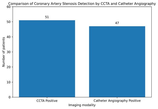
Figure 1: Comparison of coronary artery stenosis detection by CCTA and catheter angiography.
CCTA detected coronary artery stenosis in 51 patients, while catheter angiography confirmed stenosis in 47 patients.
DISCUSSION :
The present study was conducted to determine the diagnostic accuracy of coronary computed tomography angiography in diagnosing coronary artery stenosis, taking catheter angiography as the gold standard. In this study, 127 patients with suspected coronary artery disease were included. Catheter angiography confirmed coronary artery stenosis in 47 (37.0%) patients, while CCTA detected stenosis in 51 (40.2%) patients. When CCTA was compared with catheter angiography, 43 patients were true positive, 72 were true negative, 8 were false positive, and 4 were false negative. The association between CCTA and catheter angiography was statistically significant (p<0.001), showing strong agreement between the two diagnostic methods (11, 12).
In this study CCTA was highly accurate in diagnosing. Sensitivity (91.5%), specificity (90.0%), positive predictive value (84.3%), negative predictive value (94.7%) and diagnostic accuracy (90.6%) were achieved. These results indicate that CCTA is a good non-invasive imaging tool for detecting clinically important coronary artery stenosis. The high sensitivity suggests CCTA was good at identifying patients with coronary artery stenosis on catheter angiography. In a similar fashion, the high negative predictive value indicates that a negative CCTA result can be used to rule out significant coronary artery stenosis in many patients (13, 14).
The results of the current study confirm the results of previous published studies. Computed tomography angiography was also shown to have high sensitivity and high negative predictive value for the exclusion of coronary artery disease, with lower specificity when compared to other studies (15). Likewise, Xiong et al. demonstrated excellent diagnostic accuracy of coronary computed tomography angiography stenosis score, which was 91.67%, with a 90.66% sensitivity and 95.65% specificity, 98.80% positive predictive value and 72.13% negative predictive value (16). Sensitivity and overall diagnostic accuracy in the present study are similar to these published results, and this suggests that CCTA is a useful diagnostic tool for coronary artery stenosis.
In the current study, CCTA found slightly more positive cases than did the catheter angiography. The disparities could be attributed to false-positive results of CCTA, particularly for patients with coronary calcification, motion artifact, high heart rate or poor image quality. Although highly sensitive, CCTA could overestimate stenosis in very calcified coronary segments. It is possible that this can be the reason for the 8 cases of false positive in the present study. The number of false-negative cases, however, was low, which is clinically important as failure to detect important coronary stenosis may result in late presentation and poor cardiac outcomes (17).
In the present study, the coronary artery most frequently involved was the left anterior descending artery followed by the right coronary artery and left circumflex artery. This is clinically significant since the left anterior descending artery feeds a large part of the myocardium and when this artery is involved, the ischemic symptoms are common. Vessel involvement on CCTA may assist in further management and triage of which patients may require invasive catheter angiography and/or intervention (18).
Factors like age, male sex, hypertension, diabetes mellitus, and smoking were more prevalent in the coronary artery stenosis group identified by catheter angiography. Age group, hypertension, diabetes mellitus, and smoking were found to be statistically significant among the factors in the present study. This is biologically plausible as these factors all play a role in endothelial dysfunction, in development of atherosclerotic plaques and in progressive coronary artery narrowing. For this reason, such individuals should be thoroughly assessed if they present with symptoms typical of CAD (19, 20).
The major strength of this study is that catheter angiography was used as the gold standard for comparison. The study also included a fairly large number of patients (127 patients) and had well-defined diagnostic criteria (coronary artery stenosis ≥50% in one or more major coronary arteries was considered positive). The synopsis also clarified that the stenosis of the major coronary arteries was defined as CCTA positivity and occlusion of ≥50% by catheter angiography as angiography positivity, which facilitated the comparison between the two diagnostic methods.
There are also some limitations to this study. It was performed in one center and therefore the results may not apply to other populations. Coronary calcification, body habitus, image quality, patient motion and heart rate may influence the diagnostic yield of CCTA. Diagnostic accuracy was not evaluated in detail in the case of either vessel-wise or segment-wise. Moreover, although catheter angiography was used as the gold standard, visual assessment of stenosis can have some inter-observer variability. More multi-center studies are needed to verify these results with larger numbers of subjects and detailed vessel-wise analysis.
CONCLUSION :
This study concluded that coronary computed tomography angiography has high diagnostic accuracy for detecting coronary artery stenosis, using catheter angiography as the gold standard. In a sample of 127 patients, CCTA showed 91.5% sensitivity, 90.0% specificity, 84.3% positive predictive value, 94.7% negative predictive value, and 90.6% overall diagnostic accuracy, with a statistically significant association with catheter angiography findings (p<0.001). These findings support CCTA as a useful, reliable, and non-invasive diagnostic tool for evaluating suspected coronary artery disease, especially for ruling out significant coronary artery stenosis. However, positive CCTA findings should be interpreted carefully in high-risk patients and may require confirmation by catheter angiography when clinical intervention is being considered.
BIBLIOGRAPHY:
1. Gatti M, Gallone G, Poggi V, Bruno F, Serafini A, Depaoli A, et al. Diagnostic accuracy of coronary computed tomography angiography for the evaluation of obstructive coronary artery disease in patients referred for transcatheter aortic valve implantation: a systematic review and meta-analysis. 2022;32(8):5189-200.
2. Mansour HH, Alajerami YS, Abushab KM, Quffa KMJJoMI, Sciences R. The diagnostic accuracy of coronary computed tomography angiography in patients with and without previous coronary interventions. 2022;53(1):81-6.
3. Kheiri B, Simpson TF, Osman M, German DM, Fuss CS, Ferencik MJCI. Computed tomography vs invasive coronary angiography in patients with suspected coronary artery disease: a meta-analysis. 2022;15(12):2147-9.
4. Malebranche D, Hoffner MK, Huber AT, Cicovic A, Spano G, Bernhard B, et al. Diagnostic performance of quantitative coronary artery disease assessment using computed tomography in patients with aortic stenosis undergoing transcatheter aortic-valve implantation. 2022;22(1):178.
5. Abdelrahman KM, Chen MY, Dey AK, Virmani R, Finn AV, Khamis RY, et al. Coronary computed tomography angiography from clinical uses to emerging technologies: JACC state-of-the-art review. 2020;76(10):1226-43.
6. Ella T, Elsayed E, ELkersh A, Moawad MJMMJ. Diagnostic accuracy of computed tomography angiography compared with catheter angiography in coronary artery disease. 2021;34(3):1034-.
7. Elagha A, Khaled W, Gamal S, Helmy M, Kaddah AJTEHJ. Coronary computed tomography versus coronary angiography for preoperative coronary assessment before valve surgery. 2021;73(1):63.
8. Ngam PI, Ong CC, Chai P, Wong SS, Liang CR, San Teo LLJSmj. Computed tomography coronary angiography–past, present and future. 2020;61(3):109.
9. Parikh R, Patel A, Lu B, Senapati A, Mahmarian J, Chang SMJMDcj. Cardiac computed tomography for comprehensive coronary assessment: beyond diagnosis of anatomic stenosis. 2020;16(2):77.
10. Tonet E, Boccadoro A, Micillo M, Cocco M, Cossu A, Pompei G, et al. Coronary computed tomography angiography: Beyond obstructive coronary artery disease. 2023;13(5):1086.
11. Meier D, Depierre A, Topolsky A, Roguelov C, Dupre M, Rubimbura V, et al. Computed tomography angiography for the diagnosis of coronary artery disease among patients undergoing transcatheter aortic valve implantation. 2021;14(5):894-901.
12. Burch RA, Siddiqui TA, Tou LC, Turner KB, Umair MJJoCD, Disease. The cost effectiveness of coronary CT angiography and the effective utilization of CT-fractional flow reserve in the diagnosis of coronary artery disease. 2023;10(1):25.
13. Mansour HH, Alajerami YSJJCDR. Cost-effectiveness analysis of coronary computed tomography angiography and invasive coronary angiography for diagnosis of coronary artery disease. 2021;12(3):1440-53.
14. Shah NR, Pierce JD, Kikano EG, Rahnemai-Azar AA, Gilkeson RC, Gupta AJCPiDR. CT coronary angiography fractional flow reserve: new advances in the diagnosis and treatment of coronary artery disease. 2021;50(6):925-36.
15. Strong C, Ferreira A, Teles RC, Mendes G, Abecasis J, Cardoso G, et al. Diagnostic accuracy of computed tomography angiography for the exclusion of coronary artery disease in candidates for transcatheter aortic valve implantation. Scientific reports. 2019;9(1):19942 DOI: 10.1038/s41598-019-56519-3.
16. Xiong QF, Fu XR, Ku LZ, Zhou D, Guo SP, Zhang WS. Diagnostic performance of coronary computed tomography angiography stenosis score for coronary stenosis. BMC Med Imaging. 2024;24(1):39 DOI: 10.1186/s12880-024-01213-8.
17. Ghobrial M, Haley HA, Gosling R, Rammohan V, Lawford PV, Hose DR, et al. The new role of diagnostic angiography in coronary physiological assessment. 2021;107(10):783-9.
18. Jiang W, Pan Y, Hu Y, Leng X, Jiang J, Feng L, et al. Diagnostic accuracy of coronary computed tomography angiography-derived fractional flow reserve. 2021;20(1):77.
19. Pugliese L, Ricci F, Sica G, Scaglione M, Masala SJD. Non-contrast and contrast-enhanced cardiac computed tomography imaging in the diagnostic and prognostic evaluation of coronary artery disease. 2023;13(12):2074.
20. Hagar MT, Soschynski M, Saffar R, Rau A, Taron J, Weiss J, et al. Accuracy of ultrahigh-resolution photon-counting CT for detecting coronary artery disease in a high-risk population. 2023;307(5):e223305.