Appearance, Pulse, Grimace, Activity, And Respiration (APGAR) Score among Newborn with Abnormal Cardiotocography (CTG) Trace
- Bibi Maria , Postgraduate Resident, Department of Gynae and Obstetrics, Sandeman Provincial Hospital Quetta, PAK
- Zaib Un Nisa , Professor, Department of Gynae and Obstetrics, Sandeman Provincial Hospital Quetta, PAK
- Tanveer Ahmed , Consultant General Surgeon, Department of General Surgery, Sandeman Provincial Hospital Quetta,PAK
- Mehnaz Javed , Postgraduate Resident, Department of Gynae and Obstetrics, Sandeman Provincial Hospital Quetta, PAK
- Rabia Jamali , Postgraduate Resident, Department of Gynae and Obstetrics, Sandeman Provincial Hospital Quetta, PAK
Article Information:
Abstract:
Appearance, Pulse, Grimace, Activity, And Respiration (APGAR) Score among Newborn with Abnormal Cardiotocography (CTG) Trace
Keywords:
Article :
INTRODUCTION:
Perinatal asphyxia remains an important cause of neonatal morbidity and mortality, particularly in settings where timely recognition of fetal compromise and appropriate intrapartum intervention are essential. Cardiotocography (CTG) is widely used for intrapartum fetal surveillance because it continuously records fetal heart rate and uterine activity, allowing clinicians to identify patterns suggestive of fetal hypoxia and possible compromise. However, interpretation of CTG remains challenging because abnormal traces may have limited positive predictive value and can contribute to unnecessary operative deliveries.
Recent evidence demonstrates an association between abnormal fetal heart-rate patterns and adverse neonatal outcomes. In a Pakistani observational study of 470 women, 63.40% of newborns had an Apgar score below 8, while 2.34% had an Apgar score below 6; pathological or non-reactive CTG was associated with substantially reduced odds of achieving a higher Apgar score (OR 0.30, 95% CI 0.20–0.44). {1} In another study involving 500 pregnancies, CTG was abnormal in 1% of cases, and Apgar <7 occurred in 4% at 1 minute and 0.4% at 5 minutes overall. Importantly, among the five women with abnormal CTG, 100% of newborns had Apgar <7 at 1 minute and 20% remained below 7 at 5 minutes. {2}
A 2023 systematic review and meta-analysis involving 47,648 term singleton pregnancies further demonstrated that Apgar <7 at 5 minutes increased from 0.74% with category-I fetal heart-rate patterns to 1.51% with category-II and 14.63% with category-III patterns. {3} Nevertheless, evidence indicates that CTG should not be considered an isolated predictor of neonatal outcome, as its predictive performance varies according to trace characteristics and clinical context. {2,3}
Therefore, determining the frequency of poor Apgar scores among newborns with abnormal CTG traces is clinically relevant, particularly in tertiary-care settings. The present study aims to determine the frequency of poor Apgar score among newborns delivered following abnormal CTG at Bolan Medical College/Hospital, Quetta, thereby providing locally relevant evidence for intrapartum fetal surveillance and neonatal preparedness.
METHODLOGY:
A descriptive cross-sectional study was conducted in the Department of Obstetrics and Gynaecology, Bolan Medical College/Hospital, Quetta, over a period of six months after approval of the synopsis. The study population comprised booked pregnant women aged 15–49 years who were admitted for delivery, had an abnormal cardiotocography (CTG) trace at the time of admission, and had a gestational age greater than 28 weeks, irrespective of gravida or parity. The sample size was calculated using the WHO sample size calculator by taking a previously reported frequency of poor Apgar score at 5 minutes among women with abnormal CTG as 2.7%, with a margin of error of 1.7% and a 95% confidence level. The calculated sample size was 350 participants. Non-probability consecutive sampling was used for participant recruitment.
Women aged 15–49 years with abnormal CTG at admission, gestational age greater than 28 weeks, irrespective of gravida and parity, and who were booked were included in the study. Women with multiple gestation, gestational diabetes, pregnancy-induced hypertension, or other complicated pregnancies were excluded. Complicated pregnancies included conditions such as placental abruption, preterm labour, and congenital fetal anomalies, as these conditions could independently influence CTG findings and neonatal Apgar scores.
After obtaining permission from the hospital ethical committee and the College of Physicians and Surgeons Pakistan (CPSP), eligible women admitted to the gynaecology ward were enrolled. Written informed consent was obtained from the participants or their attendants after explaining the study, including its risks and benefits. Baseline demographic and clinical information was collected at enrollment. CTG was performed to confirm abnormal findings according to the predefined operational definition. An abnormal CTG was labelled when the tracing showed late decelerations after uterine contractions, variable decelerations, and beat-to-beat variability. The enrolled women were followed until delivery, and the newborn's Apgar score was assessed at 1 and 5 minutes after birth. The Apgar score consisted of five components—Appearance, Pulse, Grimace, Activity, and Respiration—with each component assigned a score from 0 to 2. An Apgar score of less than 7 at 1 minute was defined as a poor Apgar score. Maternal age, residence, gravida, parity, height, weight, body mass index (BMI), maternal education, gestational age, birth weight, mode of delivery, and Apgar scores at 1 and 5 minutes were recorded on a predesigned proforma.
Data were analyzed using SPSS version 25. Quantitative variables, including maternal age, height, weight, BMI, gestational age, birth weight, and Apgar scores at 1 and 5 minutes, were summarized using mean and standard deviation or median with interquartile range according to the distribution of the data. Qualitative variables, including residence, maternal education, parity, gravida, mode of delivery, and poor Apgar score, were expressed as frequencies and percentages. Potential effect modifiers, including maternal age, residence, maternal education, BMI, parity, gravida, birth weight, and mode of delivery, were controlled through stratification. Following stratification, the chi-square or Fisher's exact test was applied to categorical variables, while the independent t-test or Mann–Whitney U test was applied to continuous variables, as appropriate. A p-value of ≤0.05 was considered statistically significant.
The study was conducted following approval from the relevant hospital ethical committee and CPSP. Written informed consent was obtained from each participant or her attendant before enrollment. Participants were informed about the study and its risks and benefits, and confidentiality of the collected information was maintained. They were also informed that participation was voluntary and that they could withdraw from the study at any time without providing a reason and without affecting their medical care or legal rights. The information collected during the study was used solely for research purposes.
RESULT:
A total of 350 women with abnormal cardiotocography (CTG) traces were included in the analysis. The mean maternal age was 28.4 ± 5.2 years. Most participants were from urban areas (210, 60.0%), while 140 (40.0%) were from rural areas. Regarding maternal education, 72 (20.6%) participants were illiterate, 64 (18.3%) had primary education, 82 (23.4%) had secondary education, 61 (17.4%) had matric-level education, 38 (10.9%) had intermediate education, and 33 (9.4%) had graduate or higher education. Primigravida women accounted for 132 (37.7%) participants and multigravida women for 218 (62.3%). Primipara and multipara women accounted for 145 (41.4%) and 205 (58.6%), respectively. The mean BMI was 25.1 ± 3.4 kg/m², mean gestational age was 38.1 ± 1.6 weeks, and mean birth weight was 3.0 ± 0.4 kg. Vaginal delivery occurred in 132 (37.7%) cases, whereas 218 (62.3%) underwent caesarean delivery. The maternal and obstetric characteristics are presented in Table 1.
Table 1. Maternal and obstetric characteristics of the study participants (n=350).
|
Variable |
Value |
|
Maternal age (years), mean ± SD |
28.4 ± 5.2 |
|
Residence |
|
|
Urban |
210 (60.0%) |
|
Rural |
140 (40.0%) |
|
Maternal education |
|
|
Illiterate |
72 (20.6%) |
|
Primary |
64 (18.3%) |
|
Secondary |
82 (23.4%) |
|
Matric |
61 (17.4%) |
|
Intermediate |
38 (10.9%) |
|
Graduate/Higher |
33 (9.4%) |
|
Gravida |
|
|
Primigravida |
132 (37.7%) |
|
Multigravida |
218 (62.3%) |
|
Parity |
|
|
Primipara |
145 (41.4%) |
|
Multipara |
205 (58.6%) |
|
BMI (kg/m²), mean ± SD |
25.1 ± 3.4 |
|
Gestational age (weeks), mean ± SD |
38.1 ± 1.6 |
|
Birth weight (kg), mean ± SD |
3.0 ± 0.4 |
|
Mode of delivery |
|
|
Vaginal delivery |
132 (37.7%) |
|
Caesarean delivery |
218 (62.3%) |
Among the abnormal CTG traces, variable decelerations were recorded as the predominant abnormality in 145 (41.4%) cases, followed by late decelerations in 110 (31.4%) and reduced beat-to-beat variability in 95 (27.1%) cases. The mean Apgar scores were 6.6 ± 1.4 at 1 minute and 7.8 ± 1.1 at 5 minutes. A poor Apgar score (<7) at 1 minute was recorded in 95 (27.1%) newborns, while 255 (72.9%) had an Apgar score of ≥7. At 5 minutes, 30 (8.6%) newborns had an Apgar score of <7 and 320 (91.4%) had a score of ≥7. The distribution of CTG abnormalities and Apgar categories is shown in Table 2.
Table 2. CTG findings and neonatal Apgar outcomes (n=350).
|
Variable |
n (%) / Mean ± SD |
|
Predominant abnormal CTG finding |
|
|
Late decelerations |
110 (31.4%) |
|
Variable decelerations |
145 (41.4%) |
|
Reduced beat-to-beat variability |
95 (27.1%) |
|
Apgar score at 1 minute, mean ± SD |
6.6 ± 1.4 |
|
Apgar score at 5 minutes, mean ± SD |
7.8 ± 1.1 |
|
Poor Apgar score at 1 minute (<7) |
95 (27.1%) |
|
Apgar ≥7 at 1 minute |
255 (72.9%) |
|
Poor Apgar score at 5 minutes (<7) |
30 (8.6%) |
|
Apgar ≥7 at 5 minutes |
320 (91.4%) |
The frequency of poor Apgar scores at 1 and 5 minutes is shown in Figure 1. The predominant abnormal CTG findings are presented in Figure 2.
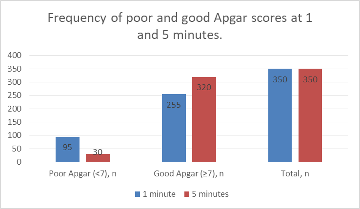
Figure 1. Frequency of poor and good Apgar scores at 1 and 5 minutes.
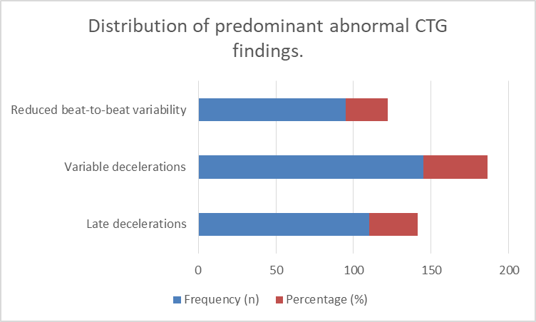
Figure 2. Distribution of predominant abnormal CTG findings.
DISCUSSION :
The present study evaluated neonatal Apgar outcomes among newborns delivered following abnormal cardiotocography (CTG) traces. The findings demonstrated that abnormal intrapartum fetal heart-rate patterns were accompanied by a clinically relevant frequency of low Apgar scores, particularly at 1 minute, with improvement in Apgar status by 5 minutes. This pattern is biologically plausible because CTG abnormalities may reflect transient or sustained impairment of fetal oxygenation during labour. Uterine contractions physiologically reduce intervillous blood flow and fetal oxygen delivery, and a fetus with limited compensatory reserve may develop progressive hypoxemia and metabolic disturbance. Recent reviews have emphasized that CTG is primarily a screening method for identifying fetuses potentially exposed to hypoxia rather than a direct measurement of fetal oxygenation or acid-base status. {5,6}
The frequency of poor Apgar scores observed in this study is broadly consistent with previous evidence linking abnormal CTG patterns with adverse neonatal outcomes. A recent systematic review of admission CTG reported that abnormal traces were associated with increased rates of low Apgar scores and neonatal intensive care admission, although diagnostic performance varied considerably between studies. {7} This variability is important because the relationship between CTG abnormalities and neonatal condition is influenced by the duration and severity of hypoxia, fetal compensatory mechanisms, gestational age, and the clinical circumstances surrounding labour. Thus, an abnormal trace should be interpreted as an indicator of potential fetal compromise rather than as definitive evidence of neonatal asphyxia. {5,7}
The improvement in Apgar status between 1 and 5 minutes is also compatible with the physiological response of many newborns to resolution of intrapartum stress following birth. Once delivery occurs, removal of uterine compression and establishment of pulmonary gas exchange can rapidly improve oxygenation and cardiovascular adaptation. Consequently, a low early Apgar score does not necessarily indicate persistent neurological injury. Nevertheless, a persistently low score may identify newborns requiring continued resuscitative or neonatal support, which reinforces the importance of preparedness of the neonatal team when an abnormal CTG is identified before delivery.
In the present study, variable decelerations constituted the most frequently documented abnormal CTG pattern. Variable decelerations are commonly related to intermittent umbilical cord compression, which can produce transient reductions in fetal venous return and cardiac output. Recurrent or prolonged abnormalities may reduce fetal oxygen delivery and, particularly when accompanied by reduced variability or other concerning features, may indicate progression toward fetal compromise. However, recent evidence indicates that individual CTG features should not be interpreted in isolation. Differences between international CTG guidelines in the relative importance assigned to variability, accelerations and decelerations demonstrate the complexity of translating individual waveform characteristics into reliable predictions of fetal hypoxia. {8,9}
Reduced beat-to-beat variability is another clinically important finding because variability reflects the interaction between the fetal autonomic nervous system and cardiovascular control. Progressive hypoxemia and metabolic disturbance can impair this autonomic activity, resulting in reduced variability. Nevertheless, reduced variability may also occur during fetal sleep or with other physiological or pharmacological influences. Recent research has therefore emphasized that the diagnostic value of CTG depends on the combined interpretation of baseline rate, variability, accelerations, decelerations, uterine activity, and the clinical context rather than reliance on a single parameter. {8,10}
The findings also need to be considered in the context of the recognized limitations of visual CTG interpretation. A recent systematic review of clinical practice guidelines identified substantial differences between recommendations regarding the interpretation and weighting of important CTG characteristics. {8} Similarly, studies evaluating clinicians' ability to identify fetal hypoxia have demonstrated only moderate predictive performance, while recent investigations of interobserver agreement have continued to demonstrate variability in interpretation of abnormal traces. {10,11} These limitations provide a possible explanation for why an abnormal CTG does not invariably correspond to a low neonatal Apgar score and why CTG should be integrated with clinical examination, labour progression, fetal risk factors, and, where available and appropriate, additional assessment of fetal acid-base status.
The clinical implications of these findings are relevant to obstetric practice in tertiary-care settings. An abnormal CTG should prompt systematic assessment and timely reassessment rather than automatic operative delivery. Excessive reliance on CTG abnormalities may increase interventions without necessarily improving neonatal outcomes. Evidence concerning adjunctive technologies supports this cautious approach. An updated meta-analysis of nine randomized trials involving 28,729 women found that adding ST waveform analysis to CTG did not significantly reduce operative delivery for fetal distress, although a reduction in metabolic acidosis was observed. {12} A separate systematic review similarly concluded that evidence for clinically important neonatal advantages of combining ST waveform analysis with CTG remained inconclusive. {13}
There is increasing interest in computerized and artificial-intelligence-assisted CTG interpretation as a means of improving recognition of fetal compromise and reducing observer variability. Recent reviews have identified promising performance of machine-learning approaches, but have also emphasized important concerns regarding dataset size, external validation, signal quality, outcome definitions, and generalizability across populations. {14,15} Such approaches may eventually provide useful decision-support tools, particularly in settings where specialist interpretation is not continuously available, but they should complement rather than replace clinical judgment until adequately validated in prospective clinical studies.
The present study has several strengths. It used a predefined sample size, consecutive recruitment, standardized assessment of Apgar scores at both 1 and 5 minutes, and a clearly specified definition of abnormal CTG. The study was conducted in a tertiary-care obstetric setting, providing locally relevant information regarding the neonatal condition associated with abnormal intrapartum fetal monitoring. The simultaneous collection of maternal, obstetric, and neonatal variables also provides a basis for examining potential effect modifiers.
Several limitations should be acknowledged. The cross-sectional design limited the ability to establish a temporal or causal relationship between individual CTG abnormalities and neonatal outcome. The study was conducted at a single tertiary-care hospital, which may limit generalizability to other institutions or lower-level healthcare facilities. Consecutive non-probability sampling may also have introduced selection bias. In addition, Apgar score is a practical clinical measure but is not a direct measure of fetal acidemia or hypoxic-ischemic injury; therefore, its use as the principal neonatal outcome cannot completely characterize the physiological significance of abnormal CTG. The study also did not incorporate umbilical cord blood gas analysis, neonatal neurological outcomes, or longer-term developmental outcomes. Future multicentre prospective studies incorporating cord blood pH, lactate, neonatal intensive care admission, encephalopathy, and longer-term neurodevelopmental outcomes would provide a more comprehensive assessment of the relationship between CTG abnormalities and neonatal health.
Overall, the findings support the continued use of CTG as an important component of intrapartum fetal surveillance while emphasizing that abnormal traces require contextual and systematic clinical interpretation. Recognition of abnormal fetal heart-rate patterns should facilitate timely assessment, appropriate obstetric decision-making, and preparation for neonatal resuscitation rather than being regarded as an isolated diagnostic endpoint. Further research in Pakistani tertiary-care populations, preferably using multicentre designs and objective biochemical neonatal outcomes, is warranted to determine which specific CTG characteristics most reliably predict clinically important neonatal compromise.
CONCLUSION :
The study demonstrated that abnormal cardiotocography was associated with an appreciable frequency of poor neonatal Apgar scores, particularly during the immediate postnatal period, with fewer newborns remaining below the poor-score threshold at 5 minutes. These findings support the clinical importance of careful intrapartum fetal surveillance and appropriate neonatal preparedness when abnormal CTG patterns are identified. CTG findings should, however, be interpreted within the overall clinical context rather than used as an isolated determinant of delivery. The study provides locally relevant evidence from a tertiary-care setting and supports further prospective, multicentre research incorporating objective neonatal outcomes to improve prediction of clinically significant fetal compromise.
BIBLIOGRAPHY:
1. Nazir L, Lakhta G, Anees K, Khan FR, Safdar S, Nazir GR, et al. Admission Cardiotocography as a Predictor of Low Apgar Score: An Observational, Cross-Sectional Study. Cureus. 2021;13(4):e14530.
2. Singh SK, Kumar R, Agarwal A, Tyagi A, Bisht SS. Intrapartum cardiotocographic monitoring and its correlation with neonatal outcome. J Family Med Prim Care. 2022;11(11):7398-7405.
3. Zullo F, Di Mascio D, Raghuraman N, Wagner S, Brunelli R, Giancotti A, et al. Three-tiered fetal heart rate interpretation system and adverse neonatal and maternal outcomes: a systematic review and meta-analysis. Am J Obstet Gynecol. 2023;229(4):377-387.
4. Tsiligkeridou S, Bolou A, Xanthos T, Gourounti K. Perinatal and Neonatal Outcomes Using Cardiotocography Versus STAN and Cardiotocography: a Systematic Review. Maedica (Bucur). 2023;18(4):684-691.
5. Francis F, Luz S, Wu H, Stock SJ, Townsend R. Machine learning on cardiotocography data to classify fetal outcomes: A scoping review. Comput Biol Med. 2024;172:108220.
6. Mendis L, Palaniswami M, Brownfoot F, Keenan E. Computerised cardiotocography analysis for the automated detection of fetal compromise during labour: a review. Bioengineering (Basel). 2023;10(9):1007.
7. Jha SP, Kumari S, Sinha A, Roy R, Sanjita, Kumari S, et al. Admission Cardiotocography in Term Pregnancies for Predicting Intrapartum Interventions and Early Neonatal Outcomes: A Systematic Review. Cureus. 2026;18(5):e109070.
8. Brown J, Kanagaretnam D, Zen M. Clinical practice guidelines for intrapartum cardiotocography interpretation: A systematic review. Aust N Z J Obstet Gynaecol. 2023;63(3):278-289. PMID: 36898674.
9. Zamora Del Pozo C, Chóliz Ezquerro M, Mejía I, et al. Diagnostic capacity and interobserver variability in FIGO, ACOG, NICE and Chandraharan cardiotocographic guidelines to predict neonatal acidemia. J Matern Fetal Neonatal Med. 2022;35(25):8498-8506.
10. Blix E, Brurberg KG, Reierth E, Reinar LM, Øian P. ST waveform analysis vs cardiotocography alone for intrapartum fetal monitoring: An updated systematic review and meta-analysis of randomized trials. Acta Obstet Gynecol Scand. 2024;103(3):437-448. PMID: 38093630.
11. Tarvonen M, Markkanen J, Tuppurainen V, Jernman R, Stefanovic V, Andersson S. Intrapartum cardiotocography with simultaneous maternal heart rate registration improves neonatal outcome. Am J Obstet Gynecol. 2024;230(4):379.e1-379.e12. PMID: 38272284.
12. Tsiligkeridou S, Bolou A, Xanthos T, Gourounti K. Perinatal and Neonatal Outcomes Using Cardiotocography Versus STAN and Cardiotocography: a Systematic Review. Maedica (Bucur). 2023;18(4):684-691. PMID: 38348066.
13. Brown J, Kanagaretnam D, Zen M. Clinical practice guidelines for intrapartum cardiotocography interpretation: A systematic review. Aust N Z J Obstet Gynaecol. 2023;63(3):278-289. PMID: 36898674.
14. Cardiotocography use for fetal assessment during labor in low- and middle-income countries: A scoping review. J Matern Fetal Neonatal Med. 2024. PMID: 38287690.
15. Zullo F, Di Mascio D, Raghuraman N, et al. Three-tiered fetal heart rate interpretation system and adverse neonatal and maternal outcomes: a systematic review and meta-analysis. Am J Obstet Gynecol. 2023;229(4):377-387. PMID: 37044237.