Diagnostic Accuracy of Magnetic Resonance Imaging in Detecting Suspected Cervical Cancer Taking Histopathology as Gold Standard
- Muhammad Junaid Tahir , MBBS, FCPS Postgraduate Trainee, Department of Radiology, Shaukat Khanum Memorial Cancer Hospital and Research Centre, Lahore, Pakistan.
- Muhammad Talha , MBBS, FCPS Postgraduate Trainee, Department of Radiology, Shaukat Khanum Memorial Cancer Hospital and Research Centre, Lahore, Pakistan.
- Awais Mustafa , MBBS, FCPS Postgraduate Trainee, Department of Radiology, Shaukat Khanum Memorial Cancer Hospital and Research Centre, Lahore, Pakistan.
- Mehreen Nawab , MBBS, FCPS, Consultant Radiologist, Department of Radiology, Shaukat Khanum Memorial Cancer Hospital and Research Centre, Lahore, Pakistan.
- Aamer Iftikhar , MBBS, FRCR, Consultant Radiologist, Department of Radiology, Shaukat Khanum Memorial Cancer Hospital and Research Centre, Lahore, Pakistan
- Anis ur Rehman , MBBS, FCPS, Consultant Radiologist, Department of Radiology, Shaukat Khanum Memorial Cancer Hospital and Research Centre, Lahore, Pakistan.
Article Information:
Abstract:
Cervical cancer is an important cause of morbidity and mortality among women. Accurate detection and characterization of cervical carcinoma are essential for appropriate management. Magnetic resonance imaging (MRI) provides detailed soft-tissue assessment and may assist in detecting cervical carcinoma, while histopathology remains the diagnostic gold standard. Objectives: To determine the diagnostic accuracy of magnetic resonance imaging in detecting cervical cancer, taking histopathology as the gold standard. Study Design & Setting: Cross-sectional validation study conducted in the Department of Radiology, Shaukat Khanum Hospital from 30 May 2025 to 30 August 2025. Methodology: A total of 113 patients aged 20–60 years with suspected cervical cancer were selected through non-probability consecutive sampling. MRI was performed and findings were assessed by a consultant radiologist. Biopsy specimens were evaluated histopathologically for the presence or absence of cervical carcinoma. Sensitivity, specificity, positive predictive value (PPV), negative predictive value (NPV), diagnostic accuracy, likelihood ratios, and area under the ROC curve (AUC) were calculated. Results: The mean age of patients was 45.6 ± 8.7 years and the mean lesion size was 3.7 ± 1.4 cm. MRI detected cervical carcinoma in 63 (55.8%) patients, while histopathology confirmed carcinoma in 65 (57.5%). MRI correctly identified 52 true-positive and 37 true-negative cases, with 11 false-positive and 13 false-negative cases. MRI demonstrated a sensitivity of 80.0%, specificity of 77.1%, positive predictive value of 82.5%, negative predictive value of 74.0%, and diagnostic accuracy of 78.8%. The positive and negative likelihood ratios were 3.47 and 0.26, respectively. The AUC was 0.86 (95% CI: 0.79–0.92; p<0.001). Among histopathologically confirmed cases, FIGO stage IIB was most frequent (27.7%), followed by IIIC1 (21.5%) and IB1 (18.5%). Tumor extension beyond the cervix was present in 59.3% of patients, while diffusion restriction was present in 61.9%. Squamous cell carcinoma was the most common histopathological type (42.5%), followed by adenocarcinoma (13.3%). Mean ADC values were 0.74 ± 0.13 × 10⁻³ mm²/s in carcinoma and 1.21 ± 0.28 × 10⁻³ mm²/s in benign cases. All MRI examinations were performed using a 1.5-Tesla unit. No significant differences in diagnostic performance were observed after stratification. Conclusion: MRI demonstrated good diagnostic accuracy for detecting cervical carcinoma when compared with histopathology.
Keywords:
Article :
INTRODUCTION:
Cervical cancer (CC) represents the fourth most frequently diagnosed cancer and the fourth leading cause of cancer death in women globally.1 Accurate staging of CC is crucial to plan the most appropriate treatment. Based on the International Federation of Gynecology and Obstetrics (FIGO) 2009 staging system, the clinical evaluation of CC has been exclusively based on physical and pelvic examination, a standard chest X-ray, and cystoscopy and proctoscopy if bladder and rectum invasion is suspected.2 However, this approach could lead to an inaccurate assessment of the local extent of tumor growth, since it is influenced by the clinician’s experience and patients’ or tumor characteristics. The major difficulties in the clinical evaluation of patients with CC are related to the assessment of parametrial invasion and lymph node involvement. Several authors reported discrepancies between clinical and pathological staging with values ranging from 25% in the early stages (stage < IIA) to 65–90% in the advanced stages (stage > IIB).3
Magnetic resonance imaging (MRI) is a noninvasive imaging method without ionizing radiation and with proven ability in evaluation of cervical tumor, its parametrial invasion, tumor size and the lymph node metastasis. Hence, MRI provides an optimal option for evaluation of the main prognostic factors and selection of therapeutic strategy.4 The sensitivity and specificity of MRI in assessing the metastases are very high, so if MRI is done, it may avoid the need for an examination under anaesthesia and the need of performing the other investigations in advanced disease.5,6 A study has shown the prevalence of cervical cancer as 57.38% and sensitivity, specificity, positive predictive value and negative predictive value of MRI for cervical carcinoma were 80%, 76.9%, 82.4% and 74.1% respectively.7 In a local study, the sensitivity and specificity of MRI in detecting cervical cancer were 90.0% and 87.0% respectively.8 Another local study has shown the sensitivity, specificity, PPV, NPV and diagnostic accuracy of MRI in diagnosing cervical cancer as 94.62%, 92.59%, 97.78%, 87.33% and 94.17% respectively.9 Heema S et al has shown the sensitivity of 50% and specificity of 90% of MRI in cervical carcinoma.10
Although previously many studies are available on this topic but as described above, these studies have shown variable diagnostic accuracy of MRI in detecting cervical cancer, so there must be re-evaluation of its diagnostic accuracy. Keeping in mind this, I have decided to conduct this study to determine the diagnostic accuracy of magnetic resonance imaging in detecting cervical cancer patients, taking histopathology as gold standard. My study will not only help to resolve the previous controversy but also if it’s diagnostic accuracy will be found high then this technique can be opted routinely in our practice for pre-operative assessment of cervical carcinoma for taking proper surgical management in order to reduce the morbidity and mortality of these particular patients. The objective of the study was to determine the diagnostic accuracy of magnetic resonance imaging in detecting cervical cancer, taking histopathology as the gold standard.
OPERATIONAL DEFINITIONS:
Diagnostic Accuracy: Diagnostic accuracy was assessed by sensitivity, specificity, PPV, NPV, and overall accuracy. True-positive and true-negative cases were positive and negative on both MRI and histopathology, respectively, while false-positive and false-negative cases were discordant between MRI and histopathology.
Suspected Cervical Cancer: Patients who had contact bleeding, particularly postcoital bleeding, and dyspareunia for more than 3 months were considered suspected cases.
MRI Findings of Cervical Carcinoma: An irregular endocervical lesion that was isointense on T1WI, hyperintense on T2WI, and enhanced on fat-saturated T2WI was considered suggestive of cervical carcinoma.
Histopathology Findings of Cervical Carcinoma: Back-to-back small glands with little or no intervening stroma, hyperchromatic nuclei, prominent nucleoli, and increased mitotic figures were considered positive for cervical carcinoma.
METHODS:
The study was a cross-sectional validation study conducted in the Department of Radiology, Shaukat Khanum Hospital, Lahore after approval of the synopsis from 30 May 2025 to 30 August 2025. A sample size of 113 cases was calculated with a 95% confidence level, prevalence of cervical cancer of 57.38%, 12% desired precision, and sensitivity and specificity of MRI for cervical carcinoma of 80% and 76.9%, respectively.⁷ Non-probability consecutive sampling was used. Patients aged 20–60 years with suspected cervical cancer, as per the operational definition, for more than 3 months, married females, and those with parity 0–4 were included. Patients with a history of abnormal cytology, previous treatment for cervical intraepithelial neoplasia (CIN), acute cervicitis, previous radiotherapy or chemotherapy, diagnosed pelvic pathologies such as pelvic tumors or congenital uterine anomalies, and contraindications to MRI such as stents, claustrophobia, metallic prostheses, or pacemakers were excluded.
After approval from the Institutional Ethical Review Committee and CPSP, 113 patients presenting to the Radiology Department of Shaukat Khanum Hospital, Lahore, who fulfilled the inclusion criteria were selected. Informed consent was obtained from each patient. Age, duration of symptoms, lesion size, parity, menopausal status, and place of living were recorded. MRI was performed using a Philips/Orient 1.5 Tesla unit. Patients were instructed to fast for 6 hours before examination to reduce small-bowel peristalsis artifacts. Axial T2-weighted images were obtained from the kidneys to the perineum using a 256 × 256 matrix, 32-cm field of view (FOV), 4-mm slice thickness, 1-mm interslice gap, and two numbers of excitations (NEX). Each MRI was reviewed by a consultant radiologist with at least 5 years of post-fellowship experience. Biopsy specimens were obtained from suspicious lesions and sent to the pathology laboratory for evaluation of the presence or absence of cervical cancer. MRI findings were correlated with histopathology reports, which were taken as the gold standard.
Collected data were analyzed using SPSS version 25.0. The Shapiro–Wilk test was used to assess the normality of the data. Age, duration of symptoms, lesion size, and parity were presented as mean ± standard deviation or median (IQR), as appropriate. Menopausal status (pre-menopause/post-menopause), place of living (rural/urban), and presence or absence of cervical carcinoma on MRI and histopathology were presented as frequencies and percentages. A 2×2 contingency table was used to calculate the sensitivity, specificity, positive predictive value, negative predictive value, and diagnostic accuracy of MRI in detecting cervical cancer, taking histopathology as the gold standard. ROC curve analysis and likelihood ratios were also calculated. Stratification was performed for age, duration of disease, lesion size, parity, menopausal status, and place of living. Post-stratification, a 2×2 contingency table was used to calculate sensitivity, specificity, positive predictive value, negative predictive value, and diagnostic accuracy. The Chi-square test was applied, and a p-value ≤0.05 was considered statistically significant.
RESULTS:
The mean age of the patients was 45.6 ± 8.7 years, while the mean duration of symptoms was 5.8 ± 1.9 months. The mean lesion size was 3.7 ± 1.4 cm and the mean parity was 2.6 ± 1.2. Among the patients, 67 (59.3%) were pre-menopausal and 46 (40.7%) were post-menopausal. Regarding place of living, 71 (62.8%) patients were from urban areas and 42 (37.2%) were from rural areas, as shown in Table 1.
Table 1. Demographic and Clinical Characteristics of Study Patients (n=113)
|
Variable |
Mean ± SD / n (%) |
|
Age (years) |
45.6 ± 8.7 |
|
Duration of symptoms (months) |
5.8 ± 1.9 |
|
Lesion size (cm) |
3.7 ± 1.4 |
|
Parity |
2.6 ± 1.2 |
|
Pre-menopausal |
67 (59.3%) |
|
Post-menopausal |
46 (40.7%) |
|
Rural residence |
42 (37.2%) |
|
Urban residence |
71 (62.8%) |
MRI detected cervical carcinoma in 63 (55.8%) patients, while 50 (44.2%) patients had no evidence of carcinoma on MRI. Histopathology confirmed cervical carcinoma in 65 (57.5%) patients and was negative in 48 (42.5%) patients, as given in Table 2.
Table 2. MRI and Histopathological Findings for Detection of Cervical Carcinoma (n=113)
|
Finding |
Present, n (%) |
Absent, n (%) |
Total, n (%) |
|
MRI |
63 (55.8%) |
50 (44.2%) |
113 (100%) |
|
Histopathology |
65 (57.5%) |
48 (42.5%) |
113 (100%) |
On comparison with histopathology, MRI correctly identified 52 true-positive and 37 true-negative cases. There were 11 false-positive and 13 false-negative cases, giving a total of 113 patients assessed, as shown in Table 3.
Table 3. Comparison of MRI Findings with Histopathology as the Gold Standard (n=113)
|
MRI findings |
Histopathology: Present |
Histopathology: Absent |
Total |
|
Present |
52 (True positive) |
11 (False positive) |
63 |
|
Absent |
13 (False negative) |
37 (True negative) |
50 |
|
Total |
65 |
48 |
113 |
The positive likelihood ratio was 3.47 and the negative likelihood ratio was 0.26. The area under the ROC curve was 0.86 (95% CI: 0.79–0.92), with a statistically significant p-value of <0.001, as given in Table 4.
Table 4. Diagnostic Accuracy of MRI in Detecting Cervical Carcinoma (n=113)
|
Parameter |
Value |
|
Positive likelihood ratio (LR+) |
3.47 |
|
Negative likelihood ratio (LR−) |
0.26 |
|
Area under the curve (AUC) |
0.86 |
|
95% Confidence interval |
0.79–0.92 |
|
p-value |
<0.001 |
MRI showed a sensitivity of 80.0%, specificity of 77.1%, positive predictive value of 82.5%, negative predictive value of 74.0%, and overall diagnostic accuracy of 78.8% for detecting cervical carcinoma, as shown in Table 5.
Table 5. Diagnostic Accuracy of MRI in Detecting Cervical Carcinoma (n=113)
|
Diagnostic measure |
Value (%) |
|
Sensitivity |
80.0 |
|
Specificity |
77.1 |
|
Positive predictive value |
82.5 |
|
Negative predictive value |
74.0 |
|
Diagnostic accuracy |
78.8 |
Among the 65 histopathologically confirmed cases, the most frequent FIGO stage was IIB, observed in 18 (27.7%) patients, followed by IIIC1 in 14 (21.5%) and IB1 in 12 (18.5%). Tumor extension beyond the cervix was present in 67 (59.3%) patients given in figure 1.
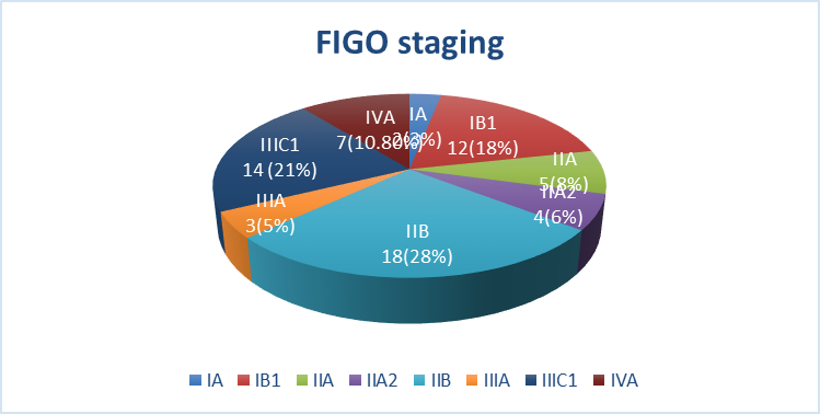
Figure 1. Distribution of Patients According to FIGO Stage of Cervical Carcinoma (n=65)
Squamous cell carcinoma was the most common histopathological type, accounting for 48 (42.5%) cases, followed by adenocarcinoma in 15 (13.3%) and other/poorly differentiated carcinoma in 2 (1.8%). Diffusion restriction was present in 70 (61.9%) patients. The mean ADC value was 0.74 ± 0.13 × 10⁻³ mm²/s in patients with cervical carcinoma and 1.21 ± 0.28 × 10⁻³ mm²/s in benign cases, with an overall mean of 0.94 ± 0.32 × 10⁻³ mm²/s. All MRI examinations were performed using a 1.5-Tesla MRI unit, as shown in Table 6.
Table 6. MRI and Histopathological Characteristics of Study Patients (n=113)
|
Variable |
Category |
n (%) / Mean ± SD |
|
Tumor extension beyond cervix |
Present |
67 (59.3%) |
|
Absent |
46 (40.7%) |
|
|
Type of cancer |
Squamous cell carcinoma |
48 (42.5%) |
|
Adenocarcinoma |
15 (13.3%) |
|
|
Other/poorly differentiated |
2 (1.8%) |
|
|
Benign |
48 (42.5%) |
|
|
Diffusion restriction |
Present |
70 (61.9%) |
|
Absent |
43 (38.1%) |
|
|
ADC value (×10⁻³ mm²/s) |
Cervical carcinoma |
0.74 ± 0.13 |
|
Benign |
1.21 ± 0.28 |
|
|
Overall |
0.94 ± 0.32 |
Diagnostic performance was comparable across age groups, symptom duration, lesion size, parity, menopausal status, and place of residence, with no statistically significant differences observed (all p>0.05), as shown in Table 7.
Table 7. Stratification of MRI Diagnostic Accuracy According to Demographic and Clinical Characteristics (n=113)
|
Variable |
Category |
Sensitivity (%) |
Specificity (%) |
PPV (%) |
NPV (%) |
Diagnostic Accuracy (%) |
p-value |
|
Age (years) |
20–40 |
77.3 |
77.8 |
81.0 |
73.7 |
77.5 |
0.742 |
|
41–60 |
81.4 |
76.7 |
83.3 |
74.2 |
79.5 |
||
|
Duration of symptoms |
≤6 months |
79.6 |
79.5 |
83.0 |
75.6 |
79.5 |
0.812 |
|
>6 months |
81.2 |
66.7 |
81.2 |
66.7 |
76.0 |
||
|
Lesion size |
≤3 cm |
72.0 |
84.0 |
81.8 |
75.0 |
78.0 |
0.293 |
|
>3 cm |
85.0 |
69.6 |
82.9 |
72.7 |
79.4 |
||
|
Parity |
0–2 |
77.4 |
81.5 |
82.8 |
75.9 |
79.3 |
0.694 |
|
3–4 |
82.4 |
71.4 |
82.4 |
71.4 |
78.2 |
||
|
Menopausal status |
Pre-menopause |
79.5 |
78.6 |
83.8 |
73.3 |
79.1 |
0.641 |
|
Post-menopause |
80.8 |
75.0 |
80.8 |
75.0 |
78.3 |
||
|
Place of living |
Rural |
78.3 |
78.9 |
81.8 |
75.0 |
78.6 |
0.958 |
|
Urban |
81.0 |
75.9 |
82.9 |
73.3 |
78.9 |
DISCUSSION :
Cervical cancer is one of the important causes of cancer-related morbidity and mortality among women. Early and accurate diagnosis is essential for timely treatment and improved outcomes. MRI provides excellent soft-tissue contrast and allows detailed assessment of cervical lesions and local tumor extension. However, MRI findings may overlap with benign and inflammatory cervical conditions. Histopathological examination remains the gold standard for confirming cervical carcinoma.11,12 Therefore, assessing the diagnostic accuracy of MRI against histopathology is clinically important.
The present study demonstrated that MRI had good diagnostic performance for detecting cervical carcinoma when histopathology was used as the gold standard. Among 113 patients, MRI identified 63 (55.8%) as positive, while histopathology confirmed carcinoma in 65 (57.5%). There were 52 true-positive, 37 true-negative, 11 false-positive, and 13 false-negative cases. The resulting sensitivity of 80.0%, specificity of 77.1%, PPV of 82.5%, NPV of 74.0%, and overall diagnostic accuracy of 78.8% indicated a relatively balanced ability of MRI to identify and exclude cervical carcinoma. The ROC analysis further supported its diagnostic performance, with an AUC of 0.86 (95% CI: 0.79–0.92; p<0.001). These findings are particularly relevant because MRI can provide detailed assessment of cervical soft tissues and local tumor extent while histopathology provides definitive tissue diagnosis.
The diagnostic estimates in the present study were remarkably similar to those observed by Shahbaz et al. (2019). Their sensitivity of 80.0%, specificity of 76.9%, PPV of 82.4%, NPV of 74.1%, and diagnostic accuracy of 78.7% differed from the present values by only 0.0, 0.2, 0.1, 0.1, and 0.1 percentage points, respectively. This close agreement suggested that MRI performance remained consistent across comparable populations and clinical settings. Their mean age of 42.4 ± 7.1 years was also relatively close to the present mean age of 45.6 ± 8.7 years. The similarity in both patient age and diagnostic estimates strengthened the evidence that MRI could provide reliable preoperative assessment of suspected cervical carcinoma.14 A somewhat higher diagnostic performance was observed in the study by Bashir et al. (2020). Their MRI sensitivity, specificity, PPV, NPV, and diagnostic accuracy were 89.29%, 86.84%, 95.24%, 73.33%, and 88.67%, respectively, compared with 80.0%, 77.1%, 82.5%, 74.0%, and 78.8% in the present study. Thus, sensitivity, specificity, PPV, and overall accuracy were higher in their study by 9.29, 9.74, 12.74, and 9.87 percentage points, respectively, whereas NPV was almost identical. Their study also included a larger sample of 150 patients and had a higher prevalence of histopathologically confirmed carcinoma (74.67% versus 57.5% in the present study). The higher disease prevalence and differences in patient selection and tumor characteristics may have contributed to the higher PPV and overall diagnostic performance.17
The findings of Sumayya et al. (2022) showed greater variability compared with the present results. Their study population was older, with a mean age of 54.46 ± 9.29 years, compared with 45.6 ± 8.7 years in the present study, and squamous cell carcinoma constituted approximately 87.5% of their cases.13
Stage IB was the predominant stage in their population, whereas stage IIB was most frequent in the present study, accounting for 27.7% of confirmed cases, followed by IIIC1 (21.5%) and IB1 (18.5%). Sumayya et al. described diagnostic estimates ranging from 47–50% sensitivity in one analysis to 92% specificity and 90% diagnostic accuracy in another analysis. In contrast, the present study produced more consistent estimates of 80.0% sensitivity, 77.1% specificity, and 78.8% diagnostic accuracy. Differences in study population, staging distribution, MRI interpretation, and methodological approach may explain the discrepancy. Importantly, both studies demonstrated the usefulness of MRI in assessing cervical carcinoma and incorporated FIGO staging into the evaluation.13
Although Shumail et al. (2025) investigated muscle-invasive urothelial carcinoma rather than cervical carcinoma, their findings provided a useful comparison regarding the general diagnostic capability of MRI for invasive malignancy. Their reported sensitivity of 93.4% and diagnostic accuracy of 85.7% were higher than the corresponding values of 80.0% and 78.8% in the present study, whereas their specificity of 52.4% was substantially lower than the 77.1% observed here. Their PPV of 89.5% was also higher than the present 82.5%, while their NPV of 64.7% was lower than the present 74.0%. Thus, the present study showed a more balanced diagnostic profile, with relatively similar sensitivity and specificity, whereas Shumail et al. demonstrated a pattern favoring sensitivity over specificity. The difference in tumor site and disease characteristics should be considered when comparing these estimates.18 More recently, Naz et al. (2026) reported sensitivity of 93.8%, specificity of 72.6%, PPV of 72.3%, NPV of 93.8%, and diagnostic accuracy of 81.7%. Compared with the present study, their sensitivity was 13.8 percentage points higher and NPV was 19.8 percentage points higher, whereas specificity was 4.5 percentage points lower and PPV was 10.2 percentage points lower. Their overall diagnostic accuracy was only 2.9 percentage points higher than the 78.8% observed in the present study. This pattern suggested that both studies demonstrated good overall MRI performance, although the balance between sensitivity and specificity differed.15 The study by Hassan et al. (2024) provided stronger diagnostic estimates, although its diagnostic endpoint was specifically parametrial invasion rather than general detection of cervical carcinoma. Among 136 patients, MRI demonstrated 89.55% sensitivity, 88.41% specificity, 88.24% PPV, 89.71% NPV, and 88.97% diagnostic accuracy, with a statistically significant p-value of 0.0001. Compared with the present study, sensitivity and specificity were higher by 9.55 and 11.31 percentage points, respectively, while PPV, NPV, and diagnostic accuracy were higher by 5.74, 15.71, and 10.17 percentage points. The more specific assessment of parametrial invasion may partly account for the stronger diagnostic estimates. Nevertheless, the results were consistent with the present finding that MRI can accurately characterize important local features of cervical malignancy.16
The present findings were also comparable with the work of Ahmed (2025), particularly with respect to patient characteristics which reported a mean age of 48.76 years, which was close to the present mean age of 45.6 years. Histopathology was positive in 74.4% of patients compared with 57.5% in the present study, while MRI was positive in 72.2% compared with 55.8% in the present study. He also reported a 93.3% success rate for MRI diagnosis, which was higher than the 78.8% overall diagnostic accuracy observed here. The higher proportion of histopathologically positive patients in their study may have increased the positive predictive performance of MRI. Despite this difference, both studies supported MRI as a useful diagnostic modality in patients with suspected cervical carcinoma.19 Altaf (2025) further demonstrated the potential advantage of incorporating diffusion-weighted imaging into MRI assessment. DWI-MRI achieved 87.5% sensitivity, 85.5% specificity, 65.6% PPV, 95.6% NPV, and 86.0% diagnostic accuracy, compared with 80.0%, 77.1%, 82.5%, 74.0%, and 78.8%, respectively, in the present study. Sensitivity, specificity, and diagnostic accuracy were therefore 7.5, 8.4, and 7.2 percentage points higher in their study, while PPV was 16.9 percentage points lower and NPV was 21.6 percentage points higher. The present study also demonstrated the relevance of diffusion characteristics, as diffusion restriction was present in 61.9% of patients and mean ADC was lower in carcinoma than benign lesions (0.74 ± 0.13 vs. 1.21 ± 0.28 ×10⁻³ mm²/s). These findings supported the role of diffusion information as an additional component of MRI evaluation.20
STUDY LIMITATIONS
The study was conducted at a single tertiary-care hospital, which may limit the generalizability of the findings. The relatively small sample size of 113 patients may have limited the precision of diagnostic estimates. The study was cross-sectional, so long-term clinical outcomes were not assessed. MRI interpretation was performed by a single consultant radiologist, so inter-observer variability could not be evaluated.
CONCLUSION :
MRI demonstrated good diagnostic accuracy for detecting cervical carcinoma compared with histopathology. It showed good sensitivity, specificity, and overall diagnostic performance. MRI may therefore serve as a useful imaging modality for the evaluation of suspected cervical carcinoma.
Acknowledgement: We sincerely acknowledge the support and guidance of our mentors, colleagues, and the staff of the participating hospital for their valuable assistance throughout this study.
Conflict of Interest: No
Funding Disclosure: None
BIBLIOGRAPHY:
1. Yogaraj K. Association between MRI findings and histopathological examination in carcinoma cervix: a retrospective study. Int J Anatomy Radiol Surg. 2021;10(2):61-4.
2. Liu X, Wang J, Hu K, Zhang F, Meng Q, Wang W, et al. Validation of the 2018 FIGO staging system of cervical cancer for stage iii patients with a cohort from China. Cancer Manag Res. 2020;12:1405-10.
3. Vani CK, Murali N, Sundari N. Role of magnetic resonance imaging in diagnosis, staging and follow up of patients with carcinoma cervix. Int J Radiol Diagnos Imaging. 2020;3(2):101-6.
4. Zubery MH, Rahman FB, Islam MN, Datta A, Rahman T, Mahdi R. A comparative study between magnetic resonance imaging and clinical figo criteria in different stages of carcinoma cervix. Mymensingh Med J. 2021;30(4):1131-8.
5. Lu H, Wu Y, Liu X, Huang H, Jiang H, Zhu C, et al. The role of dynamic contrast-enhanced magnetic resonance imaging in predicting treatment response for cervical cancer treated with concurrent chemoradiotherapy. Cancer Manag Res. 2021;13:6065–78.
6. Ditto A, Maggiore ULR, Evangelisti G, Bogani G, Chiappa V, Martinelli F, et al. Diagnostic accuracy of magnetic resonance imaging in the pre-operative staging of cervical cancer patients who underwent neoadjuvant treatment: a clinical–surgical–pathologic comparison. Cancers (Basel). 2023;15(7):2061.
7. Asad M, Tabassum S, Urooj T, Haider S, Mehmood T.. Diagnostic accuracy of magnetic resonance imaging in carcinoma of cervix taking histopathology as gold standard. Pak J Radiol. 2019;19(4):222-27.
8. Ahmad I, Habib E, Nabi M. MRI's Diagnostic reliability in cervical cancer screening detecting cancer using magnetic resonance imaging is a reliable method (MRI) a single center study. Pak J Med Health Sci. 2023;17(01):622-4.
9. Khan L, Aman N, Begum A. Diagnostic accuracy of MRI for diagnosing malignancy of cervix taking histopathology as gold standard. Pak J Med Health Sci. 2020;14(4):1330-32.
10. Heema S, Orakzai JZ, Khan M, Awan NM, Babar KS. Diagnostic accuracy of magnetic resonance imaging in carcinoma of cervix taking histopathology as gold standard: magnetic resonance imaging in carcinoma of cervix taking histopathology as gold standard. Pak BioMed J. 2022;5(6):139-43.
11. Ahmad N, Mukhtar M, Gilani A, Bukhari AA, Manzoor A, Liaqat A. Diagnostic Accuracy of MRI in Detecting Stromal Invasion in Early Cervical Cancer Patients Taking Histopathology as Gold Standard: Detecting Stromal Invasion in Cervical Cancer. Pakistan Journal of Health Sciences. 2025 Apr 30:55-9.
12. Subbarayan SK, Deepu R, Dhanasingh P. Magnetic resonance imaging evaluation of carcinoma of the cervix with histopathological correlation in a tertiary care center: emphasizing the rise of adenocarcinoma in the indian context. Cureus. 2024 Oct 18;16(10).
13. Sumayya, Heema, Orakzai ZJ, Khan M, Awan NM, Babar KS. Diagnostic accuracy of magnetic resonance imaging in carcinoma of cervix taking histopathology as gold standard. Pak Biomed J. 2022;5(6):139-143.
14. Shahbaz S. Diagnostic accuracy of magnetic resonance imaging in carcinoma of cervix taking histopathology as gold standard. Pakistan Journal of Radiology. 2019;29(4).
15. Naz F, Hussain M, Memon S, Sahito AA, Azeem N, Rani R. Diagnostic Accuracy of Magnetic Resonance Imaging (MRI) in Evaluation of Cervical Carcinoma. Mirpur Journal of Medical Sciences. 2026 Apr 7;4(1):88-93.
16. Hassan S, Khan MT, Ghafoor M, Raman R, Aslam N, Mumtaz A, Manzoor H. Diagnostic accuracy of magnetic resonance imaging in detecting parametrial invasion in cervical cancer patients, taking histopathology as the gold standard. Journal of The Society of Obstetricians and Gynaecologists of Pakistan. 2024 May 28;14(2).
17. Bashir B, Haidari FA, Ramzan M, Ahmed E, Arshad F, Rehman WU, et al. Magnetic resonance imaging: a useful tool for diagnosing malignancy—diagnostic accuracy of MRI in detection of malignancy of cervix. Pak J Med Health Sci. 2020;14(2):491-495.
18. Shumail H, Rehman AU, Sheikh AZ, Shaukat Z, Khalil S, Maryam I. Diagnostic accuracy of magnetic resonance imaging in determining T stage of urothelial carcinoma, taking histopathology as gold standard. Biol Clin Sci Res J. 2025;6(6):573-576.
19. Ahmed SN. The accuracy of magnetic resonance imaging for screening cervical cancer: magnetic resonance imaging (MRI) is a dependable tool for cancer detection. J Med Health Sci Rev. 2025;2(2).
20. Altaf R, Arooj S, Irem F, Altaf R. Diagnostic Accuracy Of Diffusion-Weighted Megnatic Resonance Imaging In Detecting Cervical Cancer Taking Histopathology As Gold Standard. Pakistan Journal of Medical & Cardiological Review. 2025 Mar 31;4(1):178-84.