Comparison of Two Rotary Instruments for Gutta Percha Removal in Extracted Root Canal Retreatment Cases; A Cone Beam Computerized Tomographic Scan Analysis
- Perkha Ali , Department of Operative Dentistry, Islamic International Dental College/Hospital, Islamabad, Pakistan
- Usman Anwer Bhatti , Department of Operative Dentistry, Islamic International Dental College/Hospital, Islamabad, Pakistan
- Ijaz ul Haq , Department of Operative Dentistry, Islamic International Dental College/Hospital, Islamabad, Pakistan
- Saad M Khan , Department of Operative Dentistry, Islamic International Dental College/Hospital, Islamabad, Pakistan
- Muhammad Daniyal Azhar , Department of Operative Dentistry, Islamic International Dental College/Hospital, Islamabad, Pakistan
Article Information:
Abstract:
In endodontics, removal of gutta-percha and sealer is necessary not only for retreatment but also to allow thorough re-instrumentation and disinfection of the root canal system. NiTi systems, such as ProTaper R and Mtwo R, have been designed to facilitate this process, but it remains challenging to eliminate it. Objective: To compare the efficacy of ProTaper R and Mtwo R rotary retreatment systems in removing gutta-percha from extracted mandibular premolars, using CBCT analysis to evaluate the amount of residual filling material. Methods: This in vitro study was carried out using 70 extracted mandibular premolars. Specimens were all obturated with gutta-percha and AH 26 sealer and incubated for 3 months to simulate clinical aging. After incubation, the samples were randomly divided into two groups with 35 samples in each group: Group A was retreated with ProTaper R and Group B with Mtwo R, with pre- and post-treatment CBCT images taken for each sample to evaluate the amount of residual filling material, and the total instrumentation time recorded for each sample. Results: These two systems had the same level of success in terms of reduction of the filling material and the ProTaper R left a lot less unused gutta-percha in the coronal and apical thirds (p<0.05). Mean overall residual length (ProTaper R) was 5.4 + 1.1 mm, whereas that of Mtwo R was 6.6 + 1.3 mm. Procedural time of (ProTaper R) was 4.5 + 0.8 min compared to that of Mtwo R, which is 5.2 + 1.0 min. The most dominant residual material was found in the apical third of both groups. Conclusion: In this in vitro study, ProTaper R was more effective and efficient at removing gutta-percha than Mtwo R in the removal of gutta-percha from mandibular premolars, but neither system was effective at removing gutta-percha completely. In this context, the CBCT was shown to be a valuable non-invasive tool for the assessment of residual filling material.
Keywords:
Article :
INTRODUCTION:
Endodontic therapy has been supported as an effective treatment modality to salvage teeth with pulpal and periapical pathologies, but failure remains common in a subset of cases because of continued or secondary intraradicular infection. Poor debridement, absent anatomy, or leakage at the coronal or apical level, which permit the survival of microbes in the root canal system, contribute to post-treatment disease (1). Nonsurgical retreatment is the more desirable treatment in such cases, as it is designed to restore periapical healing by eliminating the current filling materials, recurring cleaning, shaping, and obturation (2). It is technically demanding because of the complexity of the morphology of root canals and adhesive nature of filling material (3). The use of nickel-titanium (NiTi) rotary instruments has changed this, as it takes less time to be positioned at the chairside and the removal becomes easier than with the conventional hand files (4).
Ni-Ti rotary retreatment systems have a design that is considered one of the most important factors influencing their performance. Pro-Taper Retreatment by Pro-Taper (Pro-Taper R) and M-two Retreatment by M-two (M-two R) are the most commonly researched systems designed to cut and remove obturation materials at various levels within the canal. Pro-Taper R has three progressive taper tools (D1, D2, D3), which means it can selectively work with the coronal, middle and apical thirds, respectively. M-two R, has two files with sharp cutting ends which serve in the removal and gripping of gutta-percha (5). Several experiments have compared these systems under controlled experimental conditions. Wahane et al. have demonstrated that rotating and reciprocating instruments can effectively remove gutta-percha, although neither system completely removes it (1). Similarly, Singh et al. pointed out that the morphology and method of the operator in the canal may result in disparity in the efficiency and time taken in the process of retreatment instruments. (2). The results of Atique et al. and Buyukezer Ozkan et al. also justified that rotary systems enhance the retrievability of obturation materials, though with varying degrees of efficacy, based on file design and study parameters (3,4).
The assessment method is one of the most significant challenges in evaluating the efficacy of retreatment. Root splitting or clearing are classic methods that can lead to damage of samples and scattering of debris, reducing reliability (6). The recent introduction of imaging modalities, such as cone-beam computed tomography (CBCT), has significantly contributed to research on retreatment by providing high-quality, three-dimensional visualization of root canals in a non-invasive manner (7). CBCT enables the qualitative and quantitative study of remaining filling material, the identification of procedural mistakes, including perforations, and the accurate determination of working length (8). A survey by Madhu et al., which compared ProTaper, Mtwo, WaveOne, and Hedstrom files using CBCT, found that none of the systems demonstrated complete removal. However, there was a clear difference in efficiency among the root thirds (9). CBCT was also used by Joshi et al. to evaluate the new systems of retreatment in curved canals, supporting the importance of innovative imaging in comparing instrumentation methods (10).
In addition to conventional gutta-percha cones, obturation methods such as carrier-based systems (Thermafil, GuttaCore) further complicate retreatment procedures. Faus-Llacer et al. used micro-CT and demonstrated that these advanced filling materials can be easily extracted using specialized retreatment tools. However, there is still difficulty with the apical third (11). Atique et al.'s comparative studies in 2024 revealed that hand instruments and rotary systems differed, with NiTi files demonstrating higher efficiency in material removal and lower fatigue among operators (12). Similarly, Indi et al. compared six retreatment methods and found a significant difference in the time needed, indicating a practical difference in instrument selection and its impact on clinical efficacy (13). Kulkarni et al. added to this evidence by comparing continuous rotating single-file and multi-file designs, demonstrating that file design and amount can affect the results of removals and retreatment (14).
Adjunctive measures have also been explored to maximize the removal of gutta-percha. In their investigation, Fibryanto et al. used ultrasonics with solvents and discovered better effectiveness in the middle and apical thirds of the canals (15). Minimally invasive endodontics has also had an impact on the philosophy of retreatment, with Ribeiro suggesting that single-file rotary systems may leave more dentin intact and allow successful establishment of gutta-percha (16). Long-term outcomes are essential beyond technical efficiency. A prospective cohort study with four-year CBCT follow-up by Zhang et al. established that nonsurgical retreatment results in periapical healing in a significant proportion of cases, but the results depend on the quality of the retreatment procedures (17). These clinical views are also supported by evidence syntheses of endodontic treatment and retreatment, which have found that although rotary instruments increase efficiency, complete gutta-percha removal remains a challenge (18).
Amidst an extensive body of research, comparative data on ProTaper R and Mtwo R systems in the particular context of CBCT-based analysis are still unavailable, especially in local populations where operator competence, anatomical differences, and practice environments can impact clinical outcomes. The majority of studies agree that although rotary systems help to remove filling material more predictably and more quickly than manual equipment, dense traces of the filling material tend to be left behind, particularly in the apical third (5). This highlights the clinical significance of selecting case-specific retreatment instruments and protocols. Additionally, more clinically relevant and reliable results are obtained with standardization of evaluation using advanced imaging techniques, including CBCT (6,7). As more attention is paid to minimally invasive and effective retreatment, it is necessary to further compare the performance of ProTaper R and Mtwo R systems under standard experimental conditions. The gap of knowledge and knowledge on the best retreatment would be facilitated by the local CBCT studies (9,10). This type of research can yield evidence that can be clinically utilized through measuring the average length of residual gutta-percha, which could be used to optimize retreatment schemes and improve patient outcomes.
Objective: To compare the efficacy of ProTaper R and Mtwo R rotary retreatment systems in removing gutta-percha from extracted mandibular premolars, using CBCT analysis to evaluate the amount of residual filling material.
MATERIAL AND METHODS:
This study was completed in the Department of Operative Dentistry and Endodontics, Islamic International Dental Hospital, from 15th March, 2025 to 15th September, 2025. The sample size of 70 extracted mandibular premolars (35 specimens per group) was calculated using Open Epi version 3.01 with a 95% confidence level, 80% study power, and an effect size derived from previously published studies comparing rotary retreatment systems. Data were entered and analysed using IBM SPSS Statistics version 26.0. Descriptive statistics, including mean and standard deviation, were calculated for all quantitative variables. The primary outcome variable was the mean length of residual filling material remaining after retreatment, while instrumentation time was considered a secondary outcome variable. Baseline variables, including root length, obturation length, and canal curvature, were also recorded.
Inclusion criteria: Mandibular premolars extracted from humans with a single straight root canal, fully formed apices, intact root morphology, and no cracks, root resorption, or structural defects were included in the study. To standardize root length and canal morphology, teeth with similar root length and canal morphology were selected. Prior to retreatment procedures, the root canals were instrumented and obturated in standardized laboratory conditions.
Exclusion Criteria: Teeth with incompletely formed roots, open apices, root caries extending below the cemento-enamel junction, root fractures, internal or external resorption, calcified canals, canal curvature greater than 30°, multiple canals, or any developmental or structural abnormalities were excluded from the study.
The selected teeth were assigned to two equal groups (n = 35 each) using a computer-generated random allocation sequence. The allocation sequence was prepared before instrumentation to ensure unbiased group assignment. Group A was subjected to retreatment with ProTaper R rotary files (D1, D2, D3) in crown-down mode, and Group B to Mtwo R files (25/0.05 and 15/0.05) to the working length. Working length shall be measured using a #10 K-file, followed by standard canal preparation with the use of AH 26 sealer and gutta-percha cones. Initial CBCT scans was calculated at the end of a three-month incubation period to determine obturation lengths. Thereafter, retreatment procedures was performed using the respective instruments with 5.25% NaOCl irrigation and chloroform solvent. An electric motor, in terms of torque control, was used to perform instrumentation as per the manufacturer's guidelines. Retreatment was completed when no further filling material is apparent on the files. CBCT scans was carried out at the same parameters after operations to determine the mean residual filling material in each group.
Baseline Measurements: Prior to retreatment, baseline measurements were obtained from the preoperative CBCT scans. The root length was defined as the linear distance from cemento-enamel junction to the radiographic apex. The length of obturation was determined from the coronal reference point to the apical extension of the gutta-percha filling. The canal curvature was evaluated on the reconstructed CBCT images according to Schneider's method and only teeth that had a canal curvature of ≤30° were included in the study. The measurements were taken with the CBCT software measurement tools with standardized imaging parameters.
Evaluation of Residual Filling Material: Postoperative CBCT scans were taken with the same imaging parameters as the preoperative scans. CBCT software was used to assess the axial, coronal and sagittal reconstructed images. The radiopaque filling material was found to be gutta-percha left on the walls of the canal. The amount of residual filling material for each specimen was documented as a continuous variable (mm) and the mean values were determined for each study group.
Measurement of Instrumentation Time: Instrumentation time was measured for each specimen with a digital stopwatch. The timing started with the introduction of the first retreatment instrument into the root canal and stopped when the instrument was no longer visible and the predetermined working length was reached. The total instrumentation time was documented for each specimen and compared between the two retreatment groups and was recorded in minutes.
RESULTS:
This study used 70 extracted mandibular premolars, which were equally split into two groups that were ProTaper R (n = 35) and Mtwo R (n = 35). Baseline characteristics, including root length, obturation length, and canal curvature measured from preoperative CBCT images, were compared between the two groups to ensure baseline comparability before retreatment.
Table 1: Baseline Characteristics of Specimens in Both Groups
|
Variable |
ProTaper R (n = 35), M ± SD |
Mtwo R (n = 35), M ± SD |
p-value |
|
Root length (mm) |
21.23 ± 1.09 |
21.18 ± 1.52 |
0.869 |
|
Obturation length (mm) |
20.09 ± 1.56 |
19.91 ± 1.90 |
0.675 |
|
Canal curvature (°) |
13.92 ± 1.91 |
14.33 ± 2.33 |
0.418 |
The comparison at the baseline showed that there were no statistically significant differences in the two groups (p >.05) and thus the two groups are similar before the intervention. This homogeneity made it possible to ascertain that the outcomes differences observed could be observed to be because of the retreatment systems and not because of existing anatomical differences. Postoperative CBCT images were analyzed for the amount of residual filling material (mm) in the coronal, middle, and apical third of each root canal. Retreatment of all specimens was followed by the observation of residual filling material, but in different amounts with the two retreatment systems. Both systems proved to be effective in lessening gutta-percha, but, in all the specimens, some residual was found.
Table 2: Residual Filling Material after Retreatment (mm)
|
Canal Level |
ProTaper R (M ± SD) |
Mtwo R (M ± SD) |
p-value |
|
Coronal third |
1.23 ± 0.15 |
1.53 ± 0.36 |
< .001* |
|
Middle third |
1.83 ± 0.28 |
2.12 ± 0.32 |
< .001* |
|
Apical third |
2.41 ± 0.29 |
2.93 ± 0.38 |
< .001* |
|
Total residual |
5.47 ± 0.48 |
6.57 ± 0.57 |
< .001* |
*Note. Values are presented as mean ± standard deviation (SD). Comparisons between the ProTaper R and Mtwo R groups were performed using an independent samples t-test. A p-value < 0.05 was considered statistically significant.
Independent samples t-test demonstrated significantly lower residual filling material in the ProTaper R group compared with the Mtwo R group at the coronal, middle, and apical thirds, as well as in the total residual filling material (all p < 0.001). The highest difference was seen in the apical third which always had the highest amount of remaining material between the groups. Although both systems improved on the coronal and middle thirds, neither system was able to remove this completely. Instrumentation time was also evaluated to compare the procedural efficiency of the two retreatment systems.
Table 3: Comparison of Instrumentation Time
|
Group |
Mean Time (minutes), M ± SD |
t-value |
p-value |
|
ProTaper R |
4.63 ± 0.97 |
|
|
|
Mtwo R |
5.24 ± 0.93 |
-2.69 |
.009* |
*Note. Values are presented as mean ± standard deviation (SD). Comparisons between groups were performed using an independent samples t-test. p < 0.05 was considered statistically significant.
The mean instrumentation time was significantly lower in the ProTaper R group (4.63 ± 0.97 minutes) than in the Mtwo R group (5.24 ± 0.93 minutes) (independent samples t-test, p = 0.009), indicating greater procedural efficiency with the ProTaper R system. The review indicated that ProTaper R took much less time to retreatment process than Mtwo R (p =.009). This shortening of instrumentation time shows an increase in the efficiency of the procedure, which can be transferred to an equivalent shortening of the chairside time and optimization of the clinical workflow. A graphical comparison was also done to further demonstrate how the residual filling material was distributed.
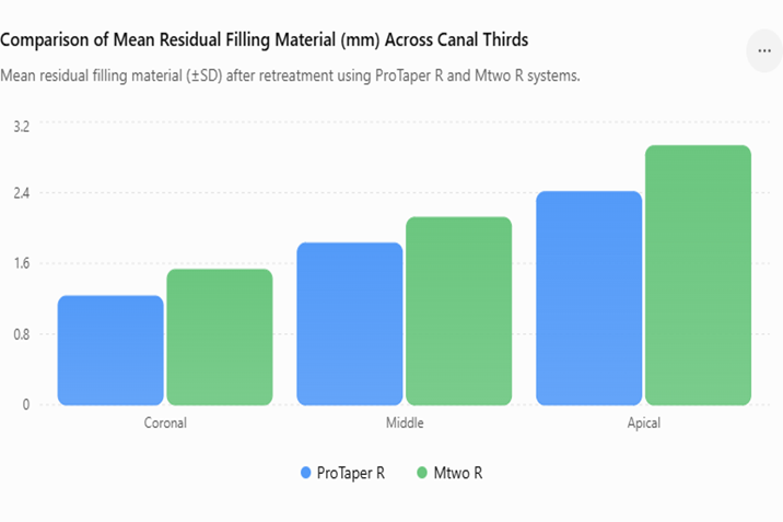
Figure 1: Comparison of Mean Residual Filling Material Across Canal Thirds
The graphical illustration brings out the highest residual material in both systems in the apical third, then the middle third and finally the coronal third. ProTaper R showed lower residual values in all the regions as opposed to Mtwo R.
Finally, the results suggest that, despite the fact that neither system has managed to get rid of gutta-percha completely, ProTaper R had a better performance both in its effectiveness and efficiency. The fact that residual material is left once the treatment process is over, especially at the apical third, reiterates the complications involved in root canal anatomy and the constraints of existing retreatment systems.
DISCUSSION :
In the present study, the efficiency and procedural efficiency of two nickel-titanium (NiTi) rotary gutta-percha removal systems (ProTaper R and Mtwo R) were compared in removing gutta-percha from extracted mandibular premolars using cone-beam computed tomography (CBCT). The present findings agree with previous studies which have demonstrated that rotary retreatment systems allow efficient removal of filling material and instrumentation time savings over conventional systems (1,2). Although these are all benefits, complete removal of gutta-percha is still difficult due to the complex anatomy of the root canal and the physical characteristics of the filling material. Significantly lower amounts of residual filling material were found in the coronal, middle, and apical thirds with ProTaper R, and the highest amount of residual material was in the apical third. This finding indicates that the anatomical challenges in the apical area remain a problem to remove all filling materials, even with the use of a retreatment system.
The apical third by all means recorded the most residual material in both groups, a fact that extends the difficulty in this area that is well documented. The intricate structure of the apical part, and the reduced canals as well as curve and irregularity of the anatomy limits access of the instruments, and minimizes cleaning. This result is in line with that of the studies which had been conducted earlier like Madhu et al and Singh et al that pointed out that full removal of filling material is hardly met particularly in the apical region (5.6). Another clinical importance of the study was that the presence of debris in this region was statistically relevant, and it could be a source of microorganisms as well as negatively affect retreatment procedures. ProTaper R took shorter periods to perform the retreatment procedure as opposed to Mtwo R. This enhancement could be attributed to the nature and arrangement of ProTaper R files, which are specifically designed to remove filling materials at a very fast rate in a crown-down manner.
This might be due to the fact that there are fewer steps involved in the procedure and the ability to cut at a greater speed. These findings have been confirmed by Indi et al. who proved the fact that various retreatment systems have a great difference in terms of time efficiency, as rotary systems have the advantage over manual methods (13). Clinically, this less instrumentation period is beneficial as it lowers the fatigue of the operator, the patient discomfort, and the efficiency of the treatment process. CBCT in this study was essential in the precise assessment of the quantity of the residual filling material. Conventional techniques like root splitting or clearing tend to be destructive and can result in loss or redistribution of debris and subsequently, interfere with the accuracy of the measurement. CBCT, on its side, offers a three-dimensional analysis of the root canal system that is non-invasive, which does not require any quantification of the residual material to be precise. CBCT as an effective instrument used in assessing the outcomes of retreatment has also been previously proven through the work by Paradkar et al. and Koç et al. (7,8). CBCT application in the current study, thus, increases validity and reliability of the findings.
Although ProTaper R showed these benefits, it is worth being noted that no specimen showed total elimination of gutta-percha. This observation is also in compliance with other researchers in the literature who have always noted that no single instrument system can be able to completely remove the filling material out of the root canal (9). Wahane et al. and Isa et al. highlighted that despite the sophisticated rotary systems, some debris remains and proposed that supplementary methods should be used to enhance the cleaning activity (10). They can involve the application of solvents, ultrasonics, or additional irrigation procedures to increase the amount of the remaining material is removed. Another conclusion made in this study is that the choice of instruments depends on the anatomy of the canals and clinical needs. Although ProTaper R showed better performance in most cases, the retreatment system type to be used ought to be a case-specific approach to the situation and one ought to consider issues like the canal curvature and complexity as well as the experience of the operator.
Research by Buyuyokzer Ozkan et al and Joshi et al has highlighted that the performance of instruments can be different with regard to the morphology and extra sensitivity of technique (11). Consequently, practitioners must follow a liberal approach and look into integrating two or more methods to get the best results. There are some drawbacks of this research that are to be mentioned. Since it is an in vitro study, the experimental conditions might not represent the clinical setting as much, where variability in patients, biological response, and skill of the operator may have an impact (17). Also, the research only considered mandibular premolars that are usually simpler in canal structure than molars. Hence, the results cannot be directly applied to more complicated cases. The research conducted in the future must cover a broader variety of teeth and must employ clinical trials to confirm these findings in practical situations. To conclude, the current research results confirm the fact that both ProTaper R and Mtwo R are effective in terms of reducing gutta-percha during retreatment but the former exhibit better efficacy and efficiency.
CONCLUSION :
Both ProTaper Universal Retreatment (ProTaper R) and Mtwo Retreatment (Mtwo R) rotary systems successfully removed the gutta-percha from extracted mandibular premolars within the limitations of this in vitro study, but neither system was able to remove the filling material completely. ProTaper R had a significantly higher retreatment efficacy with less residual filling material in the coronal, middle and apical third of the root canal than Mtwo R. It also needed much less instrumentation time, which meant it was more efficient in terms of the procedure. Although these benefits exist, there was still some filling material that was not removed, especially in the apical third, which was a problem in complete removal of filling material during retreatment. CBCT was shown to be a reliable three dimensional imaging modality for the assessment of residual filling material after retreatment. These findings need to be confirmed in further well-designed clinical and laboratory studies with various tooth types and clinical conditions to optimize retreatment protocols.
BIBLIOGRAPHY
1. Wahane KD, Kulkarni SS, Daokar S, Patil K, Patel K, Thorat T. An assessment of the efficacy of a rotary and a reciprocating retreatment file system for removal of gutta-percha from root canals: an in vitro cone-beam computed tomography study. Endodontology. 2021;33(1):20-4.
2. Singh KB, Akhtar MS, Nagar R, Agarwal A, Azhar S, Singh V, Ansari S, Singh Sr V. Comparative analysis of the efficacy of various retreatment file systems in the removal of gutta-percha in retreatment cases and time taken during the procedure: an in vitro cone beam CT study. Cureus. 2024;16(3):e55551.
3. Atique S, Ali K, Ahmed A. A comparison of effectiveness of conventional and rotary NiTi instruments in the removal of gutta-percha during root canal retreatment: a randomized ex-vivo study. Preprints. 2022. doi:10.20944/preprints202212.0340.v1.
4. Büyüközer Özkan H, Çimen T, Kaya Apaydın S, Er K. Efficacy of two different retreatment techniques in removing gutta-percha from root canals: a CBCT study. Turk Endod J. 2025;10(1):1-7.
5. Kalyankar S, Sharma A, Jadhav A, Patil R, Gujarathi N, Shelke A. Comparative evaluation of efficacy of different endodontic file systems for removal of gutta-percha from root canals using CBCT: an in-vitro study. Int J Health Sci. 2022;6(S8):2724-32.
6. Isa MM, Ramlan NA, Ayoub AA. Rotary systems for root canal gutta-percha removal: a review. Compend Oral Sci. 2023;10(1):57-75.
7. Paradkar S, Goyal K, Saha SG, Bhardwaj A, Saha MK, Nirwan AS. A comparative evaluation of retrievability of three different obturating systems using ProTaper Universal rotary retreatment files: an in vitro cone-beam computed tomography analysis. Endodontology. 2022;34(2):96-101.
8. Koç S, Harorlı H, Kuştarcı A. Comparative evaluation of the accuracy of electronic apex locators and cone-beam computed tomography in detection of root canal perforation and working length during endodontic retreatment. BMC Oral Health. 2024;24(1):953.
9. Madhu K, Karade P, Chopade R, Jadhav Y, Chodankar K, Alane U. CBCT evaluation of gutta-percha removal using ProTaper and Mtwo retreatment files, WaveOne, and Hedstrom files: an ex vivo study. Front Dent. 2021;18:19.
10. Joshi C, Hajoori M, Patel A, Somani M, Thumar S, Khunt A, Patel D, Desai N. Comparative evaluation of different retreatment files for gutta-percha removal from curved root canals accessed with novel ultra-conservative opening: an in-vitro study. J Clin Diagn Res. 2023;17(3):ZC37-42.
11. Faus-Llácer V, Pérez RL, Faus-Matoses I, Ruiz-Sánchez C, Zubizarreta-Macho Á, Sauro S, Faus-Matoses V. Efficacy of removing Thermafil and GuttaCore from straight root canal systems using a novel non-surgical root canal re-treatment system: a micro-computed tomography analysis. J Clin Med. 2021;10(6):1266.
12. Atique S, Ali K, Haroon S, Ahmed A, Javed MQ, Zafar MS, Abulhamael AM. Effectiveness of H-files and ProTaper Universal systems in removing gutta-percha during endodontic retreatment: a comparative study. J Taibah Univ Med Sci. 2024;19(3):537-44.
13. Indi S, Desai SR, Hambire A, Mustafa M, Almokhatieb AA, Abuelqomsan MA, Abdulwahed A, Alamri HM. Comparison of the time required by six different retreatment techniques for retrieval of gutta-percha: an in vitro study. Eur J Gen Dent. 2022;11(3):181-8.
14. Kulkarni PS, Patil PD, Kamat SB, Chougule PG, Sagare SV, Sathish HT. Comparative evaluation of single versus multiple continuous rotating file systems used in the retreatment of teeth obturated by cold lateral compaction and warm vertical compaction: an in vitro cone beam computed tomography study. Front Health Inform. 2024;13(3):1-7.
15. Fibryanto E, Widyastuti W, Hidayat A, Hastuty L. Effect of ultrasonic combined with solvent on gutta-percha and bioceramic sealer removal in middle and apical third of the root canal: an ex vivo study. J Dent Indones. 2024;31(1):55-60.
16. Ravi S, Kumaravadivel K, Vishwanath S, Mathew S, Thangavel B, Thangaraj DN. Evaluation of canal cleanliness of two rotary file systems with different taper systems: An in vitro scanning electron microscopic study. Endodontology. 2024 Oct 1;36(4):394-9.
17. Zhang MM, Fang GF, Chen XT, Liang YH. Four-year outcome of nonsurgical root canal retreatment using cone-beam computed tomography: a prospective cohort study. J Endod. 2021;47(3):382-90.
18. Alcalde GP, de Paula LM, de Paula EN, de Castro FP. Endodontic treatment and retreatment: a large systematic review. Sci. 2022;3:6.