Prevalence of Osteoporosis or Ostemalacia in Geriatric Population with Proximal Femoral Fracture at Tertiary Care Hospital
- Syed Wajahat Ali Zaidi , Resident Orthopaedic, Dow University of Health Sciences, Civil Hospital, Karachi, Pakistan
- Adeel Ahmed Siddiqui , Professor, Dow University of Health Sciences, Civil Hospital, Karachi, Pakistan
- Sunil Kumar , Associate Professor, Orthopaedic Surgery, Dow University of Health Sciences, Civil Hospital, Karachi, Pakistan
- Syed Muhammad Khalid Karim , Assistant Professor, Dow Medical College & Dr. Ruth K. M. Pfau Civil Hospital, Karachi, Pakistan
- Muhammad Noman , Assistant Professor, Dow University of Health Sciences, Civil Hospital, Karachi, Pakistan
- Kashif Ali Shaikh , Post Fellow, Orthopaedic Surgery, Civil Hospital, Karachi, Pakistan
- Zahid Nawaz Khan , Postgraduate Resident, Orthopaedic, Dow University of Health Sciences, Civil Hospital, Karachi, Pakistan
- Teerath Das , Postgraduate Resident, Orthopaedic, Dow University of Health Sciences, Civil Hospital, Karachi, Pakistan
- Muhammad Rukun-Uddin Siddiqui , Resident, Orthopaedic Surgeon, Dow University of Health Sciences, Civil Hospital, Karachi, Pakistan
Article Information:
Abstract:
Proximal femoral fractures are a major cause of disability, loss of independence, and mortality in older adults and are frequently associated with underlying metabolic bone disease. Osteoporosis reduces bone strength, whereas osteomalacia reflects defective mineralization, and both conditions may coexist in patients with fragility fractures. Evidence describing this overlap in South Asian populations remains limited. Identifying these abnormalities may improve secondary fracture prevention and guide more comprehensive bone health assessment after hip fracture in Pakistan. Objective: To determine the prevalence of osteoporosis, osteomalacia, and their coexistence among older adults with proximal femoral fractures in Karachi. Methods: This descriptive cross-sectional study was conducted from May to October 2025 at the Department of Orthopaedic Surgery, DUHS/Dr. Ruth K. M. Pfau Civil Hospital Karachi, after CPSP and institutional approval (CPSP/REU/OSG-2023-183-3025). Seventy-three patients aged 50–90 years with radiologically confirmed proximal femoral fractures were enrolled consecutively. Postoperative DEXA assessed bone mineral density. Serum calcium, phosphate, albumin, alkaline phosphatase, and 25-hydroxyvitamin D were measured, and intraoperative bone biopsy confirmed osteomalacia. Data were analyzed using SPSS version 18.0. Results: The mean age was 68.5 ± 8.7 years, and 46 participants (63.0%) were female. Falls accounted for 58 fractures (79.5%). Osteoporosis was present in 56 patients (76.7%), osteopenia in 12 (16.4%), and normal bone mineral density in 5 (6.9%). Histologically confirmed osteomalacia was identified in 28 patients (38.4%). Vitamin D concentrations were <10 ng/mL in 28 (38.4%), 10–20 ng/mL in 30 (41.1%), and >20 ng/mL in 15 (20.5%); overall, 58 patients (79.5%) had concentrations ≤20 ng/mL. A overlap analysis estimated coexistence of osteoporosis and osteomalacia in 22 patients (30.1%). Conclusion: Metabolic bone disease was common among older patients with proximal femoral fractures. Routine assessment of bone density and vitamin D status, with appropriate treatment of osteoporosis and mineralization abnormalities, may strengthen secondary fracture prevention.
Keywords:
Article :
INTRODUCTION:
Proximal femoral fractures (PHFs) are among the most serious injuries affecting older adults and represent an important cause of hospitalization, loss of independence, long-term disability, and mortality in ageing populations. Although these fractures frequently follow a seemingly minor fall, their occurrence often reflects underlying deterioration in bone strength rather than the severity of the trauma itself. Advancing age is accompanied by progressive changes in bone density, muscle strength, balance, mobility, and nutritional status, all of which increase susceptibility to fragility fractures (1,2). The consequences extend beyond the fracture itself, as affected individuals commonly require surgery, prolonged rehabilitation, assistance with activities of daily living, and substantial healthcare resources. Proximal femoral fractures are therefore increasingly regarded not only as an orthopedic problem but also as a clinical marker of underlying metabolic bone disease. They are among the most frequent age-related fractures, following vertebral and distal radius fractures, and occur particularly commonly in older women because of accelerated postmenopausal bone loss (3,4).
The epidemiological burden of PHFs varies considerably between populations. Reported incidence rates have ranged from approximately 60.1 to 90.8 per 100,000 person-years, although direct comparison between studies is difficult because of differences in age distribution, case definitions, study design, access to healthcare, and methods of fracture surveillance (5,6). Population ageing is expected to increase the absolute number of these fractures, particularly in low- and middle-income countries where preventive bone-health services, routine screening, and nutritional assessment may be less accessible. In addition to demographic factors, the occurrence of PHFs is influenced by physical activity, frailty, comorbid illness, medication use, fall risk, sunlight exposure, and nutritional deficiencies (7-9). Seasonal variations have also been described, with higher fracture frequencies during colder periods in some regions because of reduced sunlight exposure, changes in vitamin D status, restricted outdoor activity, and environmental conditions that increase the likelihood of falls (10,11). Such observations indicate that the burden of PHF is shaped by both skeletal health and the wider social and environmental context.
Osteoporosis is the most widely recognized metabolic bone disorder associated with fragility fractures. It is characterized by reduced bone mass and deterioration of bone microarchitecture, resulting in diminished skeletal strength and increased fracture susceptibility. Bone mineral density is most commonly assessed using dual-energy X-ray absorptiometry (DEXA), with a T-score of −2.5 or lower conventionally supporting a diagnosis of osteoporosis in appropriate populations. A markedly reduced Z-score, particularly a value of −2.5 or lower, may additionally raise suspicion of secondary causes of low bone mass and warrants further clinical evaluation (6). Osteoporosis may remain clinically silent for many years and is frequently first recognized only after a low-trauma fracture has occurred. This makes patients presenting with PHF an important group in whom underlying skeletal abnormalities should be systematically evaluated rather than attributing the injury solely to ageing or accidental falls (12).
Osteomalacia represents a different but clinically important disorder of bone metabolism. Whereas osteoporosis predominantly involves a reduction in the amount and structural quality of normally mineralized bone, osteomalacia results from defective mineralization of osteoid. Vitamin D deficiency is a major cause, although impaired vitamin D metabolism, phosphate depletion, malabsorption, chronic systemic disease, and other metabolic disturbances may also contribute (6–8). Patients may develop diffuse skeletal discomfort, muscle weakness, difficulty in mobility, and increased fracture susceptibility. Biochemical abnormalities can include reduced serum phosphate, severe deficiency of 25-hydroxyvitamin D, and compensatory elevation of parathyroid hormone, although the biochemical pattern varies according to the underlying cause and severity of disease. Bone biopsy can provide definitive confirmation in selected cases but is not routinely required for all patients (9). Importantly, osteomalacia may coexist with osteoporosis, particularly in elderly individuals with poor nutrition, limited sunlight exposure, chronic illness, or reduced mobility.
The coexistence of osteoporosis and osteomalacia has important diagnostic and therapeutic implications because the two disorders are not interchangeable. An elderly patient may have reduced BMD consistent with osteoporosis while simultaneously having impaired mineralization related to vitamin D or phosphate abnormalities. If osteomalacia is overlooked, treatment directed only toward osteoporosis may fail to address an important reversible contributor to skeletal fragility. Conversely, identifying vitamin D deficiency alone without evaluating bone density may underestimate the degree of underlying osteoporosis. This overlap is particularly relevant in patients with PHFs, in whom timely recognition of metabolic bone abnormalities may influence supplementation, pharmacological treatment, rehabilitation, secondary fracture prevention, and long-term clinical outcomes. Previous studies have reported osteoporosis in approximately three-quarters of patients presenting with PHF, with prevalence increasing further among patients aged 70 years and older. Women have also demonstrated a substantially greater burden than men, with reported prevalence rates of 86.5% and 64.1%, respectively (12). Estimates of coexisting osteomalacia have been considerably more variable, ranging from approximately 2% to 37% across different populations (13). Such variation may reflect differences in vitamin D status, dietary practices, sunlight exposure, ethnicity, socioeconomic conditions, diagnostic criteria, and laboratory thresholds.
These differences are especially relevant to Pakistan, where several factors may influence metabolic bone health in older adults. Dietary insufficiency, limited intake of calcium and vitamin D, reduced outdoor activity, cultural patterns of sun exposure, chronic comorbidities, and limited access to preventive screening may contribute to impaired skeletal health. Despite these concerns, locally generated evidence describing osteoporosis and osteomalacia among elderly patients with PHFs remains limited. Much of the available evidence originates from populations with substantially different genetic backgrounds, nutritional profiles, lifestyles, healthcare systems, and preventive practices. Consequently, prevalence estimates derived from international studies may not accurately reflect the burden or coexistence of these disorders among elderly patients in Karachi. The absence of local data also limits the development of context-specific screening strategies and may contribute to missed opportunities for secondary fracture prevention.
Determining the burden of osteoporosis and osteomalacia in patients already presenting with PHF is clinically important because the fracture provides a critical opportunity to detect previously unrecognized metabolic bone disease. Local estimates may help clinicians identify high-risk groups, improve biochemical and densitometric assessment, guide appropriate supplementation and anti-osteoporotic therapy, and support the development of fracture-prevention programs for older adults. The central research question was therefore whether osteoporosis and osteomalacia are highly prevalent and frequently coexist among elderly patients presenting with proximal femoral fractures in tertiary care hospitals in Karachi. It was hypothesized that a substantial proportion of these patients would have evidence of osteoporosis, osteomalacia, or both conditions. Accordingly, the present study aimed to determine the prevalence and coexistence of osteoporosis and osteomalacia among elderly patients with proximal femoral fractures attending tertiary care hospitals in Karachi, thereby providing locally relevant evidence to strengthen diagnostic awareness, guide clinical management, and support future strategies for geriatric bone health and secondary fracture prevention.
METHODOLOGY:
A descriptive cross-sectional study was conducted in the Department of Orthopaedic Surgery, Dow University of Health Sciences (DUHS), Dr. Ruth K. M. Pfau Civil Hospital Karachi, a tertiary-care teaching and referral center. The study was completed over a period of six months, from May to October 2025. The research synopsis was approved by the Research Evaluation Unit of the College of Physicians and Surgeons Pakistan (CPSP) under Ref. No. CPSP/REU/OSG-2023-183-3025, dated April 23, 2025. Institutional approval was also obtained from the Department of Orthopaedic Surgery, Dow Medical College/DUHS and Dr. Ruth K. M. Pfau Civil Hospital Karachi before recruitment commenced, as documented in the institutional approval letter dated 9/1/2024. The study was conducted in accordance with the ethical principles of the Declaration of Helsinki. Written informed consent was obtained from each participant before enrollment and before study-specific investigations and collection of bone specimens. Participants' clinical information was handled confidentially throughout data collection, analysis, and reporting.
A total of 73 patients aged 50–90 years with proximal femoral fractures were enrolled through non-probability consecutive sampling. The required sample size was calculated using the World Health Organization sample size calculator on the basis of a previously reported osteoporosis prevalence of 74.9% among patients with proximal femoral fractures, with a 95% confidence level and an absolute precision of 10% (12). Consecutive eligible patients presenting during the study period were recruited until the required sample size had been achieved. Male and female patients aged 50–90 years with a clinically and radiologically confirmed proximal femoral fracture, including femoral neck, intertrochanteric, and subtrochanteric fractures, were eligible for participation. Patients were excluded if they declined written informed consent or had known malignancy, metastatic bone disease, rheumatoid arthritis, advanced osteoarthritis of the hip, active systemic infection, hyperparathyroidism, or another established metabolic or endocrine disorder that could independently and substantially alter bone metabolism. Chronic kidney disease was not used as an exclusion criterion and, when present, was recorded as a clinical comorbidity.
After enrollment, demographic, anthropometric, clinical, and lifestyle information was recorded using a predesigned data collection proforma. Variables included age, sex, body weight, height, body mass index, mechanism of injury, anatomical fracture pattern, affected side, previous fragility fracture, dietary calcium intake, smoking status, habitual physical activity, relevant comorbidities, and use of medications capable of influencing bone metabolism. The interval between trauma and surgical intervention was also documented. In female participants, additional reproductive and hormonal information was obtained, including age at menarche, number of pregnancies, parity, duration of breastfeeding, menopausal status, years since menopause, and postmenopausal symptoms.
All participants underwent routine preoperative clinical assessment and biochemical investigation. Venous blood samples were obtained for measurement of serum calcium, phosphate, albumin, alkaline phosphatase, and 25-hydroxyvitamin D [25(OH)D]. A serum phosphate concentration below 3 mg/dL in conjunction with severe vitamin D deficiency, defined as a 25(OH)D concentration below 10 ng/mL, was considered biochemically suggestive of osteomalacia. Biochemical abnormalities alone were not considered sufficient for definitive classification. During operative fixation of the proximal femoral fracture, cancellous bone specimens were obtained from the fracture region and submitted to the pathology laboratory of DUHS/Civil Hospital Karachi for histopathological examination. Osteomalacia was considered present when the biochemical findings were supported by histopathological evidence compatible with defective bone mineralization (14).
Bone mineral density was assessed after surgery using dual-energy X-ray absorptiometry (DEXA) once the participant was clinically suitable for scanning. Osteoporosis was defined according to World Health Organization criteria as a DEXA T-score of ≤−2.5, osteopenia as a T-score between −1.0 and −2.5, and normal bone mineral density as a T-score of ≥−1.0 (15,16). For the primary study outcomes, osteoporosis and osteomalacia were evaluated separately. Coexistence of osteoporosis and osteomalacia was operationally defined as the presence, in the same participant, of a DEXA T-score meeting the diagnostic criterion for osteoporosis together with histopathologically confirmed osteomalacia. Thus, participants could be categorized as having osteoporosis alone, osteomalacia alone, coexistence of both disorders, or neither disorder.
Data were coded, entered, and analyzed using the Statistical Package for the Social Sciences (SPSS), version 18.0. Continuous variables, including age, BMI, biochemical measurements, and trauma-to-surgery interval, were summarized as mean and standard deviation. Categorical variables, including sex, mechanism of injury, fracture pattern, comorbidities, osteoporosis, osteopenia, osteomalacia, and coexistence of osteoporosis and osteomalacia, were expressed as frequencies and percentages. Prevalence estimates were calculated by dividing the number of participants fulfilling the relevant diagnostic criteria by the total study population. Data were stratified according to clinically relevant variables, including age, sex, BMI category, fracture type, diabetes mellitus, hypertension, and other recorded comorbidities. Associations between categorical variables were assessed using the chi-square test, while continuous variables were compared using the independent-samples Student's t-test where applicable. All statistical tests were two-sided, and a p-value of ≤0.05 was considered statistically significant.
RESULTS:
A total of 73 patients with radiologically confirmed proximal femoral fractures were included in the analysis. The mean age of the participants was 68.5 ± 8.7 years, with an age range of 50–90 years. Females constituted 63.0% (n=46) of the study population, while males accounted for 37.0% (n=27). The mean body mass index was 23.8 ± 3.5 kg/m². Falls represented the predominant mechanism of injury and were responsible for 79.5% (n=58) of fractures. Road traffic accidents accounted for 13.7% (n=10), while other low-energy mechanisms, including slips and minor trauma, accounted for 6.8% (n=5).
Extracapsular fractures were observed more frequently than intracapsular fractures, occurring in 57.5% (n=42) and 42.5% (n=31) of patients, respectively. Fracture laterality was nearly balanced, with 52.1% (n=38) involving the right side and 47.9% (n=35) involving the left side. The mean interval between trauma and surgical intervention was 5.2 ± 1.4 days.
Hypertension was the most commonly recorded comorbidity and was present in 57.5% (n=42) of patients, followed by diabetes mellitus in 47.9% (n=35). Chronic kidney disease was documented in 5.5% (n=4), while thyroid disorders were present in 4.1% (n=3). A previous fragility fracture was reported by 24.7% (n=18) of participants. Medications with a potential influence on bone metabolism, including corticosteroids and antiepileptic drugs, were being used by 30.1% (n=22).
Among the 46 female participants, the mean age at menarche was 13.2 ± 1.1 years, and the mean number of pregnancies was 4.3 ± 2.1. The mean duration since menopause was 16.4 ± 5.3 years. Regular milk consumption was reported by 60.9% (n=28) of women, whereas 39.1% (n=18) did not report regular milk intake.
Biochemical assessment demonstrated a mean serum calcium concentratiomean alkaline phosphatase level of 112 ± 28 IU/L, mean serum albumin concentration of 3.9 ± 0.5 g/dL, and mean serum 25-hydroxyvitamin D concentration of 13.7 ± 5.2 ng/mL. Severe vitamin D deficiency, defined as a concentration below 10 ng/mL, was identified in 38.4% (n=28) of patients. Vitamin D concentrations between 10 and 20 ng/mL were observed in 41.1% (n=30), while concentrations above 20 ng/mL were present in 20.5% (n=15). Overall, 79.5% (n=58) of participants had serum 25-hydroxyvitamin D concentrations of 20 ng/mL or lower.
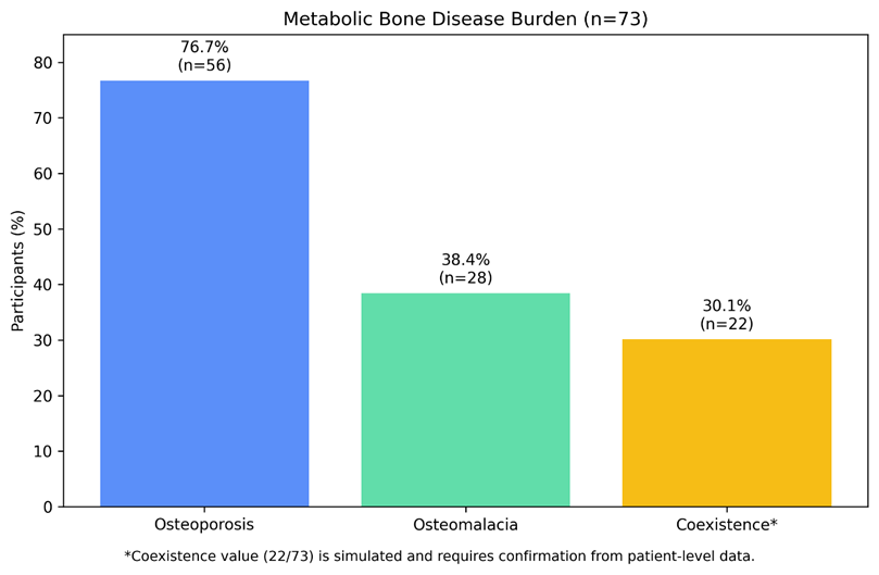
Histopathological examination demonstrated findings consistent with osteomalacia in 28 patients, yielding an osteomalacia prevalence of 38.4%. These cases corresponded to patients with severe vitamin D deficiency and biochemical findings suggestive of impaired mineralization. The remaining 45 participants showed no histopathological evidence of osteomalacia.
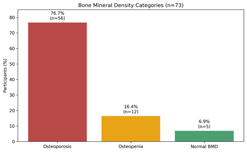
Postoperative DEXA assessment identified osteoporosis in 76.7% (n=56) of participants, based on a T-score of ≤−2.5. Osteopenia was present in 16.4% (n=12), while 6.9% (n=5) had normal bone mineral density. 22 patients were estimated to have both osteoporosis and histologically confirmed osteomalacia, corresponding to 30.1% of the total study population. On this basis, 39.3% (22/56) of patients with osteoporosis also had osteomalacia, while 78.6% (22/28) of patients with osteomalacia simultaneously fulfilled the diagnostic criteria for osteoporosis. The distribution comprised osteoporosis alone in 46.6% (n=34), osteomalacia without osteoporosis in 8.2% (n=6), coexistence of both disorders in 30.1% (n=22), and neither osteoporosis nor osteomalacia in 15.1% (n=11).
Table 1: Demographic, anthropometric, and fracture characteristics
|
Characteristic |
Result |
|
Total participants |
73 |
|
Age, years |
68.5 ± 8.7 |
|
Age range, years |
50–90 |
|
Female |
46 (63.0%) |
|
Male |
27 (37.0%) |
|
BMI, kg/m² |
23.8 ± 3.5 |
|
Fall as mechanism of injury |
58 (79.5%) |
|
Road traffic accident |
10 (13.7%) |
|
Other/minor trauma |
5 (6.8%) |
|
Extracapsular fracture |
42 (57.5%) |
|
Intracapsular fracture |
31 (42.5%) |
|
Right-sided fracture |
38 (52.1%) |
|
Left-sided fracture |
35 (47.9%) |
|
Trauma-to-surgery interval, days |
5.2 ± 1.4 |
Table 2: Clinical, female-specific, and biochemical characteristics
|
Characteristic |
Result |
|
Hypertension |
42 (57.5%) |
|
Diabetes mellitus |
35 (47.9%) |
|
Chronic kidney disease |
4 (5.5%) |
|
Thyroid disorder |
3 (4.1%) |
|
Previous fragility fracture |
18 (24.7%) |
|
Bone metabolism-affecting medication use |
22 (30.1%) |
|
Female participants |
46 |
|
Age at menarche, years |
13.2 ± 1.1 |
|
Number of pregnancies |
4.3 ± 2.1 |
|
Duration since menopause, years |
16.4 ± 5.3 |
|
Regular milk consumption |
28/46 (60.9%) |
|
No regular milk consumption |
18/46 (39.1%) |
|
Serum calcium, mg/dL |
8.4 ± 0.6 |
|
Alkaline phosphatase, IU/L |
112 ± 28 |
|
Serum albumin, g/dL |
3.9 ± 0.5 |
|
25-hydroxyvitamin D, ng/mL |
13.7 ± 5.2 |
Table 3: Vitamin D status and metabolic bone disease findings
|
Outcome |
n (%) |
|
Vitamin D <10 ng/mL |
28 (38.4%) |
|
Vitamin D 10–20 ng/mL |
30 (41.1%) |
|
Vitamin D >20 ng/mL |
15 (20.5%) |
|
Vitamin D ≤20 ng/mL overall |
58 (79.5%) |
|
Osteoporosis |
56 (76.7%) |
|
Osteopenia |
12 (16.4%) |
|
Normal bone mineral density |
5 (6.9%) |
|
Histologically confirmed osteomalacia |
28 (38.4%) |
|
Coexisting osteoporosis and osteomalacia |
22 (30.1%) |
|
Osteoporosis alone* |
34 (46.6%) |
|
Osteomalacia without osteoporosis* |
6 (8.2%) |
|
Neither condition* |
11 (15.1%) |
DISCUSSION :
The present study demonstrated a substantial burden of metabolic bone disease among older adults presenting with proximal femoral fractures, with osteoporosis identified in 76.7% of participants and histologically confirmed osteomalacia in 38.4%. Approximately 30.1% of participants had evidence of both disorders. These findings indicated that skeletal fragility in this population was unlikely to be attributable to reduced bone mineral density alone and that impaired mineralization represented an additional clinically relevant abnormality. The high prevalence of osteoporosis was broadly consistent with previous evidence showing that diminished bone strength is a dominant underlying feature among older patients presenting with hip and other fragility fractures (16). Although prevalence estimates vary across populations because of differences in age structure, nutritional status, diagnostic criteria, and access to bone-density assessment, the present findings supported the view that a proximal femoral fracture should be regarded as an important marker of underlying metabolic bone disease rather than merely the consequence of an accidental fall.
The prevalence of osteomalacia was particularly noteworthy because the condition may coexist with osteoporosis while requiring a different therapeutic approach. Previous histomorphometric research demonstrated that abnormalities compatible with defective mineralization could be present in a considerable proportion of elderly patients initially classified as having osteoporotic fractures (17). In the present study, osteomalacia was identified through a combination of biochemical abnormalities and histopathological evidence, strengthening diagnostic specificity compared with reliance on vitamin D concentration alone. Severe vitamin D deficiency below 10 ng/mL was present in 38.4% of participants, while a further 41.1% had concentrations between 10 and 20 ng/mL. Consequently, 79.5% of the study population had serum 25-hydroxyvitamin D concentrations of 20 ng/mL or lower. This high burden was consistent with previous regional reports documenting widespread vitamin D inadequacy among South Asian populations despite abundant sunlight exposure (18).
Several mechanisms may have contributed to the low vitamin D concentrations observed in this population. Reduced outdoor activity among frail older adults, limited direct sunlight exposure, dietary insufficiency, cultural clothing practices, urban air pollution, chronic illness, and use of medications affecting vitamin D metabolism may all have played contributory roles. The coexistence of osteoporosis and osteomalacia was therefore clinically plausible, as the two conditions arise through related but distinct pathways. Osteoporosis primarily reflects loss of bone mass and deterioration of skeletal microarchitecture, whereas osteomalacia reflects defective mineralization. Their simultaneous presence may further compromise bone strength and may partly explain why relatively minor trauma resulted in major proximal femoral fractures.
Women represented 63.0% of the study population, which was consistent with the established predominance of fragility fractures among postmenopausal women. Estrogen deficiency after menopause accelerates bone remodeling, cortical thinning, and trabecular deterioration, thereby increasing susceptibility to hip fractures (19). The female participants had been postmenopausal for an average of 16.4 years, indicating prolonged exposure to estrogen deficiency. In addition, 39.1% of women did not report regular milk consumption, suggesting that inadequate dietary calcium intake may have contributed to impaired skeletal health. These reproductive and nutritional findings provided useful contextual information, although the cross-sectional design did not permit determination of their independent causal contribution to osteoporosis or osteomalacia.
Falls accounted for 79.5% of fractures, while extracapsular fractures were more frequent than intracapsular fractures. The dominance of low-energy falls emphasized the interaction between skeletal fragility and age-related fall risk. A fracture in an older adult therefore represented an opportunity not only to treat the acute orthopedic injury but also to investigate the underlying bone disorder and institute secondary fracture prevention. Previous studies have shown that many patients with fragility fractures fail to undergo subsequent bone-density assessment or receive appropriate anti-osteoporotic treatment (20). This treatment gap is clinically important because pharmacological agents, including bisphosphonates, denosumab, and osteoanabolic therapies, together with correction of calcium and vitamin D deficiency, may reduce the risk of subsequent fractures in appropriately selected patients (21). The findings supported the integration of metabolic bone assessment into routine fracture care, particularly in tertiary orthopedic settings.
An important strength of the study was the combined use of DEXA, biochemical testing, and histopathological assessment. This multimodal approach permitted evaluation of both reduced bone mass and defective mineralization, conditions that may be difficult to distinguish when assessed by a single diagnostic modality. The inclusion of reproductive, nutritional, lifestyle, and comorbidity data also broadened the clinical characterization of the study population. Furthermore, histological evaluation provided additional diagnostic confidence for osteomalacia, which is often inferred solely from biochemical abnormalities in routine practice.
Several limitations required consideration. The study was conducted at a single tertiary-care hospital and included only 73 participants, limiting the precision and generalizability of prevalence estimates. Consecutive non-probability sampling may also have introduced referral and selection bias because patients managed at a tertiary center may differ from those treated in secondary or community facilities. The cross-sectional design prevented assessment of temporal relationships and did not determine whether nutritional, hormonal, or lifestyle factors directly preceded the observed skeletal abnormalities. Bone mineral density was assessed at a single postoperative time point, preventing evaluation of longitudinal changes or treatment response. In addition, bone biopsy, although diagnostically informative, is invasive and unlikely to be practical as a routine screening procedure. The coexistence estimate should also be interpreted cautiously unless confirmed directly from patient-level DEXA and histopathology records.
Future research would benefit from larger multicenter studies involving diverse populations across Pakistan, standardized DEXA protocols, comprehensive biochemical profiling, and prospective follow-up after fracture treatment. Longitudinal studies could determine the effects of vitamin D and calcium replacement, anti-osteoporotic therapy, nutritional optimization, and structured rehabilitation on recurrent fractures, functional recovery, and quality of life. Development and validation of reliable non-invasive markers for osteomalacia would also be valuable in settings where bone biopsy is impractical. Overall, the findings supported a more comprehensive approach to older patients with proximal femoral fractures in which treatment of the fracture was accompanied by systematic evaluation and management of underlying metabolic bone disease.
CONCLUSION :
The study concluded that osteoporosis and osteomalacia represented important and frequently overlapping metabolic bone disorders among older patients presenting with proximal femoral fractures. These findings emphasized that management should extend beyond surgical treatment of the fracture to include systematic assessment of underlying bone health. Incorporating DEXA-based bone mineral density evaluation, vitamin D assessment, nutritional counseling, and appropriate management of metabolic bone abnormalities into routine orthopedic and geriatric care may improve secondary fracture prevention. Greater attention to calcium and vitamin D intake, safe sunlight exposure, fall prevention, and timely use of evidence-based osteoporosis therapies could further strengthen long-term skeletal health. The study therefore highlighted the need for an integrated approach to fragility fractures in which acute fracture care and prevention of future skeletal events are addressed together.
BIBLIOGRAPHY:
1. Wang N, Chen Y, Ji J, Chang J, Yu S, Yu B. The relationship between serum vitamin D and fracture risk in the elderly: a meta-analysis. J Orthop Surg Res. 2020;15(1):81. doi:10.1186/s13018-020-01603-y.
2. Wang Q, Yu D, Wang J, Lin S. Association between vitamin D deficiency and fragility fractures in Chinese elderly patients: a cross-sectional study. Ann Palliat Med. 2020;9(4):1660-1665. doi:10.21037/apm-19-610.
3. Kanani FH, Zubairy M, Taimoor M. Frequency and trend of vitamin D deficiency in correlation with demographic and selected biochemical parameters in Karachi, Pakistan. J Pak Med Assoc. 2020;70(11):1913-1917. doi:10.5455/JPMA.28290.
4. Minisola S, Colangelo L, Pepe J, Diacinti D, Cipriani C, Rao SD. Osteomalacia and vitamin D status: a clinical update 2020. JBMR Plus. 2020;5(1):e10447. doi:10.1002/jbm4.10447.
5. Beaupre LA, Moradi F, Khong H, Smith C, Evens L, Hanson HM, et al. Implementation of an in-patient hip fracture liaison service to improve initiation of osteoporosis medication use within 1-year of hip fracture: a population-based time series analysis using the RE-AIM framework. Arch Osteoporos. 2020;15(1):83. doi:10.1007/s11657-020-00751-2.
6. Kumar K, Bhayana H, Vaiphei K, Chouhan D, Kanojia RK, Bhadada S. Coexistence of osteomalacia in osteoporotic hip fractures in more than 50 years age group. Indian J Orthop. 2021;55(3):614-620. doi:10.1007/s43465-020-00323-z.
7. Sim DS, Tay K, Howe TS, Koh SBJ. Preoperative severe vitamin D deficiency is a significant independent risk factor for poorer functional outcome and quality of life 6 months after surgery for fragility hip fractures. Osteoporos Int. 2021;32(11):2217-2224. doi:10.1007/s00198-021-05970-y.
8. Skuladottir SS, Ramel A, Eymundsdottir H, Hjaltadottir I, Launer LJ, Cotch MF, et al. Serum 25-hydroxy-vitamin D status and incident hip fractures in elderly adults: looking beyond bone mineral density. J Bone Miner Res. 2021;36(12):2351-2360. doi:10.1002/jbmr.4450.
9. Muzzammil M, Khan AS, Jahanzeb S, Qadir A, Jabbar S. Osteoporosis: prevalence, awareness and pertinent risk factors in a cohort of elderly Asian population presenting with low-energy distal radius fractures. Int J Clin Pract. 2021;75(11):e14828. doi:10.1111/ijcp.14828.
10. Arshad S, Zaidi SJA. Vitamin D levels among children, adolescents, adults, and elders in Pakistani population: a cross-sectional study. BMC Public Health. 2022;22(1):2040. doi:10.1186/s12889-022-14526-6.
11. Habibi Ghahfarrokhi S, Mohammadian-Hafshejani A, Sherwin CMT, Heidari-Soureshjani S. Relationship between serum vitamin D and hip fracture in the elderly: a systematic review and meta-analysis. J Bone Miner Metab. 2022;40(4):541-553. doi:10.1007/s00774-022-01333-7.
12. LeBoff MS, Chou SH, Ratliff KA, Cook NR, Khurana B, Kim E, et al. Supplemental vitamin D and incident fractures in midlife and older adults. N Engl J Med. 2022;387(4):299-309. doi:10.1056/NEJMoa2202106.
13. McArthur C, Lee A, Abu Alrob H, Adachi JD, Giangregorio L, Griffith LE, et al. An update of the prevalence of osteoporosis, fracture risk factors, and medication use among community-dwelling older adults: results from the Canadian Longitudinal Study on Aging. Arch Osteoporos. 2022;17(1):31. doi:10.1007/s11657-022-01073-1.
14. Mirghaderi P, Mortezaei A, Parry JA, Salimi M, Mirghaderi R, Moharrami A, et al. Osteomalacia prevalence, biochemical profile, and histology in patients with low-energy hip fractures over the age of 45. Calcif Tissue Int. 2023;113(3):257-265. doi:10.1007/s00223-023-01103-1.
15. Nadeem S, Pervez A, Abid MA, Khalid RN, Rizvi NA, Aamdani SS, et al. GRADE-ADOLOPMENT of clinical practice guideline for postmenopausal osteoporosis management—a Pakistani context. Arch Osteoporos. 2023;18(1):71. doi:10.1007/s11657-023-01258-2.
16. Manoj P, Derwin R, George S. What is the impact of daily oral supplementation of vitamin D3 (cholecalciferol) plus calcium on the incidence of hip fracture in older people? A systematic review and meta-analysis. Int J Older People Nurs. 2023;18(1): e12492. doi:10.1111/opn.12492.
17. Yan C, Chen Y, Cao J, Fang K, Shao L, Luo Y, et al. The effectiveness of fracture liaison services in patients with hip fractures: a systematic review and meta-analysis of randomized controlled trials. Heliyon. 2023;9(10): e20838. doi: 10.1016/j.heliyon. 2023.e20838.
18. Feng JN, Zhang CG, Li BH, Zhan SY, Wang SF, Song CL. Global burden of hip fracture: The Global Burden of Disease Study. Osteoporos Int. 2024;35(1):41-52. doi:10.1007/s00198-023-06907-3.
19. Kanno A, Aizawa T, Mori Y, Aizawa T. Different types of hip fragility fractures have different values of 25-hydroxyvitamin D and parathyroid hormone. J Orthop Sci. 2024;29(1):256-261. doi: 10.1016/j.jos.2022.11.003.
20. Konstantinidis C, Psoma O, Kotsias C, Panagiotopoulos V, Plakoutsis S, Tsiampas D, et al. Vitamin D deficiency in patients with low-energy hip fractures in accordance with the Mediterranean paradox. Cureus. 2024;16(4):e57583. doi:10.7759/cureus.57583.
21. Cianferotti L, Bifolco G, Caffarelli C, Mazziotti G, Migliaccio S, Napoli N, et al. Nutrition, vitamin D, and calcium in elderly patients before and after a hip fracture and their impact on the musculoskeletal system: a narrative review. Nutrients. 2024;16(11):1773. doi:10.3390/nu16111773.