Comparison of Posterior Spinal Fusion Techniques in treating Thoracic and Lumbar Spinal Tuberculosis: Titanium Mesh Cages (TMCs) vs. Autogenous Iliac Bone Grafts (AIBG)
- Wajid Akhtar , Consultant Orthopaedic & Spine Surgeon, Frontier Corps Teaching Hospital Peshawar, Pakistan.
- Khalil Ur Rehman , Orthopaedic & Spine Surgeon, Irfan General Hospital Peshawar, Pakistan,
- Asif Yousaf , Department Of Orthopedic And Spine Surgery Gujranwala Teaching Hospital Gujranwala.
- Ismail Shah Tarangi , Medical Officer Orthopaedic and Spine Surgery Unit,Frontier Corps Teaching Hospital Peshawar, Pakistan.
Article Information:
Abstract:
Objective: In this research, the treatment of thoracic and lumbar spinal TB was evaluated between posterior-only surgery with TMCs and AIBG. Study Design: The approach of a retrospective study was employed. Setting: Orthopedic and Spine Surgery Department, Khyber Teaching Hospital (KTH), Peshawar-Pakistan Subjects of the study: The investigation involved recruiting 51 participants with either lumbar or thoracic TB. Interventions: The patients go through a posterior-only procedure using a TMC (30 cases) or an AIBG (21 instances). The feasibility and usefulness of the two approaches were examined using the following metrics: intraoperative blood loss, visual analog scale (VAS), C-reactive protein (CRP), erythrocyte sedimentation rate (ESR), operation length, and associated concerns. The development of the kyphotic and neurological systems was evaluated using the Cobb angle, loss of angular correction, and Frankel grading system. Results: Compared to the preoperative condition, notable improvements were observed in CRP, VAS, ESR, Cobb angle, and Frankel grade, at the postoperative stage (p < 0.05). In comparison to the AIBG group, the TMC group outperformed it in terms of operation duration (p < .001), VAS (p <.001), intraoperative blood loss (p = 0.007), loss of angular correction (p < .001), and complications associated with surgery. The Cobb angle and Frankel grade improvement in significantly different between the AIBG and TMC groups (p > 0.05). Neither of the groups had a TB relapse. Conclusion: In posterior-only surgery treatment for the thoracic and the lumbar spinal TB, titanium mesh cages may be a better material than autogenous iliac bone transplants.
Keywords:
Article :
INTRODUCTION:
Approximately 50% of cases of bone and joint TB are caused by spinal tuberculosis (TB), a highly prevalent kind of extra-pulmonary TB.1 Paraplegia, kyphosis, spinal nerve failure, and spinal instability are common symptoms of this severe spinal illness.2 Conservative measures as well as surgical procedures are used to treat TB in the lumbar and thoracic spine. For the initial stages of thoracic and lumbar spinal TB, anti-TB chemotherapy is still the recommended treatment due to advancements in radiological technological advancements such as magnetic resonance imaging (MRI) scanning and computerized tomography, or CT.3 Surgery is nonetheless necessary for individuals who have a growing neurological deficiency, an abscess, a resistant infection, a kyphotic malformation, or segmented instability.4 Those with lumbar and thoracic spinal tuberculosis have undergone several surgical procedures 5,6 and it has been demonstrated that a posterior-only removal of debris, accompanied by an interbody combination and instrumentation, is a more successful course of action.7 For posterior-only surgeries, AIBG and TMCs have both been used to restore vertebral height. However, there hasn't been a good comparison of their clinical efficacy yet. Consequently, the current research targets to compare and assess the therapeutic effectiveness of AIBGs and TMCs in patients with lumbar and thoracic spine tuberculosis (posterior-only surgery).
Materials and methods:
Patients
A retrospective evaluation was conducted on individuals (n=51) with TB of thoracic or lumbar who received a posterior-only approach in the KTH between April 2021 and June 2024. Clinical confirmation of the tuberculosis diagnosis was obtained by clinical assessment, the lab, and radiographic investigations. All of the research participants complained of typical symptoms such as mild fever, neck discomfort, sweating throughout the night, or decreased body weight. The tuberculin test and T-SPOT findings were positive, and the lab report also showed elevated ESR and CRP. Following the completion of conventional radiography, CT scans revealed bone pathology in the spine, namely vertebral body erosion/destruction, and MRI revealed a paraspinal abscess and compression of the spinal cord. Following a biopsy, the sequestrum and inflammatory tissues were seen in the histology report. Backache measured by the utilization of the visual analog scale (VAS), and neurology was assessed using the Frankel scoring system. Thirty patients underwent TMC with instrumentation for restoring vertebral height following the removal of tissue, and twenty-one patients were subjected to instrumentation using the AIBG. Cobb angle, which is defined as the angle (º) between the first vertebra situated above and underneath the lesion, was assessed using transverse radiographs. 8 For bony fusion, Lee's standard outcomes were observed: displacement of less than 3° on the rotating radiograph, evident trabeculae of bone throughout the transplanted surface, and no gaps between neighboring bony surfaces.9
Preoperative procedure
Every patient had anti-TB chemotherapy starting at least two weeks before surgery. This included isoniazid (5–10 mg/kg daily up to a maximum of 300 mg/day), rifampicin (5–10 mg/kg daily up to a maximum of 450 mg/day), ethambutol (15 mg/kg daily up to a maximum of 750 mg/day), and pyrazinamide (25 mg/kg daily up to a maximum of 750 mg/day). Following the resolution of the common symptoms, a significant drop in ESR, CRP, and fever, and an improvement in hypoproteinemia, surgery was performed.
Surgical Techniques
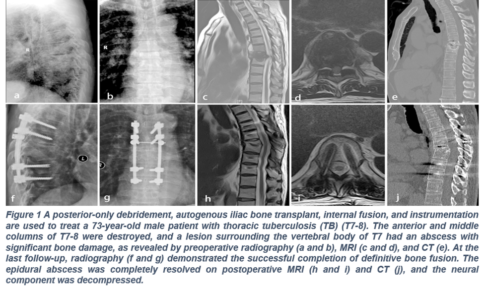
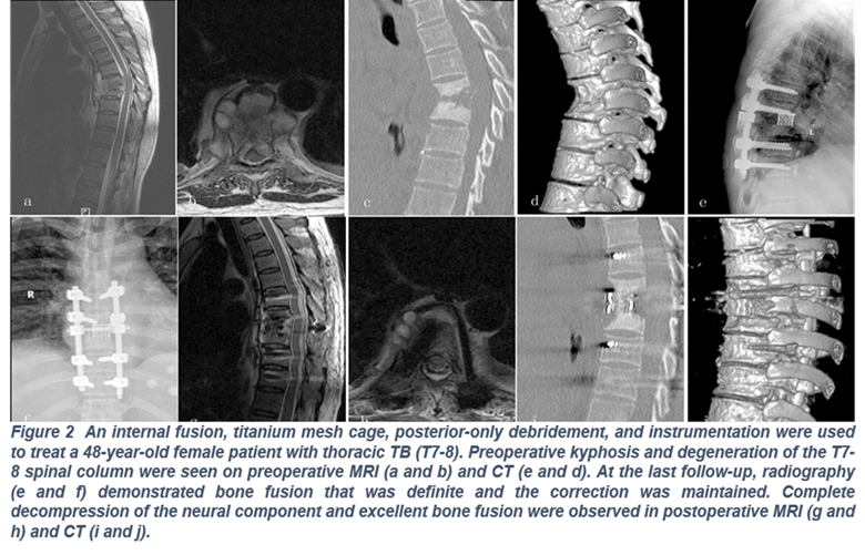
After being put under general anesthesia, patients were turned onto their backs, and the posterior centerline was cut. One vertebra above and below the sick region was revealed, as were the spinous processes, facet joints, lamina, and nerve roots of the afflicted vertebrae. On the less damaged area of the abscess, a temporary rod was inserted to support the spine and guard against damage to the spinal cord during debridement and decompression. Then, all pus, necrotic disc tissue, granulation tissues, sequestrum, and afflicted vertebral bodies were removed. After that, saline and hydrogen peroxide were used to irrigate the surgery site. Either TMC (Fig. 2) or AIBG (Fig. 1) was employed to restore intervertebral height. A bone graft (autogenous tricortical) was taken from the anterior superior iliac crest to use AIBG procedure. By using compression and disorientation, the internal fixation instrumentation was modified to rectify kyphosis. Subsequently, the surgical site was injected with streptomycin (1g) and isoniazid (0.3g) to facilitate improved and focused removal of tissues. Before the incision was closed, two drainage catheters were placed into the abscess chamber to aid in drainage and promote healing.
Postoperative care
The surgical drain catheters were removed when the amount of fluid loss was less than 30 milliliters per day. To expedite neurological recuperation and minimize any post-operative edema of the wound and nerves, postoperative patients were treated with mannitol dehydration (375 ml per day) and nerve supplement mecobalamin (1.5 mg per day). Until bone fusion was accomplished, patients were advised to put on an orthosis and train their muscles (limb) while resting for 14 days following surgery. The preoperative regimen and anti-TB chemotherapy were administered for six months following surgery, and then isoniazid, rifampicin, and ethambutol were given for an additional nine to twelve months. Following surgery, all patients had routine laboratory and radiographic testing every year for the first week, as well as every three, six, and twelve months.
Statistical analysis
For every continuous data set, the mean ± standard deviation (SD) was displayed. Software from IBM Corp., Armonk, NY, USA, called SPSS 27.0 was used to statistically analyze all of the data. The continuous parameters were compared before and after surgery using a paired-sample t-test (P<0.05). The parameters of both groups were compared using an independent-sample t-test.
Outcomes
Evaluation of Baseline Parameters
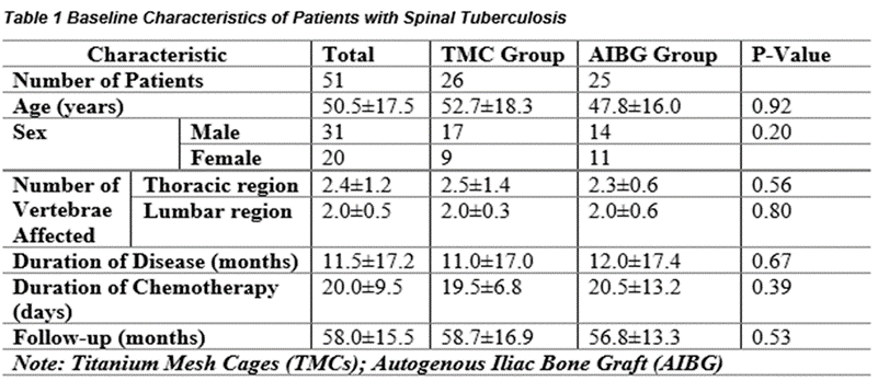
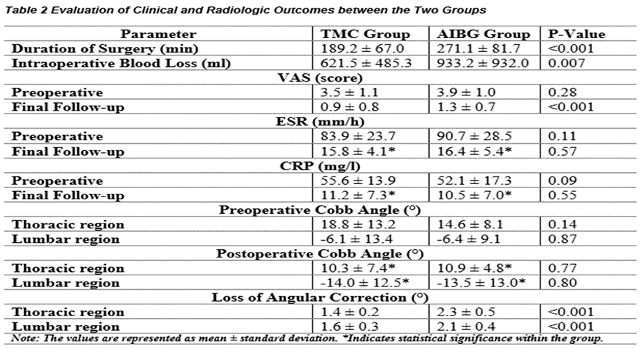
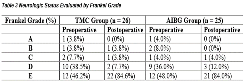
In the TMC group, there were 17 (65.0%) men and 9 (34.6%) women, with an average age of (50.5 ± 17.5 years); in the AIBG group, there were men 14 (56.0%) and women 11 (44.0%), with a mean age of (47.8±16.0 years). The damaged lumbar and thoracic vertebrae measured (2.5 ± 1.4) and (2.0 ± 0.3), respectively in the patient of TMC, while in the AIBG group, they were (2.3 ± 0.6) and (2.0 ± 0.6), respectively. The average length of tuberculosis (TB) in the TMC and AIBG patients was (11.0 ± 17.0 months) and (12.0 ± 17.4 months), respectively. In the initial stage of surgery, patients in the TMC group underwent (19.5 ±6.8 days) of chemotherapy on average, while in the AIBG group, the average length was (20.5 ± 13.2 days). The age group, gender, number of afflicted vertebrae, length of illness, and length of chemotherapy did not significantly differ between both groups (P > 0.05) as shown in Table 1. The VAS ratings (preoperative) for the patients of TMC were (3.5 ± 1.1), whereas the patients of AIBG scored (3.9 ± 1.0). In the patients of TMC, the preoperative ESR values were 83.9 ± 23.7mm/h, while in the AIBG group (90.7 ± 28.5mm/h). The preoperative CRP values in the TMC group were 55.6 ± 13.9 mg/l, whereas the AIBG group had 52.1 ± 17.3 mg/l, as given in Table 2. In the TMC, the Frankel grades; grade A (1 patient), group B (1 patient), grade C (2 patients), grade D (10 patients), and grade E (12 patients), as described in Table 3 shows that in the AIBG group, grade A (1 patient), grade B (2 patients), grade C (1 patient), grade D (9 patient), and grade E (12 patient). The VAS ratings, CRP and ESR levels (preoperative), and Frankel grading did not change significantly within the two groups (P value of 0.05), as given in Table 2-Table 3. In the TMC patients had an average follow-up time of 58.7 ± 16.9 months, whereas the AIBG had a mean follow-up time of 56.8 ± 13.3 months. There was an insignificant distinction between the groups (P value greater than 0.05) as described in Table 1.
Comparison of clinical effectiveness
In the TMC group, the average operation time was (189.2 ± 67.0 min) and the mean intraoperative blood loss was (621.5 ± 485.3ml). In contrast, patients in the AIBG group experienced an average surgical length of (271.1 ± 81.7) min and a blood loss of (933.2 ± 932.0ml), with a considerably longer operational duration (P-value of 0.001) and larger intraoperative blood loss (P-value of 0.007). The TMC group’ patients had a VAS rate (postoperative) of (0.9 ± 0.8), compared to (1.3 ± 0.7). Patients of AIBG group indicated a more substantial amount of pain reduction (P-value of 0.001) as given in Table 2. The TMC group saw a drop in ESR (15.8 ± 4.1mm/h) and CRP (11.2 ±7.3 mg/l) levels (postoperative), whereas the AIBG patients experienced a decline in ESR (16.4 ± 5.4mm/h) and CRP (10.5 ± 7.0mg/l). The before and postoperative ESR and CPR levels differed significantly (P value of 0.05). In contrast, the postoperatively ESR and CPR levels did not change considerably between the TMC and AIBG groups (P value greater than 0.05), as described in Table 2. Improvements in postoperative Frankel grading were seen in two patients (7.7%) in grade D and 22 (84.6%) in grade E in the TMC group. In a similar vein, improvements were also made for 3 (12.0%) in grade D and in grade E total 21 (84.0%) AIBG group, Table 3.
Radiological outcome comparison
The pretreatment Cobb angles of the lumbar (P value of 0.87) and thoracic (P value of 0.14) vertebrae did not change between the AIBG and TMC groups, as given in Table 2. Following surgery, the TMC group's Cobb angle of the thoracic region improved to (10.3° ± 7.4°) while the AIBG group's adjusted to (10.9° ± 4.8°). The TMC patients exhibited, a lumbar Cobb angle of (−14.0° ± 12.5°), while in the AIBG group, it was (−13.5° ± 13.0°). This indicates that the two groups' pre and postoperative thoracic and lumbar Cobb angles differed significantly (P value of 0.05). Nevertheless, there was no statistically significant variation in the postoperative Cobb angles for the lumbar (P value of 0.80) and thoracic (P value of 0.77). For the lumbar (P < 0.001) and thoracic (P < 0.001) vertebrae, the TMC group showed a lower degree of angular corrective loss than the AIBG group, as given in Table 2.
Consequences following surgery
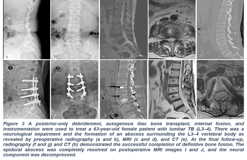
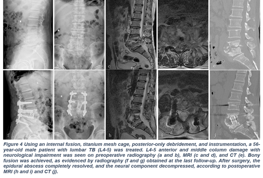
In the TMC group, there were four cases of leakage of cerebral fluid, and in the AIBG group (two cases); additionally AIBG group (three cases) and TMC (two cases) of incision site infection. In the AIBG group, ten patients (38.3%) experienced donor site problems, which included a significant hematoma in three patients, ongoing discomfort in six individuals, and an infection in one patient that was resolved with debridement, antibiotics, and symptomatic therapy. In the TMC group, six cases (24.4%) had subsidence. At the time of the investigation after the surgeries, none of the patients had any instrumentation-related problems (Figs. 3 and 4). None of the individuals had a TB recurrence.
Patients
A retrospective evaluation was conducted on individuals (n=51) with TB of thoracic or lumbar who received a posterior-only approach in the KTH between April 2021 and June 2024. Clinical confirmation of the tuberculosis diagnosis was obtained by clinical assessment, the lab, and radiographic investigations. All of the research participants complained of typical symptoms such as mild fever, neck discomfort, sweating throughout the night, or decreased body weight. The tuberculin test and T-SPOT findings were positive, and the lab report also showed elevated ESR and CRP. Following the completion of conventional radiography, CT scans revealed bone pathology in the spine, namely vertebral body erosion/destruction, and MRI revealed a paraspinal abscess and compression of the spinal cord. Following a biopsy, the sequestrum and inflammatory tissues were seen in the histology report. Backache measured by the utilization of the visual analog scale (VAS), and neurology was assessed using the Frankel scoring system. Thirty patients underwent TMC with instrumentation for restoring vertebral height following the removal of tissue, and twenty-one patients were subjected to instrumentation using the AIBG. Cobb angle, which is defined as the angle (º) between the first vertebra situated above and underneath the lesion, was assessed using transverse radiographs. 8 For bony fusion, Lee's standard outcomes were observed: displacement of less than 3° on the rotating radiograph, evident trabeculae of bone throughout the transplanted surface, and no gaps between neighboring bony surfaces.9
Preoperative procedure
Every patient had anti-TB chemotherapy starting at least two weeks before surgery. This included isoniazid (5–10 mg/kg daily up to a maximum of 300 mg/day), rifampicin (5–10 mg/kg daily up to a maximum of 450 mg/day), ethambutol (15 mg/kg daily up to a maximum of 750 mg/day), and pyrazinamide (25 mg/kg daily up to a maximum of 750 mg/day). Following the resolution of the common symptoms, a significant drop in ESR, CRP, and fever, and an improvement in hypoproteinemia, surgery was performed.
Surgical Techniques
After being put under general anesthesia, patients were turned onto their backs, and the posterior centerline was cut. One vertebra above and below the sick region was revealed, as were the spinous processes, facet joints, lamina, and nerve roots of the afflicted vertebrae. On the less damaged area of the abscess, a temporary rod was inserted to support the spine and guard against damage to the spinal cord during debridement and decompression. Then, all pus, necrotic disc tissue, granulation tissues, sequestrum, and afflicted vertebral bodies were removed. After that, saline and hydrogen peroxide were used to irrigate the surgery site. Either TMC (Fig. 2) or AIBG (Fig. 1) was employed to restore intervertebral height. A bone graft (autogenous tricortical) was taken from the anterior superior iliac crest to use AIBG procedure. By using compression and disorientation, the internal fixation instrumentation was modified to rectify kyphosis. Subsequently, the surgical site was injected with streptomycin (1g) and isoniazid (0.3g) to facilitate improved and focused removal of tissues. Before the incision was closed, two drainage catheters were placed into the abscess chamber to aid in drainage and promote healing.
Postoperative care
The surgical drain catheters were removed when the amount of fluid loss was less than 30 milliliters per day. To expedite neurological recuperation and minimize any post-operative edema of the wound and nerves, postoperative patients were treated with mannitol dehydration (375 ml per day) and nerve supplement mecobalamin (1.5 mg per day). Until bone fusion was accomplished, patients were advised to put on an orthosis and train their muscles (limb) while resting for 14 days following surgery. The preoperative regimen and anti-TB chemotherapy were administered for six months following surgery, and then isoniazid, rifampicin, and ethambutol were given for an additional nine to twelve months. Following surgery, all patients had routine laboratory and radiographic testing every year for the first week, as well as every three, six, and twelve months.
Statistical analysis
For every continuous data set, the mean ± standard deviation (SD) was displayed. Software from IBM Corp., Armonk, NY, USA, called SPSS 27.0 was used to statistically analyze all of the data. The continuous parameters were compared before and after surgery using a paired-sample t-test (P<0.05). The parameters of both groups were compared using an independent-sample t-test.
Outcomes
Evaluation of Baseline Parameters
In the TMC group, there were 17 (65.0%) men and 9 (34.6%) women, with an average age of (50.5 ± 17.5 years); in the AIBG group, there were men 14 (56.0%) and women 11 (44.0%), with a mean age of (47.8±16.0 years). The damaged lumbar and thoracic vertebrae measured (2.5 ± 1.4) and (2.0 ± 0.3), respectively in the patient of TMC, while in the AIBG group, they were (2.3 ± 0.6) and (2.0 ± 0.6), respectively. The average length of tuberculosis (TB) in the TMC and AIBG patients was (11.0 ± 17.0 months) and (12.0 ± 17.4 months), respectively. In the initial stage of surgery, patients in the TMC group underwent (19.5 ±6.8 days) of chemotherapy on average, while in the AIBG group, the average length was (20.5 ± 13.2 days). The age group, gender, number of afflicted vertebrae, length of illness, and length of chemotherapy did not significantly differ between both groups (P > 0.05) as shown in Table 1. The VAS ratings (preoperative) for the patients of TMC were (3.5 ± 1.1), whereas the patients of AIBG scored (3.9 ± 1.0). In the patients of TMC, the preoperative ESR values were 83.9 ± 23.7mm/h, while in the AIBG group (90.7 ± 28.5mm/h). The preoperative CRP values in the TMC group were 55.6 ± 13.9 mg/l, whereas the AIBG group had 52.1 ± 17.3 mg/l, as given in Table 2. In the TMC, the Frankel grades; grade A (1 patient), group B (1 patient), grade C (2 patients), grade D (10 patients), and grade E (12 patients), as described in Table 3 shows that in the AIBG group, grade A (1 patient), grade B (2 patients), grade C (1 patient), grade D (9 patient), and grade E (12 patient). The VAS ratings, CRP and ESR levels (preoperative), and Frankel grading did not change significantly within the two groups (P value of 0.05), as given in Table 2-Table 3. In the TMC patients had an average follow-up time of 58.7 ± 16.9 months, whereas the AIBG had a mean follow-up time of 56.8 ± 13.3 months. There was an insignificant distinction between the groups (P value greater than 0.05) as described in Table 1.
Comparison of clinical effectiveness
In the TMC group, the average operation time was (189.2 ± 67.0 min) and the mean intraoperative blood loss was (621.5 ± 485.3ml). In contrast, patients in the AIBG group experienced an average surgical length of (271.1 ± 81.7) min and a blood loss of (933.2 ± 932.0ml), with a considerably longer operational duration (P-value of 0.001) and larger intraoperative blood loss (P-value of 0.007). The TMC group’ patients had a VAS rate (postoperative) of (0.9 ± 0.8), compared to (1.3 ± 0.7). Patients of AIBG group indicated a more substantial amount of pain reduction (P-value of 0.001) as given in Table 2. The TMC group saw a drop in ESR (15.8 ± 4.1mm/h) and CRP (11.2 ±7.3 mg/l) levels (postoperative), whereas the AIBG patients experienced a decline in ESR (16.4 ± 5.4mm/h) and CRP (10.5 ± 7.0mg/l). The before and postoperative ESR and CPR levels differed significantly (P value of 0.05). In contrast, the postoperatively ESR and CPR levels did not change considerably between the TMC and AIBG groups (P value greater than 0.05), as described in Table 2. Improvements in postoperative Frankel grading were seen in two patients (7.7%) in grade D and 22 (84.6%) in grade E in the TMC group. In a similar vein, improvements were also made for 3 (12.0%) in grade D and in grade E total 21 (84.0%) AIBG group, Table 3.
Radiological outcome comparison
The pretreatment Cobb angles of the lumbar (P value of 0.87) and thoracic (P value of 0.14) vertebrae did not change between the AIBG and TMC groups, as given in Table 2. Following surgery, the TMC group's Cobb angle of the thoracic region improved to (10.3° ± 7.4°) while the AIBG group's adjusted to (10.9° ± 4.8°). The TMC patients exhibited, a lumbar Cobb angle of (−14.0° ± 12.5°), while in the AIBG group, it was (−13.5° ± 13.0°). This indicates that the two groups' pre and postoperative thoracic and lumbar Cobb angles differed significantly (P value of 0.05). Nevertheless, there was no statistically significant variation in the postoperative Cobb angles for the lumbar (P value of 0.80) and thoracic (P value of 0.77). For the lumbar (P < 0.001) and thoracic (P < 0.001) vertebrae, the TMC group showed a lower degree of angular corrective loss than the AIBG group, as given in Table 2.
Consequences following surgery
In the TMC group, there were four cases of leakage of cerebral fluid, and in the AIBG group (two cases); additionally AIBG group (three cases) and TMC (two cases) of incision site infection. In the AIBG group, ten patients (38.3%) experienced donor site problems, which included a significant hematoma in three patients, ongoing discomfort in six individuals, and an infection in one patient that was resolved with debridement, antibiotics, and symptomatic therapy. In the TMC group, six cases (24.4%) had subsidence. At the time of the investigation after the surgeries, none of the patients had any instrumentation-related problems (Figs. 3 and 4). None of the individuals had a TB recurrence.
Patients
A retrospective evaluation was conducted on individuals (n=51) with TB of thoracic or lumbar who received a posterior-only approach in the KTH between April 2021 and June 2024. Clinical confirmation of the tuberculosis diagnosis was obtained by clinical assessment, the lab, and radiographic investigations. All of the research participants complained of typical symptoms such as mild fever, neck discomfort, sweating throughout the night, or decreased body weight. The tuberculin test and T-SPOT findings were positive, and the lab report also showed elevated ESR and CRP. Following the completion of conventional radiography, CT scans revealed bone pathology in the spine, namely vertebral body erosion/destruction, and MRI revealed a paraspinal abscess and compression of the spinal cord. Following a biopsy, the sequestrum and inflammatory tissues were seen in the histology report. Backache measured by the utilization of the visual analog scale (VAS), and neurology was assessed using the Frankel scoring system. Thirty patients underwent TMC with instrumentation for restoring vertebral height following the removal of tissue, and twenty-one patients were subjected to instrumentation using the AIBG. Cobb angle, which is defined as the angle (º) between the first vertebra situated above and underneath the lesion, was assessed using transverse radiographs. 8 For bony fusion, Lee's standard outcomes were observed: displacement of less than 3° on the rotating radiograph, evident trabeculae of bone throughout the transplanted surface, and no gaps between neighboring bony surfaces.9
Preoperative procedure
Every patient had anti-TB chemotherapy starting at least two weeks before surgery. This included isoniazid (5–10 mg/kg daily up to a maximum of 300 mg/day), rifampicin (5–10 mg/kg daily up to a maximum of 450 mg/day), ethambutol (15 mg/kg daily up to a maximum of 750 mg/day), and pyrazinamide (25 mg/kg daily up to a maximum of 750 mg/day). Following the resolution of the common symptoms, a significant drop in ESR, CRP, and fever, and an improvement in hypoproteinemia, surgery was performed.
Surgical Techniques
After being put under general anesthesia, patients were turned onto their backs, and the posterior centerline was cut. One vertebra above and below the sick region was revealed, as were the spinous processes, facet joints, lamina, and nerve roots of the afflicted vertebrae. On the less damaged area of the abscess, a temporary rod was inserted to support the spine and guard against damage to the spinal cord during debridement and decompression. Then, all pus, necrotic disc tissue, granulation tissues, sequestrum, and afflicted vertebral bodies were removed. After that, saline and hydrogen peroxide were used to irrigate the surgery site. Either TMC (Fig. 2) or AIBG (Fig. 1) was employed to restore intervertebral height. A bone graft (autogenous tricortical) was taken from the anterior superior iliac crest to use AIBG procedure. By using compression and disorientation, the internal fixation instrumentation was modified to rectify kyphosis. Subsequently, the surgical site was injected with streptomycin (1g) and isoniazid (0.3g) to facilitate improved and focused removal of tissues. Before the incision was closed, two drainage catheters were placed into the abscess chamber to aid in drainage and promote healing.
Postoperative care
The surgical drain catheters were removed when the amount of fluid loss was less than 30 milliliters per day. To expedite neurological recuperation and minimize any post-operative edema of the wound and nerves, postoperative patients were treated with mannitol dehydration (375 ml per day) and nerve supplement mecobalamin (1.5 mg per day). Until bone fusion was accomplished, patients were advised to put on an orthosis and train their muscles (limb) while resting for 14 days following surgery. The preoperative regimen and anti-TB chemotherapy were administered for six months following surgery, and then isoniazid, rifampicin, and ethambutol were given for an additional nine to twelve months. Following surgery, all patients had routine laboratory and radiographic testing every year for the first week, as well as every three, six, and twelve months.
Statistical analysis
For every continuous data set, the mean ± standard deviation (SD) was displayed. Software from IBM Corp., Armonk, NY, USA, called SPSS 27.0 was used to statistically analyze all of the data. The continuous parameters were compared before and after surgery using a paired-sample t-test (P<0.05). The parameters of both groups were compared using an independent-sample t-test.
Outcomes
Evaluation of Baseline Parameters
In the TMC group, there were 17 (65.0%) men and 9 (34.6%) women, with an average age of (50.5 ± 17.5 years); in the AIBG group, there were men 14 (56.0%) and women 11 (44.0%), with a mean age of (47.8±16.0 years). The damaged lumbar and thoracic vertebrae measured (2.5 ± 1.4) and (2.0 ± 0.3), respectively in the patient of TMC, while in the AIBG group, they were (2.3 ± 0.6) and (2.0 ± 0.6), respectively. The average length of tuberculosis (TB) in the TMC and AIBG patients was (11.0 ± 17.0 months) and (12.0 ± 17.4 months), respectively. In the initial stage of surgery, patients in the TMC group underwent (19.5 ±6.8 days) of chemotherapy on average, while in the AIBG group, the average length was (20.5 ± 13.2 days). The age group, gender, number of afflicted vertebrae, length of illness, and length of chemotherapy did not significantly differ between both groups (P > 0.05) as shown in Table 1. The VAS ratings (preoperative) for the patients of TMC were (3.5 ± 1.1), whereas the patients of AIBG scored (3.9 ± 1.0). In the patients of TMC, the preoperative ESR values were 83.9 ± 23.7mm/h, while in the AIBG group (90.7 ± 28.5mm/h). The preoperative CRP values in the TMC group were 55.6 ± 13.9 mg/l, whereas the AIBG group had 52.1 ± 17.3 mg/l, as given in Table 2. In the TMC, the Frankel grades; grade A (1 patient), group B (1 patient), grade C (2 patients), grade D (10 patients), and grade E (12 patients), as described in Table 3 shows that in the AIBG group, grade A (1 patient), grade B (2 patients), grade C (1 patient), grade D (9 patient), and grade E (12 patient). The VAS ratings, CRP and ESR levels (preoperative), and Frankel grading did not change significantly within the two groups (P value of 0.05), as given in Table 2-Table 3. In the TMC patients had an average follow-up time of 58.7 ± 16.9 months, whereas the AIBG had a mean follow-up time of 56.8 ± 13.3 months. There was an insignificant distinction between the groups (P value greater than 0.05) as described in Table 1.
Comparison of clinical effectiveness
In the TMC group, the average operation time was (189.2 ± 67.0 min) and the mean intraoperative blood loss was (621.5 ± 485.3ml). In contrast, patients in the AIBG group experienced an average surgical length of (271.1 ± 81.7) min and a blood loss of (933.2 ± 932.0ml), with a considerably longer operational duration (P-value of 0.001) and larger intraoperative blood loss (P-value of 0.007). The TMC group’ patients had a VAS rate (postoperative) of (0.9 ± 0.8), compared to (1.3 ± 0.7). Patients of AIBG group indicated a more substantial amount of pain reduction (P-value of 0.001) as given in Table 2. The TMC group saw a drop in ESR (15.8 ± 4.1mm/h) and CRP (11.2 ±7.3 mg/l) levels (postoperative), whereas the AIBG patients experienced a decline in ESR (16.4 ± 5.4mm/h) and CRP (10.5 ± 7.0mg/l). The before and postoperative ESR and CPR levels differed significantly (P value of 0.05). In contrast, the postoperatively ESR and CPR levels did not change considerably between the TMC and AIBG groups (P value greater than 0.05), as described in Table 2. Improvements in postoperative Frankel grading were seen in two patients (7.7%) in grade D and 22 (84.6%) in grade E in the TMC group. In a similar vein, improvements were also made for 3 (12.0%) in grade D and in grade E total 21 (84.0%) AIBG group, Table 3.
Radiological outcome comparison
The pretreatment Cobb angles of the lumbar (P value of 0.87) and thoracic (P value of 0.14) vertebrae did not change between the AIBG and TMC groups, as given in Table 2. Following surgery, the TMC group's Cobb angle of the thoracic region improved to (10.3° ± 7.4°) while the AIBG group's adjusted to (10.9° ± 4.8°). The TMC patients exhibited, a lumbar Cobb angle of (−14.0° ± 12.5°), while in the AIBG group, it was (−13.5° ± 13.0°). This indicates that the two groups' pre and postoperative thoracic and lumbar Cobb angles differed significantly (P value of 0.05). Nevertheless, there was no statistically significant variation in the postoperative Cobb angles for the lumbar (P value of 0.80) and thoracic (P value of 0.77). For the lumbar (P < 0.001) and thoracic (P < 0.001) vertebrae, the TMC group showed a lower degree of angular corrective loss than the AIBG group, as given in Table 2.
Consequences following surgery
In the TMC group, there were four cases of leakage of cerebral fluid, and in the AIBG group (two cases); additionally AIBG group (three cases) and TMC (two cases) of incision site infection. In the AIBG group, ten patients (38.3%) experienced donor site problems, which included a significant hematoma in three patients, ongoing discomfort in six individuals, and an infection in one patient that was resolved with debridement, antibiotics, and symptomatic therapy. In the TMC group, six cases (24.4%) had subsidence. At the time of the investigation after the surgeries, none of the patients had any instrumentation-related problems (Figs. 3 and 4). None of the individuals had a TB recurrence.
DISCUSSION :
The most frequent extrapulmonary location of tuberculosis is the spine. Conservative chemotherapy is an effective treatment for the majority of spinal TB patients. Surgical interventions are necessary for individuals who experience kyphotic distortions or neurological dysfunction to repair the spinal intervertebral height, debride the infection site, and restore nerve function.10,11 Spinal TB has been treated using a variety of techniques, but the best approach is still up for debate.
There were few surgical alternatives available to treat spinal TB in the latter half of the 19th and early 20th century. By developing a technique known as a "lateral rhachotomy," Capener gave the surgeon greater ventral exposure via a more lateral trajectory.12 Hence, the method's shortcomings prevented it from becoming widely used. The study described the technique of anterior debridement and interbody fusion method for treating TB of the spine in the 1960s.13 Given that TB lesions often affect the spine's front and middle columns,14,15 spinal cord decompression might be accomplished and the TB focus eliminated with an anterior-only technique.16,17 Nevertheless, the anterior-only strategy had drawbacks, such as difficulties with the abdominal and thoracic cavities and poor kyphosis correction.18
The "lateral extra cavitary approach (LECA)," developed in 1976 by Larson and colleagues, gave the surgeon approach enabling posterior combination and instrumentation without thoracic or abdominal cavity complications.19 Nevertheless, it was impossible to overlook the drawbacks, which included the level of operational complexity and procedure-related issues including high blood loss. The prevalence of posterior-only and mixed anterior-posterior techniques increased with the introduction of pedicle screws.20, 21 These days, spinal tuberculosis is usually treated surgically using a single technique that uses a smaller incision for static interbody fixation.22,23 Additionally, a one-stage procedure reduces the possibility of TB spreading. Study 7 found that while a posterior-only technique had the benefits of less operational invasion and fewer procedure-related problems, it produced a more acceptable result than a combined anterior-posterior technique. Furthermore, because of the ongoing spinal cord compression, the posterior-only method was successful for victims who had evident kyphosis and neurological impairments.7 At the study's final follow-up, kyphosis correction improved for every patient, confirming that a posterior-only strategy might be a successful treatment for thoracic and lumbar spinal TB.
Larson employed an AIBG for the first time to replicate intervertebral height in 1982, however, it was utilized for odontoid fractures.24 Later, TMCs were included in the LECA for the treatment of thoracic and lumbar spinal TB by Maiman and associates.25 To restore the height of spinal intervertebral following a posterior-only debridement, TMCs, and AIBGs are now often utilized in interbody transplant procedures. The effectiveness of both of these materials hasn't been compared in many research, though. The efficacy of AIBGs and TMCs in posterior-only surgical therapies for thoracic and lumbar spinal TB was compared in the current study employing a quantitative assessment of clinical and radiological parameters along with postoperative sequelae.
AIBGs are considered the gold standard for treating bone defects because of their excellent fusion rate-promoting properties, osteogenesis, bone induction, and biocompatibility.11 Nevertheless, it's also clear that AIBGs have limits. Kemp et al.26 noted that the autografts' inability to sustain their sagittal plane alignment resulted in postoperative corrective loss and the development of progressive kyphosis. Patients in the AIBG group showed an additional serious impairment of angular correction in the current investigation. Additionally, the AIBGs showed a chance of stress fractures and fixation failure.16 Moreover, it is imperative to consider nonunion, graft failure or dislocation, and site of donor problems, including persistent discomfort and infection.27 In AIBGs, problems at the iliac crest donor site exhibited an obstacle of postoperative worry for patients as well as surgeons. Patients with chronic pain (six), a significant hematoma (three), and an infection that needed to be drained (one) were seen in the AIBG patients in the current study. The rate of complications at the donor location was 38.3% (10/25), in line with earlier research.28
Numerous studies have shown that TMCs offer significant benefits in terms of durable bone fusion, dependable spinal restoration, and a low rate of implant-related complications.27 To prevent difficulties linked to the donor location, TMCs are occupied with removed intervertebral lamina and articular processes. Furthermore, the strength of the operative sections, the maintenance and correction of the disfigurement, and the union of the bones are enforced by posterior instrumentation and TMCs. Furthermore, for the current study, the TMC group's Cobb angle loss was lower than the AIBG group's, which might be related to less bone resorption. Nonetheless, TMCs still have some drawbacks. The TMC group's subsidence rate in this research was 24.4%, which is in line with one other study.29 The quality of the bone in the vertebral end plates, the TMC approach the contact surface area with the implant-bone interface, and a three-dimensional segmented equilibrium are some of the elements that we think might be causing the sinking behavior of TMCs. To reduce settling, transpedicular studs can be inserted into the afflicted vertebra if the infection has not completely ruined the top portion of the spine. In the current investigation, the TMC had good radiographic results, strong bone fusion, and improved neurological function, but with an acceptable reduction of kyphosis.
Our research has several limitations. First of all, selection bias may have occurred since the study was retrospective and single-centered rather than prospective. Second, the very small patient population in our study may have also diminished the significance of the outcome comparison.
CONCLUSION :
Based on the findings of the study it is concluded that for the internal fusion, posterior debridement, and instrumentation of thoracic and lumbar spinal TB, titanium mesh cages may be a better material than autogenous iliac bone grafts.
Disclaimers
Contributors: None.
Funding resources: None
BIBLIOGRAPHY:
1. Jain AK, Kumar J. Tuberculosis of spine: neurological deficit. Eur Spine J. 2013;22(4):624–33.
2. Peto HM, Pratt RH, Harrington TA, LoBue PA, Armstrong LR. Epidemiology of extrapulmonary tuberculosis in the United States, 1993–2006. Clin Infect Dis. 2009;49(9):1350–7.
3. Lee TC, Lu K, Yang LC, Huang HY, Liang CL. Transpedicular instrumentation as an adjunct in the treatment of thoracolumbar and lumbar spine tuberculosis with early stage bone destruction. J Neurosurg. 1999;91(2 Suppl):163–9.
4. Zhang HQ, Li JS, Zhao SS, Shao YX, Liu SH, Gao Q, et al. Surgical management for thoracic spinal tuberculosis in the elderly: posterior only versus combined posterior and anterior approaches. Arch Orthop Trauma Surg. 2012;132(12):1717–23.
5. Ozturk C, Aydinli U, Vural R, Sehirlioglu A, Mutlu M. Simultaneous versus sequential one-stage combined anterior and posterior spinal surgery for spinal infections (outcomes and com- plications). Int Orthop. 2007;31(3):363–6.
6. He Q, Xu J. Comparison between the antero-posterior and anterior approaches for treating L5-S1 vertebral tuberculosis. Int Orthop. 2012;36(2):345–51.
7. Zhang HQ, Lin MZ, Li JS, Tang MX, Guo CF, Wu JH, et al. One- stage posterior debridement, transforaminal lumbar interbody fusion and instrumentation in treatment of lumbar spinal tubercu- losis: a retrospective case series. Arch Orthop Trauma Surg. 2013; 133(3):333–41.
8. Rajasekaran S. The natural history of post-tubercular kyphosis in children. Radiological signs which predict late increase in deform- ity. J Bone Joint Surg Br. 2001;83-B(7):954–62.
9. Lee CK, Vessa P, Lee JK. Chronic disabling low back pain syn- drome caused by internal disc derangements. The results of disc excision and posterior lumbar interbody fusion. Spine. 1995;20 (3):356–61.
10. Gong K, Wang Z, Luo Z. Single-stage posterior debridement and transforaminal lumbar interbody fusion with autogenous bone grafting and posterior instrumentation in the surgical management of lumbar tuberculosis. Arch Orthop Trauma Surg. 2011;131(2): 217–23.
11. Sundararaj GD, Amritanand R, Venkatesh K, Arockiaraj J. The use of titanium mesh cages in the reconstruction of anterior column defects in active spinal infections: can we rest the crest? Asian Spine J. 2011;5(3):155–61.
12. Capener N. The evolution of lateral rhachotomy. J Bone Joint Surg Br. 1954;36-b(2):173–9.
13. Hodgson AR, Stock FE, Fang HS, Ong GB. Anterior spinal fusion. The operative approach and pathological findings in 412 patients with Pott’s disease of the spine. Br J Surg. 1960;48:172–8.
14. Talu U, Gogus A, Ozturk C, Hamzaoglu A, Domanic U. The role of posterior instrumentation and fusion after anterior radical debridement and fusion in the surgical treatment of spinal tubercu- losis: experience of 127 cases. J Spinal Disord Tech. 2006;19(8): 554–9.
15. Zhang X, Ji J, Liu B. Management of spinal tuberculosis: a sys- tematic review and meta-analysis. J Int Med Res. 2013;41(5): 1395–407
16. Qureshi MA, Khalique AB, Afzal W, Pasha IF, Aebi M. Surgical management of contiguous multilevel thoracolumbar tuberculous spondylitis. Eur Spine J. 2013;22(Suppl 4):618–23.
17. Hassan K, Elmorshidy E. Anterior versus posterior approach in surgical treatment of tuberculous spondylodiscitis of thoracic and lumbar spine. Eur Spine J. 2016;25(4):1056–63.
18. Zeng H, Zhang P, Shen X, Luo C, Xu Z, Zhang Y, et al. One-stage posterior-only approach in surgical treatment of single-segment thoracic spinal tuberculosis with neurological deficits in adults: a retrospective study of 34 cases. BMC Musculoskelet Disord. 2015; 16:186. Doi: 10.1186/s12891-015-0640-0.
19. Larson SJ, Holst RA, Hemmy DC, Sances Jr. A. Lateral extraca- vitary approach to traumatic lesions of the thoracic and lumbar spine. J Neurosurg. 1976; 45(6):628–37.
20. Moon MS, Woo YK, Lee KS, Ha KY, Kim SS, Sun DH. Posterior instrumentation and anterior interbody fusion for tuberculous kyphosis of dorsal and lumbar spines. Spine. 1995; 20(17):1910–6.
21. Sundararaj GD, Behera S, Ravi V, Venkatesh K, Cherian VM, Lee V. Role of posterior stabilisation in the management of tuberculosis of the dorsal and lumbar spine. J Bone Joint Surg Br. 2003; 85-B(1): 100–6.
22. Sun L, Song Y, Liu L, Gong Q, Zhou C. One-stage posterior surgical treatment for lumbosacral tuberculosis with major vertebral body loss and kyphosis. Orthopedics. 2013; 36(8):e1082–90.
23. Pang X, Wu P, Shen X, Li D, Luo C, Wang X. One-stage posterior transforaminal lumbar debridement, 360 degrees interbody fusion, and posterior instrumentation in treating lumbosacral spinal tuber- culosis. Arch Orthop Trauma Surg. 2013; 133(8):1033–9.
24. Maiman DJ, Larson SJ. Management of odontoid fractures. Neurosurgery. 1982; 11(4):471–6.
25. Schmidt MH, Larson SJ, Maiman DJ. The lateral extracavitary approach to the thoracic and lumbar spine. Neurosurg Clin N Am. 2004; 15(4):437–41.
26. Kemp HB, Jackson JW, Jeremiah JD, Cook J. Anterior fusion of the spine for infective lesions in adults. J Bone Joint Surg Br. 1973;55-B(4):715–34.
27. Wang B, Lv G, Liu W, Cheng I. Anterior radical debridement and reconstruction using titanium mesh cage for the surgical treatment of thoracic and thoracolumbar spinal tuberculosis: minimium five- year follow-up. Turk Neurosurg. 2011;21(4):575–81.
28. Cho DY, Lee WY, Sheu PC. Treatment of multilevel cervical fusion with cages. Surg Neurol. 2004;62(5):378–85. discussion 385–6.
29. Moreland DB, Asch HL, Clabeaux DE, Castiglia GJ, Czajka GA, Lewis PJ, et al. Anterior cervical discectomy and fusion with implantable titanium cage: initial impressions, patient outcomes and comparison to fusion with allograft. Spine J. 2004; 4(2): 184–91; discussion 191.