Diagnostic Accuracy of Apparent Diffusion Coefficient (ADC) in Differentiating Low-and High-Grade Gliomas, Taking Histopathology as the Gold Standard
- Amna Bibi , Resident Radiologist, Department of Radiology, KRL Hospital, Sector G9/1 Islamabad, Pakistan
- Muhammad Wasim Awan , Associate Professor, Department of Radiology, KRL Hospital, Sector G9/1 Islamabad, Pakistan.
- Wajiha Arshad , Consultant Radiologist, Department of Radiology, KRL Hospital, Sector G9/1 Islamabad, Pakistan.
- Uzma Rashid , Consultant Radiologist, Department of Radiology, KRL Hospital, Sector G9/1 Islamabad, Pakistan.
- Mahnoor Nasir , Resident Radiologist, Department of Radiology, KRL Hospital, Sector G9/1 Islamabad, Pakistan.
- Nasir Usmani , Consultant Radiologist, Department of Radiology, KRL Hospital, Sector G9/1 Islamabad, Pakistan.
Article Information:
Abstract:
Gliomas are the most common primary malignant tumors of the central nervous system, and accurate preoperative differentiation between low-grade and high-grade gliomas is essential for selecting appropriate treatment strategies and predicting patient outcomes. Objective: To determine the diagnostic accuracy of the apparent diffusion coefficient in differentiating low-grade and high-grade gliomas, using histopathological examination as the gold standard. Study Design: Cross-sectional validation study. Place and Duration of Study: Department of Radiology, KRL Hospital, Islamabad, from 4 august 2025 to 4 November 2025. Methodology: A total of 207 patients with radiologically suspected glioma. All patients underwent diffusion-weighted MRI using a 1.5-Tesla scanner. ADC measurements were obtained from the solid tumor component, and gliomas were classified as high-grade when the ADC value was <1.2 × 10⁻³ mm²/s and low-grade when the ADC value was ≥1.2 × 10⁻³ mm²/s. Results: The mean age of the patients was 41.0 ± 13.9 years, and 108 (52.2%) were female. Histopathology confirmed high-grade glioma in 145 (70.0%) patients and low-grade glioma in 62 (30.0%) patients. ADC correctly identified 102 true-positive and 50 true-negative cases, with 12 false-positive and 43 false-negative results. The sensitivity, specificity, positive predictive value, negative predictive value, and overall diagnostic accuracy of ADC were 70.3%, 80.6%, 89.5%, 53.8%, and 73.4%, respectively. Conclusion: The apparent diffusion coefficient demonstrated good diagnostic performance for the preoperative differentiation of low-grade and high-grade gliomas.
Keywords:
Article :
INTRODUCTION:
Gliomas are malignant, fast-growing, highly disabling, and deadly tumors of the central nervous system (CNS). Gliomas occur in U.S. adults at a rate of about 5 cases per 100,000 people or about 4.9% of all cancer in adults [1]. These tumors develop from a variety of glial cells (oligodendrocytes, astrocytes and ependymal cells) and are graded by the WHO (grades 1-4) according to characteristics such as neoangiogenesis, nuclear pleomorphism and cellular density. Low grade gliomas (WHO grades 1 and 2) tend to be less cellular, slower growing and have a more favorable prognosis, while high grade gliomas (WHO grades 3 and 4) are more cellular, have higher mitotic activity, microvascular proliferation, and necrosis, and have a much poorer prognosis [2]. The treatment for low grade gliomas is usually maximal safe resection with observation or individualised adjuvant treatment, whereas high grade gliomas are generally treated with aggressive multimodal therapy including surgery, radiotherapy and chemotherapy. Thus, correct identification of low- and high-grade gliomas before surgery is essential for the best possible treatment [3].
Since its debut in 1985, diffusion magnetic resonance imaging (dMRI) has played a crucial role in neuroimaging for understanding the structure of brain tissue. The apparent diffusion coefficient (ADC) value is different in normal versus abnormal regions of the brain because of the heterogeneity of the brain microarchitecture [4,5]. ADC reflects water molecule diffusion in tissue and provides information on the differences between healthy and diseased brain regions. Generally, ADC values are higher in tissue with high water diffusivity and low cellularity and can be useful to distinguish tissue type [6]. A recent meta-analysis calculated the accuracy of ADC analysis to differentiate HGG from LGG and found a pooled accuracy of 0.80, 0.90 for specificity, 0.92 for AUC, and 66% for the number of true positive cases [7,8]. However, a recent study by Soliman RK et al. reported relatively low sensitivity and specificity values, and the mean ADC of HGG was found to be significantly lower than that of LGG, with a cut-off value of 1.23 × 10⁻³ mm²/s, where the sensitivity and specificity values for differentiating HGG from LGG were 70% and 80%, respectively, and the overall diagnostic accuracy was 73% with the histopathology as the gold standard [7-10].
Objective
This study aims to evaluate the diagnostic reliability of ADC values in differentiating low-grade gliomas from high-grade gliomas using histopathological analysis as the gold standard. Despite previous studies demonstrating the significance of ADC in glioma grading, current literature reveals inconsistent evidence regarding its reliability.
METHODOLOGY:
This Cross-sectional validation study was conducted at Department of Radiology, KRL Hospital, Islamabad from from 4 august 2025 to 4 November 2025. The sample size was calculated using a sensitivity and specificity sample size calculator based on a previous reference study. The calculation was based on sensitivity of 70%, specificity of 80%, expected prevalence of 70%, confidence level of 95%, precision of 10% for sensitivity, and precision of 7.5% for specificity. The final sample size calculated was 207 patients. Consecutive sampling was done non-probability sampling. Eligible patients that met the selection criteria during the study period were included up to the sample size. Patients ages 18-65 years, either male or female, with focal brain lesions with radiological characteristics that were suggestive of glioma and measure more than 5 mm were included. Exclusions included pregnant or lactating women, cardiac pacemakers, renal failure with serum creatinine > 1.5 mg/dL, purely cystic lesions (such as abscesses), demyelinating lesions, malignant infiltrates, choroid plexus tumors and tumors of the meninges of the choroid plexus, mesenchymal tumors, non-meningothelial tumors, sellar region tumors, pineal region tumors, or patients with known primary malignancy suspected of having brain metastases.
Data Collection
Patients who presented to the outpatient or emergency department who met the inclusion criteria were enrolled after obtaining ethical approval. Each patient was informed about the MRI procedure and written informed consent was received. Data on a structured proforma were collected for clinical history, physical examination, BMI, lesion size, duration of symptoms and relevant demographical data. Diffusion-weighted MRI was carried out on a 1.5-Tesla MRI scanner to determine the diffusion restriction area where the diffusion-weighted imaging signal is at its maximum and the apparent diffusion coefficient value is at its minimum in the solid tumor part. Images were analyzed on T1-weighted, T2-weighted, and post-gadolinium to exclude cystic area, hemorrhage, calcification, necrosis, large areas of degeneration, and surrounding peritumoral edema.
All relevant slices were manually segmented for regions of interest on the solid tumor part. To minimise partial volume artefacts, the first and last slices were excluded. A consultant radiologist (with ≥ 5 years of post-fellowship experience) blinded to the histopathology result interpreted the ADC values. Gliomas were considered high grade if ADC were less than 1.2 × 10⁻³ mm²/s and low grade if ADC were equal to or greater than 1.2 × 10⁻³ mm²/s. Biopsy specimens were taken mainly from the middle of the tumour and sent for histopathological examination. Histopathology was used as the reference standard and compared with ADC based grading. A structured proforma was used for all findings.
Data Analysis
IBM SPSS Statistics, version 25.0 was used for analyzing the data. Continuous variables such as age, BMI, lesion size, ADC value and duration of the symptoms were presented as mean ± SD. Categorical variables such as gender, glioma grade according to ADC, glioma grade according to histopathology, true positive/false positive, true negative/false negative were expressed in terms of frequency and percentage. Histopathology was used as the reference standard to calculate the sensitivity, specificity, positive predictive value, negative predictive value, and diagnostic accuracy in a 2 × 2 contingency table. The stratification was done based on age, sex, BMI, symptom duration and lesion size. Also post-stratification diagnostic accuracy was calculated.
RESULTS:
A total of 270 patients were included in the study, the mean age was 41.0 ± 13.9 years, with a slight female predominance (108, 52.2%) compared to males (99, 47.8%). The mean body mass index was 25.5 ± 4.2 kg/m², while the mean duration of symptoms was 28.3 ± 16.6 weeks. The average lesion size was 40.3 ± 16.7 mm, and the mean ADC value was 1.14 ± 0.31 × 10⁻³ mm²/s. Vomiting was the most common presenting symptom, occurring in 37 (17.9%) patients, followed by seizures in 33 (15.9%), focal neurological deficit and visual disturbance in 31 (15.0%) each, headache in 27 (13.0%), altered sensorium in 25 (12.1%), and memory disturbance in 23 (11.1%) (Table 1).
Table 1. Baseline Demographic, Clinical, and Radiological Characteristics of the Study Population (n = 207)
|
Variable |
Value |
|
Demographic characteristics |
|
|
Age, years |
41.0 ± 13.9 |
|
Male |
99 (47.8%) |
|
Female |
108 (52.2%) |
|
Body mass index, kg/m² |
25.5 ± 4.2 |
|
Clinical characteristics |
|
|
Duration of symptoms, weeks |
28.3 ± 16.6 |
|
Vomiting |
37 (17.9%) |
|
Seizures |
33 (15.9%) |
|
Focal neurological deficit |
31 (15.0%) |
|
Visual disturbance |
31 (15.0%) |
|
Headache |
27 (13.0%) |
|
Altered sensorium |
25 (12.1%) |
|
Memory disturbance |
23 (11.1%) |
|
Radiological characteristics |
|
|
Lesion size, mm |
40.3 ± 16.7 |
|
ADC value, ×10⁻³ mm²/s |
1.14 ± 0.31 |
|
Tumor location |
|
|
Insular region |
35 (16.9%) |
|
Thalamus |
33 (15.9%) |
|
Parietal lobe |
28 (13.5%) |
|
Cerebellum |
27 (13.0%) |
|
Corpus callosum |
26 (12.6%) |
|
Temporal lobe |
21 (10.1%) |
|
Occipital lobe |
19 (9.2%) |
|
Frontal lobe |
18 (8.7%) |
MRI evaluation showed that marked contrast enhancement was the most frequent imaging pattern, observed in 59 (28.5%) patients, followed by ring enhancement in 45 (21.7%) and heterogeneous enhancement in 41 (19.8%). Moderate peritumoral edema was the most common degree of edema, occurring in 73 (35.3%) patients, while mild and severe edema were present in 62 (30.0%) and 54 (26.1%) patients, respectively (Table 2).
Table 2. MRI Findings and Glioma Classification
|
Variable |
n (%) |
|
Marked enhancement |
59 (28.5%) |
|
Ring enhancement |
45 (21.7%) |
|
Heterogeneous enhancement |
41 (19.8%) |
|
Mild enhancement |
21 (10.1%) |
|
Patchy enhancement |
21 (10.1%) |
|
No enhancement |
20 (9.7%) |
|
Moderate peritumoral edema |
73 (35.3%) |
|
Mild peritumoral edema |
62 (30.0%) |
|
Severe peritumoral edema |
54 (26.1%) |
|
Absent peritumoral edema |
18 (8.7%) |
|
Necrosis present |
70 (33.8%) |
|
Hemorrhage present |
10 (4.8%) |
|
High-grade glioma on ADC |
114 (55.1%) |
|
Low-grade glioma on ADC |
93 (44.9%) |
|
High-grade glioma on histopathology |
145 (70.0%) |
|
Low-grade glioma on histopathology |
62 (30.0%) |
Comparison of ADC classification with histopathology demonstrated that ADC correctly identified 102 patients with high-grade glioma (true positives) and 50 patients with low-grade glioma (true negatives) (Table 3).
Table 3. Diagnostic Accuracy of ADC Compared with Histopathology
|
ADC Classification |
Histopathology High Grade |
Histopathology Low Grade |
Total |
|
|
High grade on ADC |
102 |
12 |
114 |
|
|
Low grade on ADC |
43 |
50 |
93 |
|
|
Total |
145 |
62 |
207 |
|
The diagnostic performance analysis showed that ADC achieved a sensitivity of 70.3% and specificity of 80.6% for differentiating high-grade from low-grade gliomas. The positive predictive value was 89.5%, indicating a high probability that patients classified as high-grade by ADC truly had high-grade glioma on histopathology (Table 4).
Table 4. Diagnostic Performance of ADC
|
Diagnostic Parameter |
Value |
|
Sensitivity |
70.3% |
|
Specificity |
80.6% |
|
Positive predictive value |
89.5% |
|
Negative predictive value |
53.8% |
|
Diagnostic accuracy |
73.4% |
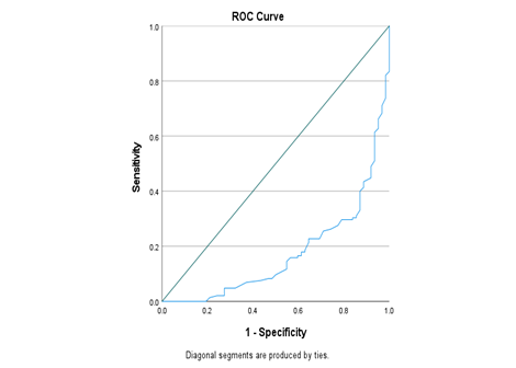
Patients with histopathologically confirmed high-grade gliomas had significantly lower mean ADC values than those with low-grade gliomas (1.00 ± 0.16 vs. 1.46 ± 0.18 × 10⁻³ mm²/s, p < 0.001). High-grade gliomas were also associated with significantly larger lesion size (44.1 ± 17.2 mm vs. 31.8 ± 10.6 mm, p < 0.001) and a shorter duration of symptoms (25.8 ± 15.2 weeks vs. 34.2 ± 18.5 weeks, p = 0.002). Moreover, moderate or marked contrast enhancement (87.6% vs. 29.0%, p < 0.001), peritumoral edema (75.9% vs. 54.8%, p = 0.004), and necrosis (43.4% vs. 11.3%, p < 0.001) (Table 5).
Table 5. Comparison of Apparent Diffusion Coefficient Values According to Histopathological Glioma Grade
|
Variable |
Low-grade Glioma (n = 62) |
High-grade Glioma (n = 145) |
p-value |
|
ADC value (×10⁻³ mm²/s) |
1.46 ± 0.18 |
1.00 ± 0.16 |
<0.001 |
|
Lesion size (mm) |
31.8 ± 10.6 |
44.1 ± 17.2 |
<0.001 |
|
Duration of symptoms (weeks) |
34.2 ± 18.5 |
25.8 ± 15.2 |
0.002 |
|
Moderate/Marked contrast enhancement |
18 (29.0%) |
127 (87.6%) |
<0.001 |
|
Peritumoral edema present |
34 (54.8%) |
155 (75.9%) |
0.004 |
|
Necrosis present |
7 (11.3%) |
63 (43.4%) |
<0.001 |
|
Area Under the Curve |
||||
|
Area |
Std. Errora |
Asymptotic Sig.b |
Asymptotic 95% Confidence Interval |
|
|
Lower Bound |
Upper Bound |
|||
|
.174 |
.030 |
.000 |
.115 |
.233 |
|
The test result variable(s): ADCvalue10mms has at least one tie between the positive actual state group and the negative actual state group. Statistics may be biased. |
||||
|
a. Under the nonparametric assumption |
||||
|
b. Null hypothesis: true area = 0.5 |
||||
DISCUSSION:
The present study aimed to assess the diagnostic value of apparent diffusion coefficient (ADC) in distinguishing low grade and high grade gliomas with the reference standard of histopathology. These findings revealed that ADC was found to be a reliable non-invasive imaging biomarker overall in terms of diagnostic accuracy (73.4%), sensitivity (70.3%), specificity (80.6%), PPV (89.5%) and NPV (53.8%). These results suggest that DW-MRI can be used to gather useful pre-operative information for the grading of gliomas and can be helpful to the clinician in the planning of the appropriate management before histopathological diagnosis. Patients' baseline demographic characteristics included a mean age of 41.0 ± 13.9 years and a slight female predominance (52.2%). The mean ADC was 1.14 ± 0.31 × 10⁻³ mm²/s, and the mean lesion size was 40.3 ± 16.7 mm, which is consistent with previous studies. The mean ADC was 1.14 × 10⁻³ mm²/s ± 0.31, and the mean lesion size was 40.3 ± 16.7 mm, which is similar to previous studies. It is noted that there is a similarity in these findings, and that the present study population is similar to those already published [11].
As far as the clinical presentation was concerned, vomiting and seizures were the most common presenting complaints, and the most common tumor location was the insular region and thalamus. It has previously been consistently observed that the symptoms of gliomas are greatly dependent on the location of the tumor, its size, and intracranial pressure, the most common being headache, seizures, vomiting, and focal neurological deficits. In the present study, the distribution of tumor locations mirrors the heterogeneous nature of gliomas in the brain. Magnetic resonance imaging revealed that there was a greater likelihood of finding high-grade tumors with marked contrast enhancement, moderate peritumoral edema, and necrosis [12]. Histopathology detected 70.0% high-grade gliomas, while ADC detected 55.1% high-grade gliomas. High-grade gliomas have been shown to have higher levels of angiogenesis, blood-brain barrier permeability, cellular proliferation, and necrosis, resulting in the typical MRI appearance of heterogeneous enhancement and surrounding edema. But there remains overlap between imaging appearances of low-grade and high-grade lesions, even with conventional MRI sequences alone [13].
In the present study, 102 true positive and 50 true negative cases were found, while 12 false positive and 43 false negative cases were observed. In this research, the sensitivity and specificity values that were achieved are comparable with the ones that were reported in the literature. The sensitivity of ADC in glioma grading was previously reported to be around 70% and the specificity around 80%, while the pooled sensitivity of ADC was recently shown to be close to 80% and the pooled specificity is close to 90% [14]. The slight difference between studies may be due to variations in MRI protocols, field strength, tumor heterogeneity, region of interest selection, and patient characteristics. The positive predictive value of 89.5% in the present study suggests that the most likely explanation for these types of lesions being identified as high-grade by ADC was that they would likely be confirmed by histopathology [15]. The relatively low negative predictive value (53.8%) also indicates that, due to intratumoral necrosis, cystic degeneration, mixed histological components, or sampling variability, a certain proportion of high-grade gliomas may show relatively high ADC values [16]. Similarly, previous studies have shown that ADC is not a sufficient marker alone to increase diagnostic confidence but needs to be used with conventional MRI parameters and clinical assessment [17]. Histopathology is still the most accurate method to grade gliomas but needs invasive sampling of tissue and can sometimes be influenced by sampling error due to tumour heterogeneity [18]. ADC measurement, on the other hand, is a fast, non-invasive, repeatable, and easily available imaging biomarker, which can assess the whole tumor before surgery. Incorporating ADC into the routine preoperative MRI process could potentially enhance surgical planning, patient counselling, and decrease diagnostic uncertainty, especially in health care environments with limited access to advanced molecular diagnostics [19,20].
Limitations
The present study is limited in that it was conducted at a single center, which may limit the generalizability of the results. While the sample size was reasonable, larger multicenter studies are necessary to validate further. The study focused only on ADC values derived from the diffusion-weighted MRI and did not involve advanced imaging modalities like perfusion MRI, MR spectroscopy, diffusion tensor imaging or radiomics. Only one ADC threshold was applied, which does not completely represent the heterogeneity of gliomas. Selection bias can also occur when consecutive sampling is used, which is a type of non-probability sampling. In addition, there may have been sampling error as a result of intratumoral heterogeneity, and molecular markers, including IDH mutation and 1p/19q codeletion, were not assessed.
CONCLUSION:
The present study demonstrated that the apparent diffusion coefficient is a reliable non-invasive imaging biomarker for differentiating low-grade and high-grade gliomas. Using histopathology as the gold standard, ADC showed good sensitivity, specificity, and overall diagnostic accuracy, supporting its value in preoperative tumor grading. Incorporating ADC into routine MRI evaluation may improve diagnostic confidence, facilitate treatment planning, and assist in clinical decision-making.
REFERENCES:
Sharma A, Graber JJ. Overview of prognostic factors in adult gliomas. Ann Palliat Med. 2021;10(1):863-74.
2. Chen B, Chen C, Zhang Y, Xu J. Recent incidence trend of elderly patients with glioblastoma in the United States, 2000-2017. BMC Cancer. 2021;21(1):54-7.
3. Malla S, Bavikar R, Gore C, Chugh A, Gurwale S. Histopathological Spectrum of Gliomas and Its Immunohistochemical Correlation in a Tertiary Care Setup. Cureus. 2024;16(7):e65036-8.
4. Bihan DL. From Brownian motion to virtual biopsy: a historical perspective from 40 years of diffusion MRI. Japanese J Radiol. 2024;42:1357-71.
5. Bušić M, Rumboldt Z, Ćerina D, Bušić Ž, Dolić K. Prognostic Value of Apparent Diffusion Coefficient (ADC) in Patients with Diffuse Gliomas. Cancers (Basel). 2024;16(4):681-4.
6. Wang QP, Lei DQ, Yuan Y, Xiong NX. Accuracy of ADC derived from DWI for differentiating high-grade from low-grade gliomas: Systematic review and meta-analysis. Medicine (Baltimore). 2020;99(8):e19254-7.
7. Soliman RK, Essa AA, Elhakeem AAS, Gamal SA, Zaitoun MMA. Texture analysis of apparent diffusion coefficient (ADC) map for glioma grading: Analysis of whole tumoral and peri-tumoral tissue. Diagn Interv Imaging. 2021;102(5):287-95.
8. Phuttharak W, Thammaroj J, Wara-Asawapati S, Panpeng K. Grading Gliomas Capability: Comparison between Visual Assessment and Apparent Diffusion Coefficient (ADC) Value Measurement on Diffusion-Weighted Imaging (DWI). Asian Pac J Cancer Prev. 2020 Feb 1;21(2):385-390. doi: 10.31557/APJCP.2020.21.2.385. PMID: 32102515; PMCID: PMC7332154.
9. Khan R, Selehria A, Aquil H, Sheraz A, Khan S, et al. Diagnostic accuracy of apparent diffusion coefficient (ADC) in differentiating low- and high-grade gliomas, taking histopathology as the gold standard. J Radiol Oncol. 2023; 7: 013-019.
10. Kamimura K, Nakano T, Nakajo M, Kamizono J, Hasegawa T, Tobo D, Mukai A, Kamimura Y, Ejima F, Nagano H, Takumi K, Nakajo M, Higa N, Yonezawa H, Hanaya R, Kirishima M, Tanimoto A, Otsuka H, Hirahara D, Imai H, Feiweier T, Yoshiura T. Time-dependent diffusion-weighted imaging assessment of tumor grading and isocitrate dehydrogenase genotypes in adult-type diffuse gliomas. Jpn J Radiol. 2026 May;44(5):882-894. doi: 10.1007/s11604-025-01936-w. Epub 2026 Jan 5. PMID: 41489694; PMCID: PMC13144230.
11. Yao R, Cheng A, Zhang Z, Jin B, Yu H. Correlation Between Apparent Diffusion Coefficient and the Ki-67 Proliferation Index in Grading Pediatric Glioma. J Comput Assist Tomogr. 2023 Mar-Apr 01;47(2):322-328. doi: 10.1097/RCT.0000000000001400. PMID: 36957971; PMCID: PMC10045956.
12. Azizova A, Prysiazhniuk Y, Cakmak M, Kaya E, Petr J, Barkhof F, Wamelink IJHG, Keil VC. Visual versus region-of-interest based diffusion evaluation and their diagnostic impact in adult-type diffuse gliomas. Neuroradiology. 2026 Feb;68(2):503-515. doi: 10.1007/s00234-025-03832-6. Epub 2025 Nov 8. PMID: 41204956; PMCID: PMC13021811.
13. Dung LT, Hung ND, Hung NK, Khuong NH, Thien LQ, Duy NQ, Duc NM. Magnetic resonance imaging characteristics of glioblastoma of the optic pathway during adulthood. Radiol Case Rep. 2023 May 26;18(8):2628-2632. doi: 10.1016/j.radcr.2023.05.010. PMID: 37273722; PMCID: PMC10238590.
14. Iqbal B, Gul N, Mehmood K, Jawwad S, Afzal K, Yousaf M. Diagnostic Accuracy of Apparent Diffusion Coefficient Value in Differentiating Benign and Malignant Brain Lesions Keeping Histopathology as Gold Standard. 2023; 4(2):87-92. doi: http://doi.org/10.37185/LnS.1.1.299
15. Wen PY, van den Bent M, Youssef G, Cloughesy TF, Ellingson BM, Weller M, Galanis E, Barboriak DP, de Groot J, Gilbert MR, Huang R, Lassman AB, Mehta M, Molinaro AM, Preusser M, Rahman R, Shankar LK, Stupp R, Villanueva-Meyer JE, Wick W, Macdonald DR, Reardon DA, Vogelbaum MA, Chang SM. RANO 2.0: Update to the Response Assessment in Neuro-Oncology Criteria for High- and Low-Grade Gliomas in Adults. J Clin Oncol. 2023 Nov 20;41(33):5187-5199. doi: 10.1200/JCO.23.01059. Epub 2023 Sep 29. PMID: 37774317; PMCID: PMC10860967.
16. Whitfield BT, Huse JT. Classification of adult-type diffuse gliomas: Impact of the World Health Organization 2021 update. Brain Pathol. 2022 Jul;32(4):e13062. doi: 10.1111/bpa.13062. Epub 2022 Mar 14. PMID: 35289001; PMCID: PMC9245936.
17. Feraco P, Franciosi R, Picori L, Scalorbi F, Gagliardo C. Conventional MRI-Derived Biomarkers of Adult-Type Diffuse Glioma Molecular Subtypes: A Comprehensive Review. Biomedicines. 2022 Oct 5;10(10):2490. doi: 10.3390/biomedicines10102490. PMID: 36289752; PMCID: PMC9598857.
18. Hangel G, Schmitz-Abecassis B, Sollmann N, Pinto J, Arzanforoosh F, Barkhof F, Booth T, Calvo-Imirizaldu M, Cassia G, Chmelik M, Clement P, Ercan E, Fernández-Seara MA, Furtner J, Fuster-Garcia E, Grech-Sollars M, Guven NT, Hatay GH, Karami G, Keil VC, Kim M, Koekkoek JAF, Kukran S, Mancini L, Nechifor RE, Özcan A, Ozturk-Isik E, Piskin S, Schmainda KM, Svensson SF, Tseng CH, Unnikrishnan S, Vos F, Warnert E, Zhao MY, Jancalek R, Nunes T, Hirschler L, Smits M, Petr J, Emblem KE. Advanced MR Techniques for Preoperative Glioma Characterization: Part 2. J Magn Reson Imaging. 2023 Jun;57(6):1676-1695. doi: 10.1002/jmri.28663. Epub 2023 Mar 13. Erratum in: J Magn Reson Imaging. 2024 Apr;59(4):1468. doi: 10.1002/jmri.28934. PMID: 36912262; PMCID: PMC10947037.
19. Prysiazhniuk Y, Server A, Leske H, Bech-Aase Ø, Helseth E, Eijgelaar RS, Fuster-García E, Brandal P, Bjørnerud A, Otáhal J, Petr J, Nordhøy W. Diffuse glioma molecular profiling with arterial spin labeling and dynamic susceptibility contrast perfusion MRI: A comparative study. Neurooncol Adv. 2024 Jul 5;6(1):vdae113. doi: 10.1093/noajnl/vdae113. PMID: 39036439; PMCID: PMC11259011.
20. Thust SC, Heiland S, Falini A, Jäger HR, Waldman AD, Sundgren PC, Godi C, Katsaros VK, Ramos A, Bargallo N, Vernooij MW, Yousry T, Bendszus M, Smits M. Glioma imaging in Europe: A survey of 220 centres and recommendations for best clinical practice. Eur Radiol. 2018 Aug;28(8):3306-3317. doi: 10.1007/s00330-018-5314-5. Epub 2018 Mar 13. PMID: 29536240; PMCID: PMC6028837.