Incidence And Common Pathogens Associated with Ventilator-Associated Pneumonia in Oncological Critical Care Patients.
- Jalil Ur Rehman Butt , Fellow in Critical Care, SKMCH-RC
- Muhammad Imran , Resident in Anesthesia, SKMCH-RC
- Muhammad Asif , Resident in Anesthesia, SKMCH-RC
- Sadia Sadaqat Ali , Consultant Anaesthetist and Critical Care, SKMCH-RC
- Saad Ur Rehman , Consultant Anaesthetist and Critical Care, SKMCH-RC
- Ahsun Waqar Khan , Consultant Anaesthetist and Critical Care, SKMCH-RC
Article Information:
Abstract:
Objective: To determine the incidence of ventilator-associated pneumonia and identify the common pathogens causing VAP among oncological patients admitted to the ICU.Study design: Retrospective observational study. Study place and duration: Intensive Care Unit, Shaukat Khanum Memorial Cancer Hospital and Research Centre, over a 12-month period, from 29th Oct, 2024 to 28th Oct, 2025Methods: Medical records of 140 oncological patients admitted to the ICU who required mechanical ventilation for more than 48 hours during the study period were reviewed. Diagnosis of VAP was confirmed using the Clinical Pulmonary Infection Score (CPIS) based on documented clinical, radiological, and microbiological data. Respiratory culture results (endotracheal aspirates and BAL samples) were assessed and recorded. Results: The overall median age of the patients enrolled in the study was 30 (40) years. Ventilator-associated pneumonia was found in 39 (27.9%) patients. The commonest pathogens isolated in VAP patients were Escherichia coli (5%), Pseudomonas aeruginosa (1.4%), Staphylococcus aureus (1.4%), Staphylococcus epidermidis (1.4%), and Streptococcus species (1.4%). Conclusion: In oncological patients admitted in critical care, VAP was seen in 27.9%. The most common causal pathogens are gram-negative bacteria, including Escherichia coli and Pseudomonas aeruginosa
Keywords:
Article :
INTRODUCTION:
Over the past several decades, the prognosis of malignancies has markedly improved, accompanied by an increase in overall survival rates.1,2Despite these advances, patients with cancer remain at heightened risk of infections and treatment-related complications, particularly those associated with chemotherapy, central venous catheters, extensive surgical interventions, and other factors contributing to increased morbidity and mortality.3,4
Moreover, oncologic patients exhibit multiple predisposing factors for respiratory failure arising from both infectious and noninfectious etiologies, including pneumonia, pulmonary thrombosis, sepsis, transfusion-related acute lung injury (TRALI), and pulmonary edema.5,6 Consequently, such patients frequently require mechanical ventilation (MV) and admission to the intensive care unit (ICU)7.8.
Ventilator-Associated Pneumonia (VAP) represents the most prevalent ICU-acquired infection, occurring in approximately 25%–30% of patients intubated for more than 48 hours. with a progressively increasing risk during the first 14 days of mechanical ventilation9. The estimated incidence of VAP varies between 2 and 16 cases per 1000 ventilator-days10. Furthermore, the emergence of multidrug-resistant bacteria (MDRB) has evolved into a major public health concern, imposing an additional burden on hospital care systems, particularly among patients admitted to the ICU.
Despite this increased risk, little is known about the prevalence and microbiological characteristics of VAP, particularly in Pakistani oncology patients hospitalized in critical care units. Because hospitals and areas have different microbial patterns and profiles of antibiotic resistance, understanding the local range of causal microorganisms is crucial. Therefore, the current study aimed to determine the incidence of ventilator-associated pneumonia and identify the common pathogens causing VAP among oncological patients admitted to the ICU. Our study also identified the length of ICU stay and mortality risk in these patients.
MATERIALS AND METHODS:
This was a retrospective observational study conducted at the Intensive Care Unit, Shaukat Khanum Memorial Cancer Hospital and Research Centre. Approval was obtained from the Institutional Review Board prior to commencement. Data collection was performed over a 12-month period, from 29th Oct, 2024 to 28th Oct, 2025. All eligible oncological ICU patients meeting inclusion criteria during the study period were included by non-probabiliy consecutive sampling.
Adult oncological patients (>18 years) requiring mechanical ventilation >48 hours were included in the study. Patients with pneumonia prior to intubation, patients intubated for <48 hours, and those with incomplete or missing medical records were excluded from the study.
Ventilator-associated pneumonia was labeled as a type of hospital-acquired pneumonia that occurred more than 48 hours after endotracheal intubation and mechanical ventilation. This was further classified into early onset (within the first 96 hours of MV) and late onset (more than 96 hours after the initiation of MV), which was more commonly attributable to multidrug–resistant pathogens. An oncological critical patient was defined as any patient admitted to the ICU with an underlying diagnosis of malignancy (solid or hematological). Incidence was defined as the number of new cases of VAP per 1000 ventilator-days. The primary outcome was the incidence of VAP per 1000 ventilator days, and the secondary outcomes were the spectrum of pathogens, length of ICU stay, and mortality risk.
Following ethical approval, medical records of all oncological patients admitted to ICU who required mechanical ventilation for more than 48 hours during the study period were reviewed. Diagnosis of VAP was confirmed using the Clinical Pulmonary Infection Score (CPIS) based on documented clinical, radiological, and microbiological data. A score of more than 6 was associated with VAP. Respiratory culture results (endotracheal aspirates and BAL samples), demographic characteristics, duration of ventilation, comorbidities, malignancy type, antimicrobial sensitivity patterns, and outcomes were derived from electronic medical records.
Data were analyzed using IBM SPSS version 25.0. The Shapiro-Wilk test was used for assessing the data normality, and it was found that the data was non-normal in distribution; therefore, numerical variables such as age, duration of MV, temperature, leucocyte count, and PaO2/FiO2 ratio were presented as median and interquartile range, and the Mann-Whitney U test was used to compare these variables in patients with and without VAP, and a p-value of 0.05 was considered significant. Frequency of VAP and the pathogens isolated were presented as frequency and percentage. The chi-square test was used for categorical variables, and a p-value of 0.05 was considered significant. Logistic regression was used to identify risk factors.
RESULTS:
A total of 140 patients' records were assessed. Ventilated associated pneumonia was found in 39 (27.9%) patients (Figure 1).
The overall median age of the patients enrolled in the study was 30 (40) years, the median duration of MV was 5 (6) days, and the median overall temperature of patients recorded was 37 (0.70) degrees Celsius. The overall median leukocyte count was 8.28 (11.622) cells/mm3. The PaO2/FiO2 ratio was 2.75 (1.70). The baseline demographic and clinical features of the patients with and without VAP are shown in Table I.
The frequency of sociodemographic, clinical, and microbiological findings in patients with or without VAP is shown in Table 2. It was found that the culture findings, diabetes status, and radiological findings on chest X-ray differed significantly in patients with VAP and those without it, as indicated by a p-value of <0.05.
Factors such as gender, hypothyroidism, SIADH, hepatic encephalopathy, and cord compression had a protective role against the occurrence of VAP, as indicated by the odds ratio of <1. Patients with factors such as the presence of comorbidity, malignancy, acute kidney injury, cardiac dysfunction, diabetes, hypertension, and ischemic heart disease were associated with higher odds of developing VAP (Table II).
Figure 1: Frequency of ventilator-associated pneumonia (n=140)
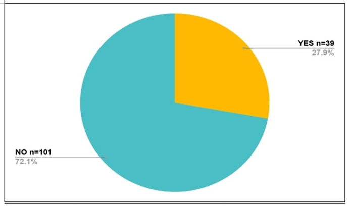
Table I: Baseline demographic and clinical scores of the scales (n=140)
|
Variable |
Overall |
In patients without VAP (n=101) |
In patients with VAP (n=39) |
p Value* |
|
Age (in years) |
30 (40) |
26 (40) |
34 (27) |
0.404 |
|
Duration of MV (in days) |
5 (6) |
5 (5) |
7 (8) |
0.045 |
|
Temperature (in degrees Celsius) |
37 (0.70) |
36.9 (0.70) |
37.2 (0.60) |
0.098 |
|
Leucocyte count (cells/mm3) |
8.28 (11.622) |
9 (11.705) |
6.04 (11.850) |
0.298 |
|
Oxygenation status (PaO2/FiO2) |
2.75 (1.70) |
2.8 (1.6) |
2.35 (2.66) |
0.555 |
Abbreviations: VAP—ventilator-associated pneumonia, MV—mechanical ventilation
*p value 0.05 was considered statistically significant after comparing patients with VAP with those without it
Table II: Frequency of sociodemographic, clinical and microbiological findings in patients with or without VAP (n=140)
|
Variable |
In patients without VAP (n=101) |
In patients with VAP (n=39) |
Total |
p Value* |
Odds ratio** |
|
Gender: Male Female |
58 (41.4%) 43 (30.7%) |
29 (20.7%) 19 (7.2%) |
87 (62.1%) 53 (37.9%) |
0.064 |
0.465 |
|
Age: <19 years 19 to 40 years 41 to 60 years >60 years |
36 (25.7%) 32 (22.9%) 22 (15.7%) 9 (6.4%) |
9 (6.4%) 17 (12.1%) 9 (6.4%) 4 (2.9%) |
45 (32.1%) 49 (35%) 31 (22.1%) 13 (9.3%) |
0.464 |
-
|
|
Malignancy: Yes No |
91 (65%) 10 (7.2%) |
37 (26.4%) 2 (1.4%) |
128 (91.4%) 12 (8.6%) |
0.366 |
2.033 |
|
Origin of malignancy: None Hematological Solid organ malignancy CBS/Brain tumor Germ cell tumors/pediatric tumors |
11 (7.9%) 41 (29.3%) 29 (20.7%) 14 (10%) 6 (4.3%) |
2 (1.4%) 19 (13.6%) 13 (9.3%) 4 (2.9%) 1 (0.7%) |
13 (9.3%) 60 (42.9%) 42 (30%) 18 (12.9%) 7 (5%) |
0.633 |
- |
|
Type of malignancy: No malignancy ALL AML Burkitt’s lymphoma CA bladder CA breast CA colon CA Esophagus CA stomach CA pancreas CA prostate Cholangiocarcinoma CML Brain tumor DLBCL Ewing's sarcoma GCT HNC Hodgkin’s lymphoma Lymphoma Pediatric adenocarcinoma Penile tumor Prostate hyperplasia Renal cell carcinoma |
11 (7.9%) 15 (10.7%) 6 (4.3%) 4 (1.4%) 0 (0%) 6 (4.3%) 5 (3.6%) 1 (0.7%) 1 (0.7%) 1 (0.7%) 0 (0%) 1 (0.7%) 4 (2.8%) 14 (10%) 8 (5.8%) 1 (0.7%) 6 (4.3%) 8 (5.8%) 4 (2.8%) 0 (0%) 1 (0.7%) 1 (0.7%) 1 (0.7%) 2 (1.4%) |
2 (1.4%) 4 (2.8%) 4 (2.8%) 3 (2.1%) 2 (1.4%) 2 (1.4%) 1 (0.7%) 1 (0.7%) 0 (0%) 1 (0.7%) 1 (0.7%) 0 (0%) 0 (0%) 4 (2.8%) 2 (1.4%) 0 (0%) 1 (0.7%) 3 (2.1%) 5 (3.6%) 1 (0.7%) 0 (0%) 0 (0%) 0 (0%) 2 (1.4%) |
13 (9.3%) 19 (13.6%) 10 (7.1%) 7 (5%) 2 (1.4%) 8 (5.7%) 6 (4.3%) 2 (1.4%) 1 (0.7%) 2 (1.4%) 1 (0.7%) 1 (0.7%) 4 (2.9%) 18 (12.8%) 10 (7.1%) 1 (0.7%) 7 (5%) 11 (7.9%) 9 (6.4%) 1 (0.7%) 1 (0.7%) 1 (0.7%) 1 (0.7%) 4 (2.8%) |
0.392 |
- |
|
Pneumonia before MV: Yes No |
51 (36.4%) 50 (35.7%) |
19 (13.6%) 20 (14.3%) |
70 (50%) 70 (50%) |
0.852 |
- |
|
Pathogens identified: None Acinobacter species Escherichia coli Pseudomonas aeruginosa Staphylococcus aureus Staphylococcus epidermidis Aspergillus species Burkholderia cepacia Enterococcus faecalis Klebsiella pneumoniae Listeria species Staphylococcus hominis Stenotrophomonas species Streptococcus species Mixed infection |
58 (41.4%) 3 (2.1%) 8 (5.7%) 3 (2.1%) 9 (6.4%) 3 (2.1%) 1 (0.7%) 2 (1.4%) 4 (2.8%) 0 (0%) 1 (0.7%) 1 (0.7%) 1 (0.7%) 2 (1.4%) 5 (3.6%) |
16 (11.4%) 1 (0.7%) 7 (5%) 2 (1.4%) 2 (1.4%) 2 (1.4%) 0 (0%) 1 (0.7%) 1 (0.7%) 1 (0.7%) 0 (0%) 0 (0%) 0 (0%) 2 (1.4%) 2 (1.4%) |
74 (52.8%) 4 (2.8%) 15 (10.7%) 5 (3.5%) 4 (2.8%) 5 (3.5%) 1 (0.7%) 3 (2.1%) 5 (3.6%) 1 (0.7%) 1 (0.7%) 1 (0.7%) 1 (0.7%) 6 (4.4%) 7 (5%) |
0.437 |
- |
|
Culture findings: None Gram-negative bacteria Gram-positive bacteria Fungal Mixed infection |
55 (39.3%) 31 (22.1%) 8 (5.7%) 4 (2.8%) 3 (2.1%) |
4 (2.8%) 24 (17.2%) 5 (3.6%) 2 (1.4%) 4 (2.8%) |
59 (42.1%) 55 (39.3%) 13 (9.3%) 6 (4.3%) 7 (5%) |
<0.001 |
- |
|
Comorbidity: Yes No |
18 (12.8%) 83 (59.3%) |
8 (5.8%) 31 (22.1%) |
26 (18.6%) 114 (81.4%) |
0.714 |
1.190 |
|
Acute kidney injury: Yes No |
3 (2.1%) 98 (70%) |
3 (2.1%) 36 (25.8%) |
6 (4.2%) 134 (95.8%) |
0.216 |
2.722 |
|
Cardiac dysfunction: Yes No |
2 (1.4%) 99 (70.7%) |
1 (0.7%) 38 (27.1%) |
3 (2.1%) 137 (95.8%) |
0.831 |
1.303 |
|
Diabetes: Yes No |
5 (3.6%) 96 (68.6%) |
6 (4.3%) 33 (23.5%) |
11 (7.9%) 129 (92.1%) |
0.04 |
3.491 |
|
Hypertension: Yes No |
4 (2.9%) 97 (69.3%) |
2 (1.4%) 37 (26.4%) |
6 (4.3%) 134 (95.7%) |
0.760 |
1.311 |
|
Ischemic heart disease: Yes No |
3 (2.2%) 98 (70%) |
2 (1.4%) 37 (26.4%) |
5 (3.6%) 135 (96.4%) |
0.537 |
1.766 |
|
Hypothyroidism: Yes No |
5 (3.6%) 96 (68.5%) |
0 (0%) 39 (27.9%) |
5 (3.6%) 135 (96.4%) |
0.157 |
0.711 |
|
Cord compression/spina bifida: Yes No |
1 (0.7%) 100 (71.4%) |
0 (0%) 39 (27.9%) |
1 (0.7%) 139 (99.3%) |
0.533 |
0.719 |
|
Hepatic encephalopathy: Yes No |
1 (0.7%) 100 (71.4%) |
0 (0%) 39 (27.9%) |
1 (0.7%) 139 (99.3%) |
0.533 |
0.719 |
|
Syndrome of inappropriate antidiuretic hormone secretion (SIADH): Yes No |
1 (0.7%) 100 (71.4%) |
0 (0%) 39 (27.9%) |
1 (0.7%) 139 (99.3%) |
0.533 |
0.719 |
|
Outcome measure: Discharged Death |
33 (23.6%) 68 (48.6%) |
17 (12.1%) 22 (15.7%) |
50 (35.7%) 90 (64.3%) |
0.227 |
- |
|
Radiographic findings on chest X-ray: Normal/unremarkable Bilateral infiltrates Unilateral/focal infiltrates Lobar consolidation/pneumonia pattern Pleural / other complications |
38 (27.1%) 50 (35.7%) 8 (5.7%) 3 (2.1%)
2 (1.4%) |
5 (3.6%) 28 (20%) 5 (3.6%) 0 (0%)
1 (0.7%) |
43 (30.7%) 78 (55.7%) 13 (9.3%) 3 (2.1%)
3 (2.1%) |
0.039 |
- |
*p value 0.05 was considered statistically significant after comparing patients with VAP with those without it
**Odds ratio of value 1 denoted no association, <1 denoted protective effect, >1 denoted increased risk/ odds, >2 denoted strong positive association
DISCUSSION :
The current study findings revealed that in oncological patients in the critical care unit, the frequency of VAP was 27.9%. The most common pathogens identified in patients who had VAP were Escherichia coli, Pseudomonas aeruginosa, Staphylococcus aureus, Staphylococcus epidermidis and Streptococcus species. Culture findings revealed that gram-negative bacteria were among the commonest pathogens. The results show that VAP is a serious complication among cancer patients on MV, highlighting the necessity of close observation, prompt diagnosis, and efficient infection control procedures in critical care environments.
Due to weakened immune systems brought on by cancer, chemotherapy, radiation, malnourishment, and extended hospital stays, cancer patients are more vulnerable to VAP11,12. By circumventing natural airway defenses and promoting microbial colonization of the lower respiratory tract, mechanical breathing raises the risk even further13,14. Our study reported that the frequency of VAP in patients admitted to critical care, i.e., 27.9%. In an Indian study, the authors found that the frequency of VAP in cancer patients was 15.2%15. In a study conducted in Southeast Asia, the rate of VAP ranged from 2.13 to 116 per thousand days16. In a study conducted in Mexico, the rate of VAP in oncological patients was 12.2%17. In a study from a developing country, i.e., Italy, Colaneri et al. revealed that the frequency of VAP was 9.8%18. The study's observed rate of VAP is in line with studies from other developing nations, where rates are still greater than those found in many high-income countries.
In our study, Escherichia coli, a gram-negative bacteria, was the commonest pathogen found in patients with VAP, followed by Pseudomonas aeruginosa, and among the gram-positive bacteria were the Staphylococcus species and Streptococcus species. Malik et al. similarly revealed that gram-negative bacteria were the commonest pathogens associated with VAP15. A study conducted by Kharel et al. found that Gram-positive bacteria, including Staphylococcus aureus and Enterococcus species, as well as Gram-negative bacteria like Acinetobacter species, Pseudomonas aeruginosa, and Klebsiella pneumoniae, were commonly discovered16. Colaneri et al. revealed that Pseudomonas species (21.0%), Staphylococcus aureus (20.2%), and Klebsiella species (20.1%) were shown to be the most common pathogens in microbiological profiling of VAP17. Cornejo-Juárez et al. revealed that the commonest pathogens in patients with VAP were Pseudomonas aeruginosa, Escherichia coli, and Klebsiella spp18. These findings are supportive of our study findings that the majority of the cases of VAP are associated with gram-negative bacteria. Long-term antibiotic exposure, invasive operations, cross-transmission in intensive care units, and environmental contamination may all contribute to the prevalence of Gram-negative microbes.
Early infection detection is especially crucial for cancer patients in critical care since underlying immunosuppression can cause fast clinical deterioration. Clinicians might potentially lower complications and mortality by starting more appropriate empirical antibiotic therapy while awaiting culture results when they are aware of the distribution of local pathogens.
This study's focus on a specific and understudied group of critically ill cancer patients in Pakistan is one of its main advantages. The results provide important local epidemiological information that can help medical practitioners create focused infection-control and treatment plans.
Nonetheless, a number of limitations must be noted. The results of a single-center study could not apply to all Pakistani healthcare facilities. The prevalence and microbiological range of VAP may also be impacted by differences in ICU procedures, patient characteristics, and antibiotic prescription patterns. To provide a more thorough picture of VAP epidemiology among cancer patients, future multicenter studies with higher sample sizes are advised.
CONCLUSION :
The current study concluded that among Pakistani cancer patients on MV, VAP is still a frequent and dangerous side effect, as indicated by a frequency of 27.9%. The most common causal pathogens are gram-negative bacteria, including Escherichia coli and Pseudomonas aeruginosa. Reducing the incidence of VAP and improving outcomes in this susceptible patient population requires ongoing surveillance, adherence to VAP prevention methods, and prudent use of antibiotics.
ACKNOWLEDGEMENTS:
We sincerely thank all of our seniors and coworkers who assisted us in gathering information, conducting the necessary literature search, and assembling this work.
Conflict of interest: None
Funding: None
BIBLIOGRAPHY:
1. Belenguer-Muncharaz A, Albert-Rodrigo L, Ferrandiz-Sellés A, Cebrián-Graullera G. Evolución de 10 años de aplicación de la ventilación mecánica en la insuficiencia respiratoria aguda del paciente hematológico ingresado en la unidad de cuidados intensivos [Ten-year evolution of mechanical ventilation in acute respiratory failure in the hematogical patient admitted to the intensive care unit]. Med Intensiva. 2013 Oct;37(7):452-460. Doi: 10.1016/j.medin.2012.12.011.
2. Park SA, Cho SS, Kwak GJ. Factors influencing ventilator-associated pneumonia in cancer patients. Asian Pac J Cancer Prev. 2014;15(14):5787-91. Doi: 10.7314/apjcp.2014.15.14.5787.
3. Aly NY, Al-Mousa HH, Al Asar el SM. Nosocomial infections in a medical-surgical intensive care unit. Med Princ Pract. 2008;17(5):373-7. Doi: 10.1159/000141500.
4. Oliveira VF, Lopes DD, Cabral VS, Junior LD, Matos SS, Almeida LA, et al. Mortality in Ventilator‐Associated Tracheobronchitis and Pneumonia in Oncology Patients: The Impact of Microbiological Aspects. Can J Infect Dis Med. Microbiol. 2026;2026(1):5887462. Doi: https://doi.org/10.1155/cjid/5887462
5. Medjedovic S, Jurca T, Povšič MK. Ventilator-associated Pneumonia in the Intensive Care Unit of the Institute of Oncology Ljubljana in 2021 and the Role of Nurses in its Prevention. Open Access Maced J Med Sci. 2023;11(G):69-77. Doi: https://doi.org/10.3889/oamjms.2023.11470
6. Miron M, Blaj M, Ristescu AI, Iosep G, Avădanei AN, Iosep DG, et al. Hospital-acquired pneumonia and ventilator-associated pneumonia: a literature review. Microorganisms. 2024;12(1):213. Doi: https://doi.org/10.3390/microorganisms12010213
7. Kreitmann L, Gaudet A, Nseir S. Ventilator-associated pneumonia in immunosuppressed patients. Antibiotics. 2023;12(2):413. Doi: https://doi.org/10.3390/antibiotics12020413
8. Halat DH, Moubareck CA. Hospital-acquired and ventilator-associated pneumonia caused by multidrug-resistant Gram-negative pathogens: Understanding epidemiology, resistance patterns, and implications with COVID-19. F1000Research. 2024;12:92. Doi: https://doi.org/10.12688/f1000research.129080.2
9. Patil HV, Patil VC. Incidence, bacteriology, and clinical outcome of ventilator-associated pneumonia at tertiary care hospital. J Nat Sci Biol Med. 2017 Jan-Jun;8(1):46-55. Doi: 10.4103/0976-9668.198360.
10. Sarda C, Fazal F, Rello J. Management of ventilator-associated pneumonia (VAP) caused by resistant gram-negative bacteria: which is the best strategy to treat? Expert Rev Respir Med. 2019 Aug;13(8):787-798. Doi: 10.1080/17476348.2019.1632195.
11. Ramadan RA, Bedawy AM, Negm EM, Hassan TH, Ibrahim DA, ElSheikh SM, et al. Carbapenem-resistant Klebsiella pneumoniae among patients with ventilator-associated pneumonia: Evaluation of antibiotic combinations and susceptibility to new antibiotics. Infect Drug Resist. 2022;15:3537-3548. Doi: https://doi.org/10.2147/IDR.S371248
12. Kao HH, Peng CK, Sheu CC, Lin YC, Chan MC, Wang SH, et al. Mortality and ventilator dependence in critically ill patients with ventilator-associated pneumonia caused by carbapenem-resistant Acinetobacter baumannii. J Microbiol Immunol Infect. 2023;56(4):822-832. Doi: https://doi.org/10.1016/j.jmii.2023.04.004
13. Alnimr A. Antimicrobial resistance in ventilator-associated pneumonia: predictive microbiology and evidence-based therapy. Infect Dis Ther. 2023;12(6):1527-1552. Doi: https://doi.org/10.1007/s40121-023-00820-2
14. Velasquez-Garcia L, Mejia-Sanjuanelo A, Viasus D, Carratala J. Causative agents of ventilator-associated pneumonia and resistance to antibiotics in COVID-19 patients: a systematic review. Biomed. 2022;10(6):1226. Doi: https://doi.org/10.3390/biomedicines10061226
15. Malik S, Mukherjee S, Ghosh PS, Bagchi S, Goel G, Chatterji S, et al. A prospective cohort study of the incidence, etiology and outcome of pneumonia among cancer patients in an oncology intensive care unit from Eastern India. Ecancer. 2025;19:1970.
16. Kharel S, Bist A, Mishra SK. Ventilator-associated pneumonia among ICU patients in WHO Southeast Asian region: A systematic review. PLoS One. 2021;16(3):e0247832. Doi: 10.1371/journal.pone.0247832.
17. Colaneri M, Montrucchio G, Scaglione G, Monti G, Tricella G, Genovese C, et al. Incidence, microbiology, and mortality of ventilation-associated pneumonia in a large Italian cohort of critically ill patients: results from the PROSAFE project. Clinical Microbiology and Infection. 2025;31(9):1491-1499. Doi: https://doi.org/10.1016/j.cmi.2025.05.026
18. Cornejo-Juárez P, González-Oros I, Mota-Castañeda P, Vilar-Compte D, Volkow-Fernández P. Ventilator-associated pneumonia in patients with cancer: Impact of multidrug resistant bacteria. World J Crit Care Med. 2020;9(3):43-53. Doi: 10.5492/wjccm.v9.i3.43.