Tracheobronchopathia Osteochondroplastica: A Rare Cause of Chronic Respiratory Symptoms
- Arun Kumar , Senior resident Department of Respiratory Medicine Dhanalakshmi Srinivasan Medical College and Hospital, Sirvachur, Perambalur
- Rajasekar Selvaraj , Junior Resident, Department of Respiratory Medicine Dhanalakshmi Srinivasan Medical College and Hospital, Sirvachur, Perambalur
- Priyadharshini.T , Junior Resident, Department of Respiratory Medicine, Dhanalakshmi Srinivasan Medical College and Hospital, Sirvachur, Perambalur
- B.Anju , Junior Resident, Department of Respiratory Medicine, Dhanalakshmi Srinivasan Medical College and Hospital, Sirvachur, Perambalur
- Anusiya Elangovan , Junior Resident, Department of Respiratory Medicine, Dhanalakshmi Srinivasan Medical College and Hospital, Sirvachur, Perambalur
Article Information:
Abstract:
Tracheobronchopathia osteochondroplastica (TBPO) is a rare, benign disorder of the tracheobronchial tree characterized by the presence of multiple submucosal osseous and/or cartilaginous nodules projecting into the anterior and lateral walls of the airways, characteristically sparing the posterior membranous wall. The exact etiology remains unknown, and the disease is often diagnosed incidentally during bronchoscopy or imaging performed for unrelated indications. We present the case of a 55-year-old female with a history of systemic hypertension who presented with chronic breathlessness, recurrent cough with mucoid expectoration for two years, and progressive dysphagia for solids for six months. Contrast-enhanced computed tomography of the chest revealed calcified nodules along the anterior wall of the trachea and a foregut duplication cyst in the subcarinal region. Flexible bronchoscopy demonstrated characteristic nodular lesions involving the tracheal rings in the anterior wall, with sparing of the posterior membranous wall. Endobronchial biopsy revealed subepithelial mature chondro-osseous material without evidence of dysplasia or malignancy. The patient was managed conservatively with inhaled corticosteroids and bronchodilators, with significant symptomatic improvement. This case highlights the importance of recognizing TBPO as a differential diagnosis in patients with chronic respiratory symptoms and emphasizes the role of bronchoscopy in establishing the diagnosis. As there are no established treatment guidelines, conservative management remains the mainstay for asymptomatic or mildly symptomatic patients.
Keywords:
Article :
INTRODUCTION:
Congenital melanocytic nevi (CMN) are pigmented Tracheobronchopathia osteochondroplastica (TBPO) is an uncommon benign disease of unknown etiology, characterized by the presence of multiple sessile, bony or cartilaginous submucosal nodules measuring 1–3 mm in diameter, projecting into the lumen of the tracheobronchial tree. These nodules typically extend selectively from the anterolateral walls of the trachea and bronchi, characteristically sparing the posterior membranous wall. The disease was first described macroscopically by Rokitansky in 1855 and microscopically by Wilks in 1857. The exact pathophysiology of TBPO remains a mystery; however, the origin of the nodules is postulated to be from metaplasia of the submucosal connective tissue or ecchondrosis and exostosis of tracheal cartilage originating from a tracheal ring. Other proposed etiologies include metabolic abnormalities, ageing-related degenerative processes, amyloidosis, malignancy, inheritance, silicosis, chronic irritation, and infection, but these hypotheses lack significant supporting evidence.
TBPO is a rare condition, with approximately 400 cases reported worldwide. The estimated incidence ranges from 0.01 to 4.2 per 100,000 population, with autopsy studies reporting an incidence of approximately 0.3%. The condition is more common in males, with a male-to-female ratio of approximately 3:1, and is usually diagnosed in the fifth to seventh decades of life. Clinical manifestations are diverse, variable, and non-specific; most patients present with chronic cough, dyspnea, wheezing, or recurrent respiratory infections. Dysphagia, as seen in our patient, is an unusual presentation and may be related to compression from associated mediastinal pathology. Diagnosis is often incidental during bronchoscopy, difficult intubation, or autopsy.
There are no specific guidelines for the management of TBPO. Treatment is generally conservative, focusing on symptomatic relief and management of infections. Severe cases with significant airway obstruction may warrant advanced bronchoscopic interventions such as laser ablation, mechanical debridement, or surgical resection. We present a case of TBPO in a 55-year-old female with associated foregut duplication cyst, highlighting the diagnostic challenges and management considerations in this rare condition. This is a case report and doesnt require a ethics approval and has been waived off
CASE PRESENTATION:
A 55-year-old female, a known case of systemic hypertension for five years, presented to the Department of Respiratory Medicine with complaints of breathlessness (grade 2 on the modified Medical Research Council dyspnea scale), recurrent cough with mucoid expectoration for two years which had aggravated over the preceding two weeks, and progressive dysphagia (more for solids) for six months. She gave a history of using nebulizations and inhalers for the past two years with only partial relief of symptoms. There was no history of fever, hemoptysis, chest pain, or weight loss. She was a non-smoker and did not consume alcohol. There was no significant family history of respiratory illnesses or malignancies.
On general examination, the patient was afebrile, with stable vitals. Her pulse rate was 88 beats per minute, respiratory rate 20 breaths per minute, and blood pressure 140/90 mmHg (on antihypertensive medication). Oxygen saturation was 98% on room air. There was no clubbing, cyanosis, or lymphadenopathy. Respiratory system examination revealed bilateral normal vesicular breath sounds with no added sounds. Examination of the cardiovascular, abdominal, and neurological systems was unremarkable. The patient's body mass index was 26.4 kg/m².
Investigations
Chest radiography (posteroanterior view) showed increased bronchovascular markings with no other significant findings. A contrast-enhanced computed tomography (CECT) of the chest performed outside the institution (film not available) had suggested a mediastinal lesion. Repeat CECT chest at our institution revealed multiple calcified nodular lesions along the anterior wall of the trachea. Additionally, a foregut duplication cyst was identified in the subcarinal region, extending into the middle and posterior mediastinum (Figure 1).
Flexible bronchoscopy was performed for further evaluation. The procedure revealed multiple nodular lesions involving the tracheal rings along the anterior and lateral walls of the trachea, with characteristic sparing of the posterior membranous wall (Figure 2). The appearance was consistent with the classic "cobblestone" or "rock-garden" appearance described in TBPO. Bronchoalveolar lavage (BAL) was negative for bacterial culture and sensitivity, no acid-fast bacilli were seen on smear, KOH mount was negative for fungal elements, and Truenat assay was negative for Mycobacterium tuberculosis. BAL cytology and endobronchial brush cytology were negative for malignancy.
Endobronchial biopsy was performed from the nodular lesions. Histopathological examination revealed subepithelial mature chondro-osseous material with no evidence of dysplasia or malignancy (Figure 3). The overlying respiratory epithelium showed pseudo-stratified ciliated columnar epithelium. No amyloid deposits were identified on Congo red staining. Upper gastrointestinal endoscopy (UGIScopy) was performed to evaluate the dysphagia and revealed antral gastritis without any obstructive lesions in the esophagus or gastroesophageal junction.
Treatment
Given the benign nature of the condition and the absence of significant airway obstruction, the patient was managed conservatively. She was started on inhaled formoterol and budesonide via nebulization during the hospital stay, which was subsequently changed to a dry powder inhaler (DPI) formulation of formoterol and budesonide upon discharge. The patient was advised to avoid airway irritants, maintain adequate hydration, and seek prompt treatment for any intercurrent respiratory infections. She was also continued on her antihypertensive medications. The foregut duplication cyst was managed conservatively as it was asymptomatic and incidentally detected. The patient was discharged in stable condition and has been on regular follow-up. At six-month follow-up, she reported significant improvement in her symptoms, with reduced cough, expectoration, and breathlessness. Dysphagia had also improved, likely due to resolution of associated inflammation.
DISCUSSION:
Tracheobronchopathia osteochondroplastica is a rare, benign disease of the tracheobronchial tree, first described by Rokitansky in 1855 and later characterized microscopically by Wilks in 1857. The disease is characterized by multiple submucosal cartilaginous and osseous nodules that project into the tracheobronchial lumen, typically involving the anterior and lateral walls while sparing the posterior membranous wall. The exact etiology remains unknown, though several hypotheses have been proposed including chronic infection, congenital anomaly, chemical or mechanical irritation, degenerative or metabolic abnormalities, and genetic predisposition. The most widely accepted hypothesis is that chronic inflammation leads to cartilaginous and osseous metaplasia of the tracheobronchial submucosa.
The epidemiology of TBPO reflects its rare nature. Approximately 400 cases have been reported worldwide. The estimated incidence ranges from 0.01 to 4.2 per 100,000 population. Autopsy studies report an incidence of approximately 0.3%, while bronchoscopy studies show a wide variation ranging from 1 in 125 to 1 in 5000 procedures. The disease is more common in males, with a male-to-female ratio of approximately 3:1, and is typically diagnosed in the fifth to seventh decades of life. Our patient, a 55-year-old female, is consistent with the typical age at diagnosis, though the female sex is less commonly reported.
Clinical manifestations of TBPO are diverse and non-specific. Most patients present with chronic cough, which is reported in up to 54% of cases. Other symptoms include dyspnea, wheezing, hemoptysis, and recurrent respiratory infections. The presence of dysphagia, as seen in our patient, is unusual and may be related to the associated foregut duplication cyst causing extrinsic compression or to the presence of extensive tracheal involvement. The severity of symptoms often does not correlate with the extent of airway involvement, and many patients remain asymptomatic. The nonspecific nature of symptoms frequently leads to misdiagnosis as asthma, chronic bronchitis, or other chronic respiratory conditions.
The diagnosis of TBPO is primarily based on imaging, bronchoscopy, and histopathological examination. Chest CT is often the first clue to the diagnosis, demonstrating irregular, nodular thickening of the anterolateral tracheal wall with frequent calcification. However, the characteristic findings may be subtle and are often missed by radiologists unfamiliar with the condition. Bronchoscopy is the gold standard for diagnosis, revealing characteristic nodular lesions with a "cobblestone," "rock-garden," or "stalactite cave" appearance. The nodules typically involve the cartilaginous rings while sparing the posterior membranous wall, a feature that helps distinguish TBPO from other tracheal pathologies. Histopathological confirmation of the diagnosis may be obtained through endobronchial biopsy, though biopsy may be challenging due to the hard, calcified nature of the lesions. The classic histological finding is subepithelial mature chondro-osseous material, as seen in our patient. The differential diagnosis includes amyloidosis, endobronchial sarcoidosis, tuberculosis, papillomatosis, bronchial and tracheal tumours, and relapsing polychondritis.
The association of TBPO with a foregut duplication cyst, as seen in our patient, is an unusual finding. Foregut duplication cysts are rare congenital anomalies of the alimentary tract that can occur in the mediastinum. While the coexistence of these two conditions may be coincidental, the possibility of a common embryological or inflammatory pathway cannot be excluded. The foregut duplication cyst in our patient was asymptomatic and managed conservatively.
Treatment of TBPO is generally conservative, as the disease is benign and often slowly progressive. Management focuses on symptomatic relief through the use of bronchodilators, inhaled corticosteroids, and prompt treatment of respiratory infections. Avoidance of airway irritants and maintenance of airway humidity are also recommended. In patients with significant airway obstruction or debilitating symptoms, advanced bronchoscopic interventions such as mechanical debridement, laser ablation, cryotherapy, or balloon dilatation may be considered. Surgical resection is reserved for severe, localized disease. There are no established guidelines for treatment or follow-up, and management decisions are individualized based on symptom severity and disease extent. In our patient, conservative management with inhaled corticosteroids and bronchodilators resulted in significant symptomatic improvement.
CONCLUSION :
Tracheobronchopathia osteochondroplastica is a rare, benign condition that should be considered in the differential diagnosis of patients presenting with chronic cough, dyspnea, and recurrent respiratory infections, particularly when symptoms are refractory to standard asthma or bronchitis therapy. The characteristic bronchoscopic appearance of nodular lesions involving the anterior and lateral tracheal walls with sparing of the posterior membranous wall is almost pathognomonic and should prompt consideration of this diagnosis. While biopsy may be challenging due to the nature of the lesions, histopathological confirmation is valuable to exclude other more serious conditions. Management is primarily conservative, focusing on symptom control and prevention of complications. Increased awareness of this condition among clinicians, radiologists, and pulmonologists is essential to ensure timely diagnosis and appropriate management, avoiding unnecessary invasive procedures and improving patient outcomes.
Figure Legends
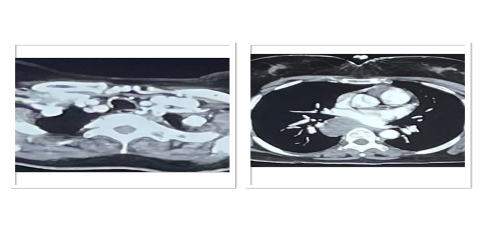
Figure 1: Contrast-enhanced computed tomography (CECT) of the chest showing calcified nodular lesions along the anterior wall of the trachea (arrows). A foregut duplication cyst is also seen in the subcarinal region extending into the middle and posterior mediastinum.
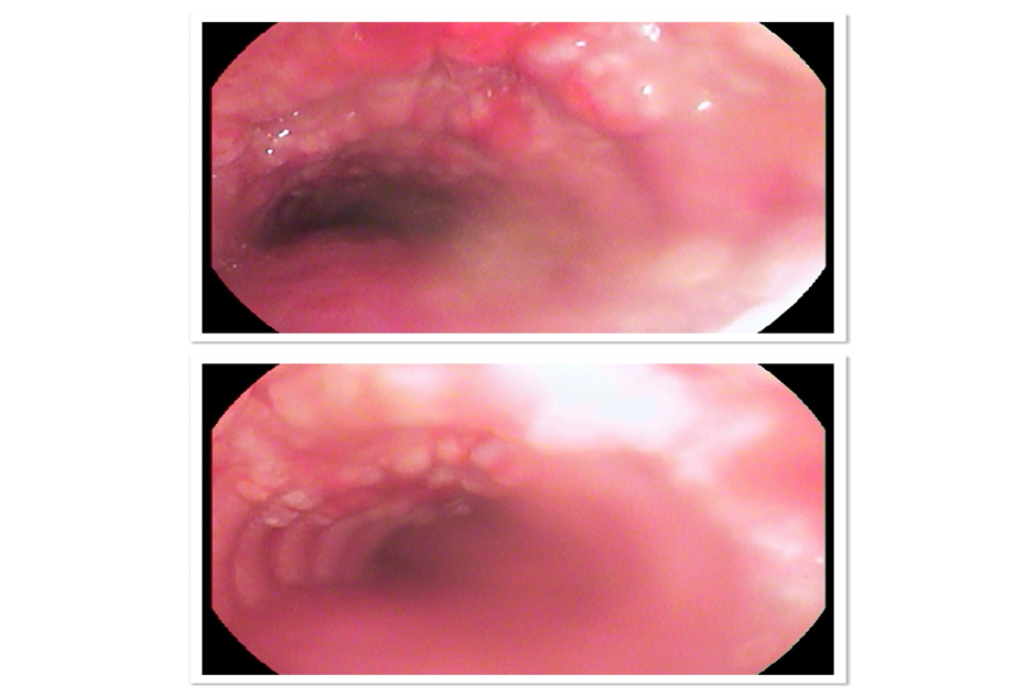
Figure 2: Flexible bronchoscopy image showing multiple nodular lesions involving the tracheal rings along the anterior wall of the trachea. Note the characteristic sparing of the posterior membranous wall, giving a "cobblestone" appearance.
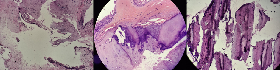
Figure 3: Histopathological examination of the endobronchial biopsy specimen showing subepithelial mature chondro-osseous material with overlying pseudo-stratified ciliated columnar epithelium (H&E stain, 40×). No evidence of dysplasia or malignancy was seen.
BIBLIOGRAPHY:
1. García CA, Sangiovanni S, Zúñiga-Restrepo V, et al. Tracheobronchopathia Osteochondroplastica-Clinical, Radiological, and Endoscopic Correlation: Case Series and Literature Review. J Investig Med High Impact Case Rep. 2020;8:2324709620921609. 10.1177/2324709620921609
2. Gaddam M, Tangutoori S, Gullapalli D, et al. A Bumpy Ride Through a Turbulent Airway: But Not Always—A Case Report of Tracheobronchopathia Osteochondroplastica. Journal of Investigative Medicine High Impact Case Reports. 2025 Jul;13:23247096251357757.. 10.1177/23247096251357757
3. Dall'Amico GF, Sharma D, Ritchie L, Sandhu G. Severe tracheobronchopathia osteochondroplastica in an asymptomatic patient. BMJ Case Rep. 2023 Apr 13;16(4):e249976. doi: 10.1136/bcr-2022-249976.
4. Prakash UBS. Tracheobronchopathia osteochondroplastica. Semin Respir Crit Care Med. 2002;23(2):167-175. https://pubmed.ncbi.nlm.nih.gov/16088609/
5. Sun J, Xie L, Su X, Zhang X. Tracheobronchopathia osteochondroplastica: case report and literature review. Respiratory Medicine Case Reports. 2015 Jan 1;15:14-7. [DOI: 10.1016/j.rmcr.2015.03.003
6. Barthwal MS, Chatterji RS, Mehta A. Tracheobronchopathia osteochondroplastica. Indian J Chest Dis Allied Sci. 2004 Jan-Mar;46(1):43-6. https://pubmed.ncbi.nlm.nih.gov/14870868/
7. Xu C, Wang X, Yin X, Wang X, Liu X, Zhou L, Du F, Jiang Y, Su D, Wang Y. Tracheobronchopathia osteochondroplastica: A rare case report. Medicine. 2026 Jan 23;105(4):e47049. 10.1097/MD.0000000000047049
8. Zack JR, Rozenshtein A. Tracheobronchopathia osteochondroplastica: report of three cases. J Comput Assist Tomogr. 2002 Jan-Feb;26(1):33-6. doi: 10.1097/00004728-200201000-00006.
9. Luo T, Zhou H, Meng J. Clinical Characteristics of Tracheobronchopathia Osteochondroplastica. Respir Care. 2019 Feb;64(2):196-200. doi: 10.4187/respcare.05867.
10. Sakaguchi Y, Matsumoto K, Nishioka K, Iizuka N, Hirayama Y, Kitaoka A, Tanimura K, Takahashi KI, Kato M. Bronchoscopic Surgery for a Solitary Tracheal Tumor of Tracheobronchopathia Osteochondroplastica. Ann Thorac Surg. 2020 Jun;109(6):e419-e421. doi: 10.1016/j.athoracsur.2019.09.014.
11. Devaraja K, Surendra VU. Clinicopathological Features and Management Principles of Tracheobronchopathia Osteochondroplastica - A Scoping Review. Indian J Otolaryngol Head Neck Surg. 2023 Dec;75(4):3798-3814. doi: 10.1007/s12070-023-03998-6.