COMPARISON OF INTRAVENOUS DEXMEDETOMIDINE AND REMIFENTANIL AS AN ADJUVANT TO ANESTHESIA AND THEIR EFFECTS ON HEMODYNAMIC PARAMETERS IN PATIENTS UNDERGOING ELECTRO CONVULSIVE THERAPY (ECT): A BIS GUIDED STUDY.
- Uday Gollamudi , Associate Professor, Dept. of Anaesthesia, Apollo Institute of Medical Sciences & Research, Hyderabad, Telangana, India
- Pravallika Rallapalli , Senior Resident, Dept. of Anaesthesia, Government Medical College, Sangareddy, Telangana, India
- Sheetal Meena , Associate Professor, Dept. of Anaesthesia, Apollo Institute of Medical Sciences & Research, Hyderabad, Telangana, India
- Neha Zeenath , Junior Resident, Dept. of Anaesthesia, Apollo Institute of Medical Sciences & Research, Hyderabad, Telangana, India
- R.S.Sachidanand , Head Of the Department, Dept. of Anaesthesia, Apollo Institute of Medical Sciences & Research, Hyderabad, Telangana, India
Article Information:
Abstract:
Electroconvulsive therapy (ECT) is widely used in the management of severe psychiatric disorders and is associated with significant hemodynamic fluctuations. Attenuation of this hyperdynamic response is essential to improve patient safety. Dexmedetomidine and remifentanil are commonly used adjuncts to anaesthesia for this purpose. Aim: To compare the effects of intravenous dexmedetomidine and remifentanil as adjuncts to anaesthesia on hemodynamic parameters, depth of anaesthesia, seizure duration, and recovery characteristics in patients undergoing ECT. Materials and Methods: This prospective, randomized, single-blinded study included 20 patients (ASA I/II) aged 18–70 years undergoing ECT. Patients were divided into two groups: Group D received dexmedetomidine (1 µg/kg IV over 10 minutes), and Group R received remifentanil (1 µg/kg IV prior to induction). Standard anaesthesia with propofol and succinylcholine was administered. Hemodynamic parameters (HR, SBP, DBP), oxygen saturation, and bispectral index (BIS) were recorded at predefined intervals. Sedation was assessed using the Ramsay Sedation Scale. Statistical analysis was performed using paired t-test and Mann–Whitney U test. Results: Dexmedetomidine showed significantly better attenuation of heart rate (p = 0.03) and systolic blood pressure (p = 0.04) compared to remifentanil. BIS values were more stable in the dexmedetomidine group (p = 0.02). Sedation scores were significantly higher in Group D during the early recovery period (p = 0.04). There was no significant difference in diastolic blood pressure, oxygen saturation, or seizure duration between the groups. Conclusion: Dexmedetomidine provides superior hemodynamic stability, better sedation, and more controlled depth of anaesthesia compared to remifentanil in patients undergoing ECT, without affecting seizure duration. It may be considered a preferable adjunct in ECT anaesthesia, particularly in patients at cardiovascular risk.
Keywords:
Article :
INTRODUCTION:
Electroconvulsive therapy (ECT) is indicated in patients with treatment-resistant depression or severe major depression that impairs activities of daily living. ECT is a psychiatric treatment that causes a generalized seizure by passing electrical current through the brain.[1] ECT is often used as an intervention for mental disorders when other treatments are inadequate. Conditions responsive to ECT include major depressive disorder, mania, and catatonia.[2] The general physical risks of ECT are similar to those of brief general anaesthesia.[3] Immediately following treatment, the most common adverse effects are confusion and transient memory loss.[2][4] Among treatments for severely depressed pregnant women, ECT is one of the least harmful to the fetus.[5] The usual course of ECT involves multiple administrations, typically given two or three times per week until the patient no longer has symptoms. ECT is administered under anaesthesia with a muscle relaxant.[6] ECT can differ in its application in three ways: electrode placement, treatment frequency, and the electrical waveform of the stimulus. Differences in these parameters affect symptom remission and adverse side effects. Placement can be bilateral, where the electric current is passed from one side of the brain to the other, or unilateral, in which the current is solely passed across one hemisphere of the brain. High-dose unilateral ECT has some cognitive advantages compared to moderate-dose bilateral ECT while showing no difference in antidepressant efficacy.[7]In patients with severe depression we can see reduced activity and volumetric reductions in the dorsal areas of the frontal lobes. Areas of the ventral and orbital frontal cortex have altered the processing of emotional stimuli.[8] In addition to the frontal lobe, functional alterations and volumetric reductions are apparent in the hippocampus, parahippocampal gyri, and amygdala.[9] The hypothalamic-pituitary-adrenal (HPA) axis becomes hypersensitive to stressors and exhibits chronically elevated levels of stress hormones and impaired feedback regulation.[10]
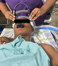
Figure: 1- Electro Convulsive Therapy
Bispectral index (BIS) is used for assessing the depth of anaesthesia remains a persistent challenge for clinical anaesthesiologists. Conventional monitoring of anaesthetic depth is primarily assessed by the patient’s clinical signs and symptoms, such as changes in heart rate, blood pressure, and limb movements.[11,12] Lacking objective data support, these methods also face challenges in continuous monitoring due to low specificity and sensitivity.[11] Such limitations may lead to inaccurate and untimely assessments, potentially resulting in either excessive or insufficient anaesthesia, which significantly impacts patients’ mental health, disease recovery, and long-term survival rates.[11] The Bispectral Index (BIS) offers an objective and precise method for monitoring the depth of anaesthesia,[13] which is a crucial component of some Enhanced Recovery After Surgery (ERAS) guidelines.[14,15] ERAS is an evidence-based approach to surgical care aimed at improving the quality of perioperative care and supporting quick recovery.[15, 16]
The BIS monitor processes EEG signals to obtain a value reflecting the patient's consciousness level. It collects this data through its sensors and uses an algorithm to analyse and interpret it. The data displays as a number on the BIS view monitor and ranges from 0 to 100. A value of 0 represents the absence of brain activity, and 100 represents an awake state. Values less than 40 represent a deep hypnotic state. BIS values between 40 and 60 represent adequate general anaesthesia for surgery and prevent awareness under anaesthesia. BIS monitoring involves the application of 4 electrodes on the forehead. The skin on the forehead is first cleaned with an alcohol swab, and then 2 to 5 seconds of digital pressure is applied over the sensor leads.[17] The sensor is comprised of disposable wet gel electrodes. EMG activity of the frontalis muscle is measured by lead 4, which is the ground electrode as well. The 2-channel monitor includes a user-configurable display. The 4-channel monitor has enhanced bihemispheric capabilities. BIS-extended sensors are available for use in ICU where patients require long-term monitoring.
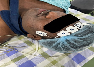
Figure:2- Placement of BIS (Bispectral Index) Electrodes on the Patient’s Forehead for Monitoring Depth of Anaesthesia
The structure of remifentanil, like alfentanil and sufentanil, is based on its parent drug fentanyl. The crucial difference is the addition of an ester group (highlighted) allowing it to be rapidly metabolized by non-specific plasma and tissue esterases. This gives rise to its characteristic ultra-fast offset and allows for rapid titration. Despite being broken down by esterases, remifentanil can be used safely in patients with pseudocholinesterase deficiency[18] . Its major metabolite, remifentanil acid, undergoes renal excretion and accumulates in patients with reduced renal function.[18,19]Despite this, the dosing of remifentanil does not need to be adjusted for renal dysfunction as remifentanil acid is almost entirely inactive. [18,19] When comparing remifentanil to other short acting opioids (fentanyl, alfentanil and sufentanil), it is associated with deeper anaesthesia and analgesia intra-operatively.[20] This manifests as a lower blood pressure and heart rate. Higher doses of remifentanil are associated with an increased risk of hypotension and bradycardia as well as apnoea.[20] It is prudent to have vasopressors and anticholinergics to hand when using remifentanil. High intra-operative doses of remifentanil have been associated with post-operative hyperalgesia,[21] although this may be due in-part to inadequate provision of post op-analgesia. Remifentanil is an excellent choice for TIVA; it is safe in malignant hyperthermia and reduces the need for higher doses of propofol due to a synergistic interaction between the two drugs.[22]
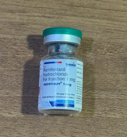
Figure:3- Remifentanil vial
Dexmedetomidine, a potent sedative, alleviates anxiety and stress by suppressing central nervous system activity and modulating neurotransmitters.[23] During the preoperative anaesthesia induction phase, dexmedetomidine can facilitate a smooth and rapid transition to anaesthesia in conjunction with other anaesthetic agents, minimizing patient discomfort.[24]Prior studies demonstrate that dexmedetomidine pretreatment effectively attenuates acute hyperdynamic responses during MECT.[25,26] However, dose-dependent prolongation of recovery times has been reported, which may compromise therapeutic efficacy and elevate adverse event risks.[27,28] Drugs such as alpha-2 agonists and beta-blockers have been used to attenuate this response.[29,30,31] Alpha-2 agonistic activity results in augmentation of cardiovagal activity in brain leading to bradycardia and hypotension. Clonidine is the prototypical alpha-2 agonist that has been reported to have a beneficial effect on the hyperdynamic response to ECT.[32] Dexmedetomidine, the newer alpha-2 agonist, is being studied for many procedural sedations.[33,34,35,36,37,38] Dexmedetomidine decreases the stress-induced sympathoadrenal responses to painful stimuli, improves intraprocedural hemodynamics, and reduces the anaesthetic requirements.
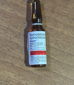
Figure: 4 – Dexmedetomidine Ampoule
MATERIAL AND METHODS::
A total of 20 patients undergoing ECT were randomly selected into two groups using computer generated randomization list and included in the study. The inclusion criteria were patients scheduled to undergo ECT, with ASA physical status I or II, of either sex and aged between 18 to 70 years. The exclusion criteria were patients unwilling to provide written informed consent. Known allergy or contraindication to Dexmedetomidine or Remifentanil, Severe cardiac disease (e.g., recent MI, heart block, unstable angina), Pheochromocytoma or severe hypertension, Pregnancy or lactation, Neuromuscular disorders contraindicating succinylcholine use, Raised intracranial pressure with mass effect and patients younger than 18 or older than 70 years.
This study was a prospective, randomized, comparative single-blinded observation study. Patients in group D (Dexmedetomidine) received Dexmedetomidine 1 µg/kg diluted in 10 mL saline, administered IV over 10 minutes prior to induction, those in group R (Remifentanil) received Remifentanil 1 µg/kg diluted in 10 mL saline, administered IV 1 minute prior to induction.
Pre anaesthetic checkup was done for all patients a day before procedure and patients will be kept nil per oral (NPO) for at least 6 hours prior to ECT. Routine laboratory investigations like haemoglobin concentration, platelet count, leucocyte count, blood sugar, electrocardiogram, urea, creatine, bleeding time, clotting time, blood grouping, Once shifted to the operation theatre an intravenous line (20G) will be secured, all non-invasive Standard monitoring like ECG, pulse, blood pressure, saturation, respiratory rate, temperature and BIS were attached.
Pre anaesthetic procedure was standardized in both the groups, both groups received glycopyrrolate 0.2 mg IV and ondansetron 0.1 mg/kg was given as premedication. Group D patients received dexmedetomidine at 1 µg/kg and group R patients received remifentanil at 1 µg/kg. Preoxygenation with 100% O₂ for 3 minutes will be performed.
Anaesthesia Protocol:
● Induction: propofol 1-2 mg/kg IV will be administered slowly until loss of eyelid reflex.
● Muscle Relaxation: Succinylcholine 0.5–1 mg/kg IV will be given to facilitate seizure and prevent injury.
● Bite block will be inserted to prevent tongue injury.
ECT Procedure:
● Electrical stimulus (60–90 Hz, pulse width 1 ms, duration 1–3 seconds) will be delivered via bitemporal electrodes.
● EEG seizure duration will be recorded using the ECT machine monitor.
● After seizure termination, ventilation will be assisted with 100% oxygen until spontaneous breathing resumes.
● HR, SBP, DBP, SpO₂, and BIS will be recorded at:
1. Baseline (before study drug)
2. After study drug administration
3. After induction
4. Immediately post-ECT stimulus
5. 1, 2, 3,5,10 and 20 minutes post-stimulus
6. Sedation score after every 10 mins for 1 hour in recovery room.
● Recovery times will be noted (time to eye-opening, following verbal command, and orientation).
Data Collection:
● Primary outcome: Changes in HR and BP during the peri-ECT period.
● Secondary outcomes: BIS trends, seizure durations (EEG), recovery time, and incidence of side effects
● Ramsay sedation score was used for monitoring sedation levels in the patients post procedure.
● Data will be entered into structured case record forms and later analysed statistically.
RAMSAY EDATION SCALE
|
Score |
Level of Sedation |
|
1 |
Patient is anxious and agitated or restless |
|
2 |
Patient is cooperative, oriented and tranquil |
|
3 |
Patient responds to commands only |
|
4 |
Patient exhibits brisk response to light tactile or loud auditory stimulus |
|
5 |
Patient exhibits sluggish response to light tactile stimuli or loud auditory stimulus |
|
6 |
Patient exhibits no response |
Exclusion Criteria:
● Known allergy or contraindication to Dexmedetomidine or Remifentanil
● Severe cardiac disease (e.g., recent MI, heart block, unstable angina)
● Pheochromocytoma or severe hypertension
● Pregnancy or lactation
● Neuromuscular disorders contraindicating succinylcholine use
● Raised intracranial pressure with mass effect
● History of allergy to opioids
SAMPLE SIZE / STATISTICAL ANALYSIS :
(Zα/2+Z1−β )2 2σ2
d2
● Sample size estimation was performed using the formula for comparing two independent means. For calculation, the standard deviation (σ) was assumed to be 5.0, and the minimum clinically significant difference (Δ) between the two groups was taken as 7.0. The level of significance (α) was set at 0.05 (two-tailed), with a desired statistical power (1–β) of 80%.
● Based on these assumptions, the required sample size was determined to be 9 participants per group. To account for an anticipated 10% drop-out rate, the final sample size was adjusted to 10 participants per group.
Sample size: 20 (10/group)
RESULTS::
A total of 20 patients were randomly divided into 2 groups. Males were 14 and females were 6. The demographic profile was comparable between the 2 groups and found no statistically significant difference (P > 0.05) as shown in table 1.
Table 1: Demographic profile of the patients
|
Characterteristics |
Group D |
Group R |
P-Value |
|
Age in years |
30.2 + 5 |
30.4 + 4 |
P > 0.05 |
|
Sex (M/F) |
7/3 |
7/3 |
P > 0.05 |
|
ASA Grade I/II |
8/2 |
9/1 |
P > 0.05 |
|
Weight |
50.4 + 10 |
52.2 + 8 |
P > 0.05 |
|
Heart rate |
80.6 + 8 |
80.1 + 8.4 |
P > 0.05 |
|
SBP |
125.3 + 5 |
124.9 + 6 |
P > 0.05 |
|
DBP |
81.2 + 7.1 |
80.8 + 8.2 |
P > 0.05 |
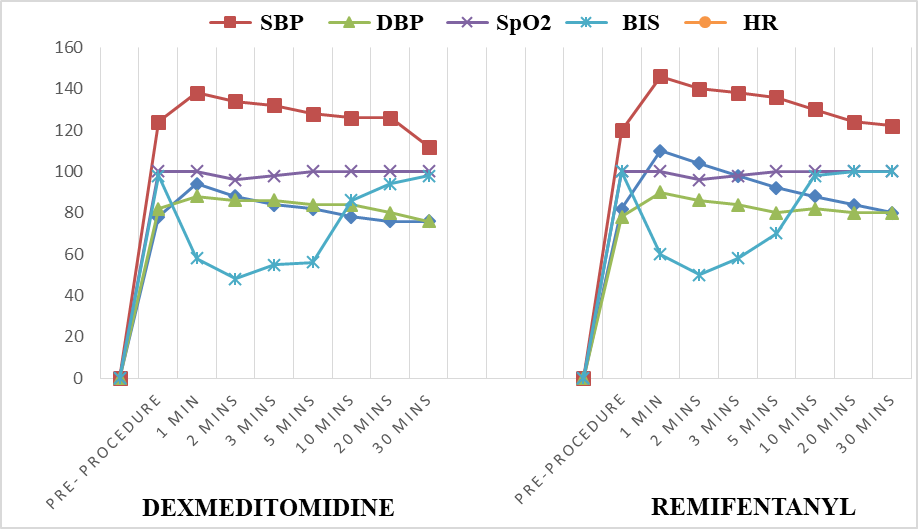
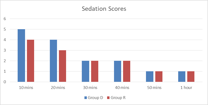
The mean demographic data of vitals in both the groups is mentioned in table 2 and table 3. The graphical data of vitals in both the groups is shown in graph 1. The mean sedation score is mentioned in table 4. The graphical representation of sedation score is mentioned in graph 2. The sedation score is calculated for every 10 mins post shifting the patient to the recovery room for a period of 1 hour.
Table 2: Mean demographic data of patients in group D
|
Time |
HR |
SBP |
DBP |
SpO2 |
BIS |
|
Pre- procedure |
78 |
124 |
82 |
100 |
98 |
|
1 min |
94 |
138 |
88 |
100 |
58 |
|
2 mins |
88 |
134 |
86 |
96 |
48 |
|
3 mins |
84 |
132 |
86 |
98 |
55 |
|
5 mins |
82 |
128 |
84 |
100 |
56 |
|
10 mins |
78 |
126 |
84 |
100 |
86 |
|
20 mins |
76 |
126 |
80 |
100 |
94 |
|
30 mins |
76 |
112 |
76 |
100 |
98 |
Table 3: Mean demographic data of patients in group R
|
Time |
HR |
SBP |
DBP |
SpO2 |
BIS |
|
Pre-procedure |
82 |
120 |
78 |
100 |
100 |
|
1 min |
110 |
146 |
90 |
100 |
60 |
|
2 mins |
104 |
140 |
86 |
96 |
50 |
|
3 mins |
98 |
138 |
84 |
98 |
58 |
|
5 mins |
92 |
136 |
80 |
100 |
70 |
|
10 mins |
88 |
130 |
82 |
100 |
98 |
|
20 mins |
84 |
124 |
80 |
100 |
100 |
|
30 mins |
80 |
122 |
80 |
100 |
100 |
From table 2 and table 3 which are group D ( dexmedetomidine) and group R (remifentanil) respectively, the data was compared between both the groups at various time intervals using the formula paired t- test and the results can be seen in table 4.
d = mean of the differences between paired observations
Sd = standard deviation of the differences
n = number of pairs
t = t-statistic
Table 4: Comparison of P-Value between both the groups
|
PARAMETERS |
P-Value |
Significance |
|
HR |
0.03 |
Significant |
|
SBP |
0.04 |
Significant |
|
DBP |
0.09 |
Not significant |
|
SpO2 |
0.60 |
Not significant |
|
BIS |
0.02 |
Significant |
From the results (table 4) of comparison of both the groups using paired t-test we can clearly see that there is significant difference between both the groups in 3 parameters.
Table 5: Mean Sedation score in both the groups
From table 5 the Ramsey’s sedation score is calculated between groups that is group D (dexmedetomidine) and group R (remifentanil) using Mann Whitney U test. The formula is
n1 = sample size of group D
n2 = sample size of group R
R1 = sum of ranks of group 1
The result of this test between the groups is 0.04, which shows that there is significant difference between the groups. The sedation is more in group D than in group R for first 20 mins and this can be even seen by the difference between the groups.
Graph 1: Graphical comparison between the two groups.
Graph 2: Graphical comparison of sedation scores between the groups
There was no significant change in seizure duration in both the groups. The average seizure duration in remifentanil group (R) was 30.33 + 6.18 and in dexmedetomidine group (D) was 29.55 + 6.22. The comparative p value was < 0.05 between both groups after using paired t test which means that there is no significant change.
DISCUSSION :
Electroconvulsive therapy (ECT) is well known to produce a biphasic autonomic response characterized by an initial parasympathetic surge followed by a pronounced sympathetic discharge, resulting in transient tachycardia and hypertension. Attenuation of this hyperdynamic response remains a key anaesthetic goal, particularly in patients with cardiovascular vulnerability. In the present study, both dexmedetomidine and remifentanil were evaluated as adjuncts to propofol-based anaesthesia for their effects on hemodynamic parameters, depth of anaesthesia (BIS), seizure duration, and recovery characteristics.
The findings of this study demonstrate that dexmedetomidine provided superior attenuation of heart rate and systolic blood pressure responses compared to remifentanil, with statistically significant differences observed in HR (p = 0.03) and SBP (p = 0.04). This is consistent with the pharmacological profile of dexmedetomidine as a selective α2-adrenergic agonist, which reduces sympathetic outflow and norepinephrine release, thereby stabilizing cardiovascular responses. Similar observations were reported in a study by Li et al., where dexmedetomidine significantly blunted the hyperdynamic response to ECT without compromising seizure quality.[39]Furthermore, a randomized controlled trial comparing remifentanil, dexmedetomidine, and metoprolol also concluded that dexmedetomidine was more effective in maintaining hemodynamic stability during ECT, particularly in controlling post-stimulus tachycardia and hypertension.[40]
Remifentanil, owing to its ultra-short-acting opioid profile, also attenuated hemodynamic responses but to a lesser extent compared to dexmedetomidine. The transient rise in HR and BP observed immediately after ECT stimulus in the remifentanil group aligns with findings from previous studies, where remifentanil reduced but did not completely abolish the sympathetic surge. A double-blinded study comparing dexmedetomidine, remifentanil, and labetalol reported that while remifentanil was effective in moderating hemodynamic changes, dexmedetomidine provided more consistent control across all time intervals.[41] This suggests that although remifentanil is beneficial, its short duration of action may limit sustained sympatholytic effects during the peri-ECT period.
In terms of depth of anaesthesia, BIS values showed a statistically significant difference between the two groups (p = 0.02), with dexmedetomidine demonstrating a smoother and more controlled decline and recovery pattern. This reflects its sedative and hypnotic properties, which complement propofol anaesthesia. The ability of dexmedetomidine to reduce anaesthetic requirements and maintain stable BIS levels has been supported in earlier studies, indicating improved intraoperative control and reduced anaesthetic fluctuations.[39]
Sedation scores assessed using the Ramsay Sedation Scale revealed significantly higher sedation levels in the dexmedetomidine group during the initial 20 minutes post-procedure (p = 0.04). This finding is in agreement with previous literature demonstrating that dexmedetomidine produces a dose-dependent sedation resembling natural sleep, which may prolong early recovery but enhances patient comfort. Similar results were observed in studies where dexmedetomidine premedication led to deeper sedation without respiratory compromise.[41]However, this prolonged sedation should be interpreted cautiously, as excessive sedation may delay recovery room discharge in certain clinical settings.
Importantly, seizure duration—a critical determinant of ECT efficacy—was not significantly different between the two groups (p > 0.05). This indicates that neither dexmedetomidine nor remifentanil adversely affected seizure quality. Preservation of adequate seizure duration while achieving hemodynamic stability is essential, and our findings are consistent with prior studies demonstrating that dexmedetomidine does not significantly shorten seizure duration at clinically used doses.[39,40] This supports its safety profile as an adjunct in ECT anaesthesia.
The absence of significant differences in oxygen saturation between groups suggests that both drugs are safe from a respiratory standpoint when used in controlled settings. This is particularly relevant given the known respiratory depressant effects of opioids like remifentanil, although its rapid metabolism minimizes prolonged effects.
Overall, the results of this study suggest that dexmedetomidine is more effective than remifentanil in attenuating the hemodynamic response to ECT, providing better sedation and more stable BIS profiles without compromising seizure duration. These findings are in concordance with existing literature and reinforce the role of dexmedetomidine as a valuable adjunct in ECT anaesthesia. However, the relatively small sample size and single-centre design of this study may limit the generalizability of the findings. Larger, multicentric trials are warranted to further validate these results and determine optimal dosing strategies.
In conclusion, dexmedetomidine appears to offer superior hemodynamic stability and sedation compared to remifentanil, making it a preferable adjunct in patients undergoing ECT, particularly those at risk of cardiovascular complications.
CONCLUSION :
Both dexmedetomidine and remifentanil are effective adjuncts to propofol anaesthesia in patients undergoing electroconvulsive therapy. However, dexmedetomidine demonstrated superior control of hemodynamic responses, with better attenuation of heart rate and blood pressure fluctuations. It also provided more consistent sedation and stable BIS values, without compromising seizure duration, which is essential for the therapeutic efficacy of ECT.
Although remifentanil was effective in reducing the sympathetic response, its effects were comparatively less sustained. Overall, dexmedetomidine appears to be a more reliable and advantageous agent for achieving hemodynamic stability and adequate sedation during ECT, particularly in patients with increased cardiovascular risk. Further studies with larger sample sizes are recommended to validate these findings.
LIMITATIONS OF THE STUDY:
The study’s sample size was limited, and follow-up duration was short, which restricts the ability to generalise the findings to a larger population. Additionally the study relied on subjective score values reported by the patients, making it susceptible to participant bias that could not be fully controlled.
CONFLICT OF INTEREST:
There are no conflict of interests.
FUNDING SOURCE:
The patients are managed according to the protocol laid down for the management of the patient. There are no financial implications. There was no financial support or sponsorship.
ETHICAL AND LEGAL CONSIDERATIONS:
A patient information and consent was given to the patients in their local language, all there queries were satisfactorily answered and when they were willing to participate, signature of the patient or her husband/guardian was obtained and only after that the study was initiated. Institutional ethical approval was also obtained before starting the study.
BIBLIOGRAPHY:
1. Rudorfer MV, Henry ME, Sackeim HA (2003). "Electroconvulsive therapy" (PDF). In Tasman A, Kay J, Lieberman JA (eds.). Psychiatry (Second ed.). Chichester: John Wiley & Sons Ltd. pp. 1865–1901. Archived (PDF) from the original on 2007-08-10.
2. FDA. FDA Executive Summary. Prepared for the January 27–28, 2011 meeting of the Neurological Devices Panel Meeting to Discuss the Classification of Electroconvulsive Therapy Devices (ECT). Quote, p. 38: "Three major practice guidelines have been published on ECT. These guidelines include: APA Task Force on ECT (2001); Third report of the Royal College of Psychiatrists' Special Committee on ECT (2004); National Institute for Health and Clinical Excellence (NICE 2003; NICE 2009). There is significant agreement between the three sets of recommendations."
3. "- Reports of the Surgeon General - Profiles in Science Search Results". profiles.nlm.nih.gov. Retrieved 2025-04-10.
4. American Psychiatric Association, Committee on Electroconvulsive Therapy, Richard D. Weiner (chairperson), et al. (2001). The practice of electroconvulsive therapy: recommendations for treatment, training, and privileging (2nd ed.). Washington, DC: American Psychiatric Publishing. ISBN 978-0-89042-206-9.
5. Pompili M, Dominici G, Giordano G, Longo L, Serafini G, Lester D, et al. (December 2014). "Electroconvulsive treatment during pregnancy: a systematic review". Expert Review of Neurotherapeutics. 14 (12): 1377–1390. doi:10.1586/14737175.2014.972373. PMID 25346216. S2CID 31209001.
6. "5 Outdated Beliefs About ECT". Psych Central.com. Archived from the original on 2015-04-25. Retrieved 2025-04-10.
7. Kolshus E, Jelovac A, McLoughlin DM (February 2017). "Bitemporal v. high-dose right unilateral electroconvulsive therapy for depression: a systematic review and meta-analysis of randomized controlled trials" (PDF). Psychological Medicine. 47 (3): 518–530. doi:10.1017/S0033291716002737. PMID 27780482. S2CID 10711085. Archived (PDF) from the original on 2021-06-16.
8. Mayberg HS, Lewis PJ, Regenold W, Wagner HN. Paralimbic hypoperfusion in unipolar depression. J Nucl Med. 1994 Jun;35(6):929-34. [PubMed]
9. Botteron KN, Raichle ME, Drevets WC, Heath AC, Todd RD. Volumetric reduction in left subgenual prefrontal cortex in early onset depression. Biol Psychiatry. 2002 Feb 15;51(4):342-4. [PubMed]
10. de Kwaasteniet B, Ruhe E, Caan M, Rive M, Olabarriaga S, Groefsema M, Heesink L, van Wingen G, Denys D. Relation between structural and functional connectivity in major depressive disorder. Biol Psychiatry. 2013 Jul 01;74(1):40-7. [PubMed]
11. Dutta H., Suchismita M., Baisakhi L., Sarbari S., Uday S. M., and Sarmila G., Monitoring of general anesthesia by qCON and qNOX indices versus conventional clinical parameters in urological surgery: a randomized controlled clinical trial, Asian Journal of Medical Sciences. (2024) 15, no. 3, 20–25, https://doi.org/10.3126/ajms.v15i3.59955.
12. Bajaj P., Depth of anaesthesia: clinical applications, Indian Journal of Anaesthesia. (2007) 51.
13. Mathur S., Patel J., Goldstein S., Hendrix J. M., and Jain A., Bispectral Index, 2023, StatPearls Publishing, St. Petersburg, FL, USA.
14. BISTM for Enhanced Recovery after Surgery Medtronic, 2021, https://www.medtronic.com/covidien/en-us/clinical-solutions/bis-enhanced-recovery-after-surgery.html.
15. Feldheiser A., Aziz O., Baldini G., Cox B. P. B. W., Fearon K. C. H., Feldman L. S., Gan T. J., Kennedy R. H., Ljungqvist O., Lobo D. N., Miller T., Radtke F. F., Ruiz Garces T., Schricker T., Scott M. J., Thacker J. K., Ytrebø L. M., and Carli F., Enhanced Recovery after Surgery (ERAS) for gastrointestinal surgery, part 2: consensus statement for anaesthesia practice, Acta Anaesthesiologica Scandinavica. (2016) 60, no. 3, 289–334, https://doi.org/10.1111/aas.12651, 2-s2.0-84956819211.
16. Altman A. D., Helpman L., McGee J., Samouëlian V., Auclair M. H., Brar H., and Nelson G. S., Enhanced recovery after surgery: implementing a new standard of surgical care, Canadian Medical Association Journal. (2019) 191, no. 17, E469–E475, https://doi.org/10.1503/cmaj.180635, 2-s2.0-85065468610.
17. Johansen JW. Update on bispectral index monitoring. Best Pract Res Clin Anaesthesiol. 2006 Mar;20(1):81-99. [PubMed]
18. Egan TD. Pharmacokinetics and pharmacodynamics of remifentanil: an update in the year 2000. Current Opinion in Anaesthesiology. 2000, 13:449-455.
19. Breen D, Wilmer A, Bodenham A, Bach V, Bonde J, Kessler P, Albrecht S, Shaikh S. Offset of pharmacodynamic effects and safety of remifentanil in intensive care unit patients with various degrees of renal impairment. Critical Care. 2004, 8:R21-R30.
20. Komatsu R, Turan AM, Orhan-Sungur M, McGuire J, Radke OC, Apfel CC. Remifentanil for general anaesthesia: a systematic review. Anaesthesia 2007; 62: 1266-1280.
21. Fletcher D, Martinez V. Opioid-induced hyperalgesia in patients after surgery: a systematic review and a metaanalysis. British Journal of Anaesthesia 2014; 112(6): 991 – 1004.
22. Mertens MJ, Olofsen E, Engbers FH, Burm AG, Bovill JG, Vuyk J. Propofol reduces perioperative remifentanil requirements in a synergistic manner: response surface modeling of perioperative remifentanil-propofol interactions. Anesthesiology. 2003; 99(2): 347-59.
23. Moller MH, et al. Use of dexmedetomidine for sedation in mechanically ventilated adult ICU patients: a rapid practice guideline. Intensive Care Med. 2022;48:801–10. 10.1007/s00134-022-06660-x. [DOI] [PubMed] [Google Scholar]
24. Subsoontorn P, et al. Premedication with dexmedetomidine for prevention of hyperdynamic response after electroconvulsive therapy: a cross-over, randomized controlled trial. BMC Psychiatry. 2021;21:408. 10.1186/s12888-021-03406-9. [DOI] [PMC free article] [PubMed] [Google Scholar]
25. Sannakki D, et al. Effectiveness of dexmedetomidine as premedication prior to electroconvulsive therapy, a randomized controlled cross over study. Indian J Psychiatry. 2017;59:370–4. 10.4103/psychiatry.IndianJPsychiatry_33_17. [DOI] [PMC free article] [PubMed] [Google Scholar]
26. Li X, et al. Dexmedetomidine combined with intravenous anesthetics in electroconvulsive therapy: a meta-analysis and systematic review. J ECT. 2017;33:152–9. 10.1097/YCT.0000000000000398. [DOI] [PubMed] [Google Scholar]
27. Zhang K, Gao Y, Zhu G. Effect of dexmedetomidine on stress response and myalgia in the treatment of modified electroconvulsive therapy. J Clin Anesthesiol. 2018;34:140–3. [Google Scholar]
28. Li X, Zhang L, Li T. Effect of ultra-low dose dexmedetomidine on cough during anesthesia recovery period in elderly patients. J Clin Anesthesiol. 2024;02:119–23. [Google Scholar]
29. Wells DG, Davies GG, Rosewarne F. Attenuation of electroconvulsive therapy induced hypertension with sublingual nifedipine. Anaesth Intensive Care. 1989;17:31–3. doi: 10.1177/0310057X8901700107. [DOI] [PubMed] [Google Scholar]
30. Figiel GS, DeLeo B, Zorumski CF, Baker K, Goewert A, Jarvis M, et al. Combined use of labetalol and nifedipine in controlling the cardiovascular response from ECT. J Geriatr Psychiatry Neurol. 1993;6:20–4. doi: 10.1177/002383099300600103. [DOI] [PubMed] [Google Scholar]
31. Castelli I, Steiner LA, Kaufmann MA, Alfillé PH, Schouten R, Welch CA, et al. Comparative effects of esmolol and labetalol to attenuate hyperdynamic states after electroconvulsive therapy. Anesth Analg. 1995;80:557–61. doi: 10.1097/00000539-199503000-00022. [DOI] [PubMed] [Google Scholar]
32. Fu W, Stool LA, White PF, Husain MM. Is oral clonidine effective in modifying the acute hemodynamic response during electroconvulsive therapy? Anesth Analg. 1998;86:1127–30. doi: 10.1097/00000539-199805000-00042. [DOI] [PubMed] [Google Scholar]
33. Venn RM, Grounds RM. Comparison between dexmedetomidine and propofol for sedation in the Intensive Care Unit: Patient and clinician perceptions. Br J Anaesth. 2001;87:684–90. doi: 10.1093/bja/87.5.684. [DOI] [PubMed] [Google Scholar]
34. Dere K, Sucullu I, Budak ET, Yeyen S, Filiz AI, Ozkan S, et al. A comparison of dexmedetomidine versus midazolam for sedation, pain and hemodynamic control, during colonoscopy under conscious sedation. Eur J Anaesthesiol. 2010;27:648–52. doi: 10.1097/EJA.0b013e3283347bfe. [DOI] [PubMed] [Google Scholar]
35. Gupta K, Jain M, Gupta PK, Rastogi B, Saxena SK, Manngo A. Dexmedetomidine premedication for fiberoptic intubation in patients of temporomandibular joint ankylosis: A randomized clinical trial. Saudi J Anaesth. 2012;6:219–23. doi: 10.4103/1658-354X.101211. [DOI] [PMC free article] [PubMed] [Google Scholar]
36. Cooper L, Candiotti K, Gallagher C, Grenier E, Arheart KL, Barron ME. A randomized, controlled trial on dexmedetomidine for providing adequate sedation and hemodynamic control for awake, diagnostic transesophageal echocardiography. J Cardiothorac Vasc Anesth. 2011;25:233–7. doi: 10.1053/j.jvca.2010.06.006. [DOI] [PubMed] [Google Scholar]
37. Koroglu A, Demirbilek S, Teksan H, Sagir O, But AK, Ersoy MO. Sedative, haemodynamic and respiratory effects of dexmedetomidine in children undergoing magnetic resonance imaging examination: Preliminary results. Br J Anaesth. 2005;94:821–4. doi: 10.1093/bja/aei119. [DOI] [PubMed] [Google Scholar]
38. Aantaa RE, Kanto JH, Scheinin M, Kallio AM, Scheinin H. Dexmedetomidine premedication for minor gynecologic surgery. Anesth Analg. 1990;70:407–13. doi: 10.1213/00000539-199004000-00011. [DOI] [PubMed] [Google Scholar]
39. Effect of adding dexmedetomidine or remifentanil to thiopental in patients with mood disorder candidate for electroconvulsive therapy.
40. Tajabadi, Nastaran & Kamali, Alireza & Alaghmand, Anita & Jamilian, Hamidreza & Pazooki, Shirin & Tajerian, Amin. (2023). The Effects of Remifentanil, Dexmedetomidine, and Metoral as Adjuncts to Thiopental on Hemodynamic Status After Electroconvulsive Therapy in Patients with Major Depressive Disorder: A Randomized Controlled Clinical Trial. Anesthesiology and Pain Medicine. 13. 10.5812/aapm-139383.
41. Modir, Hesameddin & Bahramsari, Sina & Moshiri, Esmail & Jamilian, Hamidreza & Mohammadbeigi, Abolfazl. (2020). Comparing the premedication effects of dexmedetomidine, remifentanil and labetalol before electroconvulsive therapy on haemodynamic responses and seizure duration in psychotic patients: A double-blinded clinical trial. Advances in Human Biology. 10. 65. 10.4103/AIHB.AIHB_121_19.