In vitro and in vivo evaluation of antidiabetic potential of Flavopiridol in HFD-Streptozotocin-induced diabetes in rats
- Shallu Sharma , School of Pharmacy and Research Centre, Sanskriti University, Chhata, Mathura, 281401, India
- Sarda Ruhil , Amity Institute of Pharmacy, Amity University Uttar Pradesh, Noida, UP- 201303, India
- Vishal M Balaramnavar , Amity Institute of Pharmacy, Amity University Uttar Pradesh, Noida, UP- 201303, India
Article Information:
Abstract:
Type 2 diabetes mellitus (T2DM) is a chronic and progressive metabolic disorder characterized by hyperglycemia, insulin resistance, oxidative stress, and impaired functionality of pancreatic β-cell. The aim of the present study was to evaluate the antidiabetic properties of flavopiridol on in vitro and in vivo experimental models. Flavopiridol inhibition against α-amylase and α-glucosidase enzymes were evaluated to check its postprandial glucose inhibitory activity. In vivo antidiabetic activity was carried out in high-fat diet (HFD) and streptozotocin (STZ)-induced diabetic Wistar rats treated orally with flavopiridol (1.25 and 5 mg/kg) along with standard drugs like metformin and pioglitazone. Fasting blood glucose level, body weight, serum insulin, antioxidant parameters (GSH, GPx, CAT, SOD), hepatic markers (AST, ALT) and histopathology of pancreatic tissues were examined. Flavopiridol caused dose-dependent inhibition of both α-amylase and α-glucosidase enzymes, revealing significant in vitro antidiabetic potential. In diabetic animals, administration of flavopiridol dose-dependently ameliorated fasting blood glucose, gained body weight, improved serum insulin, increased activities of antioxidant enzymes, suppressed lipid peroxidation, attenuated elevated liver enzymes and histopathology of pancreas and liver revealed the recovery in regeneration of islets, conservation of architecture of β-cells and restoration of normal architecture in both pancreas and liver tissues, especially at the higher dose. Flavopiridol demonstrated considerable antidiabetic and antioxidant activities, which indicate its potential as a promising multifunctional therapeutic candidate for type 2 diabetes mellitus.
Keywords:
Article :
INTRODUCTION:
Diabetes mellitus (DM) is a group of metabolic diseases in which the person has elevated blood sugar levels, as a result of a defect in insulin secretion, in insulin action, or in both. As a major public health problem, the incidence of diabetes mellitus is rising rapidly in the world due to its high prevalence of diabetes mellitus-associated complications, such as microvascular and macrovascular diabetes complications. Out of various types of DM, type 2 DM (T2DM) is estimated to comprise 90-95% of all diagnosed cases of diabetes and is associated with defects in insulin resistance, progressive pancreatic β-cell dysfunction, obesity, sedentariness, and genetic susceptibility (1,2). Current statistics suggest that an estimated 589 million adults aged 20-79 years will have diabetes worldwide in 2025, and this figure could be projected to go over 850 million by 2050 in the absence of proper preventative measures (3). Increasing prevalence and global health impact of T2DM pose considerable social, health and economic burden, especially in developing countries like India, which has experienced rapid urbanisation leading to increased incidence of diabetes mellitus. Pathophysiology of T2DM is very multifactorial, involving altered insulin sensitivity, deficient insulin secretion, and also chronic low-grade inflammation, oxidative stress, mitochondrial dysfunction, disrupted lipid metabolism, and disordered glucose homeostasis. During the initial stages of the disease, target tissues such as the skeletal muscle, liver, and adipose tissues develop insulin resistance, resulting in diminished peripheral glucose uptake and increased hepatic glucose production. The pancreatic β-cells try to compensate for the resistance by hypersecretion of insulin, but due to persistent metabolic stress and stress-induced exhaustion of the β-cells, these eventually undergo dysfunction, apoptosis and are no longer able to meet the glucose requirements and a state of hyperglycemia prevails (4,5). Increased chronic hyperglycemia further promotes an increase in oxidative stress and inflammatory pathways in a cyclic fashion, escalating the disease process which in turn results in diabetic complications of various organs such as cardiovascular system, kidney, eye, peripheral nerve and liver (6).
The active chemical moiety, Flavopiridol (Alvocidib) is a semisynthetic flavonoid isolated from the natural alkaloid rohitukine that was originally sourced from Dysoxylum binectariferum. The first attempt in the development of Flavopiridol was a highly potent inhibitor of CDK for the treatment of cancer since it stops the cell-cycle progress and causes the cells to undergo apoptosis in the cases of highly dividing cells (7). Various in vivo and in vitro pharmacological studies reported several biological actions of flavopiridol apart from anticancer activities, such as anti-inflammatory, antioxidant, immunomodulatory, and anti-fibrotic effects (8). In addition to the regulation of cell-cycle progress, there is emerging evidence that CDKs are also involved in glucose metabolism, insulin signalling, inflammatory response, mitochondrial functions and lipid metabolism. Therefore, inhibition of CDKs has become a strategy for treating metabolic diseases, such as T2DM (9). Since the chronic inflammatory pathway is responsible for much insulin resistance and dysfunctional β-cells, NF-κB, TNF-α, IL-6, and oxidative stress play a significant role (10).
Flavopiridol was found to reduce the expression of inflammatory cytokines, suppress NF-κB activation, diminish oxidative stress and adjust multiple transcriptional pathways regulating the metabolic processes of cells (11). Additionally, in many animal models of chronic metabolic disorder, CDK inhibitors are demonstrated to enhance insulin sensitivity, diminish the inflammatory responses and inhibit tissue damage (12). Thus, it is plausible to explain the anti-diabetic effect of flavopiridol with some mechanisms other than controlling glucose levels. The literature clearly indicates that the antidiabetic potential of flavopiridol has been largely overlooked and most studies reported its anticancer property. More importantly, it has never been investigated systematically for its effects on carbohydrate-metabolising enzymes, oxidative stress markers, insulin secretion, pancreatic histological alterations, and liver damage in a T2DM animal model. To add to that, the lack of any comparative study between flavopiridol and some other potent antidiabetic agents, such as metformin and pioglitazone in a relevant T2DM setting is also noticeable.
Materials and Methods:
2.1 In Vitro Antidiabetic Assays
Type 2 diabetes mellitus (T2DM) is a complex chronic metabolic disease that is characterized by persistently elevated levels of glucose in the blood due to defective insulin secretion, insulin resistance and impaired carbohydrate metabolism. One of the therapeutic approaches to manage postprandial hyperglycemia is by retarding the digestion and absorption of dietary carbohydrates by inhibiting important digestive enzymes. Both α-amylase and α-glucosidase are two important enzymes, responsible for stepwise degradation of carbohydrates into absorbable monosaccharides.
The inhibition of α-amylase and α-glucosidase therefore serves as an established approach to control postprandial blood glucose levels and to improve glycemic status. Flavopiridol, a synthetic flavonoid-derived cyclin-dependent kinase (CDK) inhibitor, displays diverse pharmacological properties such as anti-inflammatory, antioxidant and metabolic modulating activities. Recent evidence indicates that alterations of intracellular oxidative stress and cell signaling pathway are linked with its therapeutic application for the metabolic disorders like T2DM. In addition to these intracellular effects, flavopiridol is known to impact carbohydrate metabolism by inhibition of enzymes responsible for glucose production during digestion. Consequently, in this present study, we investigated the in vitro antidiabetic activity of flavopiridol by its inhibition of α-amylase and α-glucosidase enzymes with acarbose as a positive control. Inhibition was determined at various concentrations and respective IC50 values were calculated to determine enzyme inhibition efficiency of flavopiridol compared to reference standard. These enzyme assays suggest potential for flavopiridol as effective carbohydrate digestion inhibiting agent and supporting potential multifunctional treatment modality for type 2 diabetes mellitus.
α-Amylase Inhibition Assay
Determination of α-amylase inhibitory activity of flavopiridol A modified starch-iodine colorimetric method was applied to assay α-amylase inhibitory activity of flavopiridol. Briefly, 0.5 mL flavopiridol solution at different concentrations (10, 25, 50, 100, 200, 400, and 800 g/mL) was prepared in DMSO. Then, the flavopiridol solution was incubated with 0.5 mL porcine pancreatic α-amylase solution (1 U/mL) in 20 mM phosphate buffer (pH 6.9 containing 6 mM NaCl) for 10 min at 37°C.The mixture was treated with 0.5 mL 1% soluble starch solution as substrate, and then further incubated for 15 min at 37°C. The reaction was terminated by adding 1 mL of dinitrosalicylic acid (DNSA) reagent and heating in a boiling water bath for 5 min. The solution was then cooled to room temperature, diluted with distilled water, and the absorbance was measured at 540 nm on a UV-Visible spectrophotometer (13,14). Acarbose served as positive control. In negative control, the reaction mixture was prepared without adding inhibitors. All experiments were done in triplicate.
Percentage inhibition of α-amylase activity was determined by the formula:
%Inhibition = [(Ac-As)/Ac] *100
where Ac refers to the absorbance of the control, and As is the absorbance of the sample. The IC50 value was calculated from the dose-response curve.
α-Glucosidase Inhibition Assay
The inhibitory effect of flavopiridol on α-glucosidase activity was assessed with the use of p-nitrophenyl-α-D-glucopyranoside (pNPG) as substrate. Various concentration of flavopiridol (10–800 g/mL) was prepared in DMSO. A 50 L of sample solution was mixed with 100 L of phosphate buffer (100 mM, pH 6.8) and 50 L of -glucosidase enzyme solution (1 U/mL). The reaction mixture was pre-incubated for 15 min at 37°C. The reaction was initiated by the addition of 50 L of 5 mM pNPG solution and subsequently incubated for 20 min at 37°C. The reaction was terminated with addition of 100 L of 0.1 M sodium carbonate and the absorbance was measured at 405 nm (15,16). Acarbose was used as a positive control whereas the enzyme without the sample was employed as a negative control. Each experiment was performed in three replicates.
The percentage inhibition was computed with following formula:
%Inhibition = [(Ac-As)/Ac] *100
where Ac refers to the absorbance of the control, and As is the absorbance of the sample. The IC50 value was calculated from the dose-response curve.
2.2 In Vivo Antidiabetic Study
Chemicals and Drugs
Flavopiridol (alvocidib) was employed as test agent at three doses. Metformin was applied as conventional insulin-sensitizing standard. Pioglitazone was considered as the PPAR-γ pathway mechanistic standard. Streptozotocin (STZ) for diabetes induction was prepared fresh just before each administration in 0.1M citrate buffer, pH4.5.
Animals
Adult male Wistar rats (180-220g, 8-10weeks old) were used in this study after a 7-day period of acclimatization under conventional husbandry conditions (light-dark: 12h: 12h, temperature 22 2°C, ad libitum food and water). All protocols of the study were approved by the institutional Animal Ethics Committee in compliance with CPCSEA/OECD guidelines.
Induction of Experimental Type 2 Diabetes
Insulin resistance was developed using the standard high-fat diet (HFD) which comprised of 58% of total kcal from fat for four weeks. Following an overnight fast, a single low dose of STZ (35-40mg/kg, i.p.) was injected and 5% glucose solution was given to the animals for 24 h after injection to prevent any chance of death from hypoglycemia. Diabetes was confirmed after 72h by measuring the blood glucose level>250mg/dL (17,18). The HFD feeding was continued for entire duration of the study in order to sustain the insulin resistant background (Table 1).
Table 1: HFD-STZ-induced diabetic rats.
|
Component |
Ingredient |
High-Fat Diet g/kg |
|
Protein |
Casein |
258.25 |
|
L-Cystine |
3.88 |
|
|
Carbohydrate |
Corn Starch |
0 |
|
Maltodextrin 10 |
161.53 |
|
|
Sucrose, fine granulated |
94.08 |
|
|
Fiber |
Cellulose |
64.61 |
|
Fat |
Soybean Oil |
32.31 |
|
Lard |
316.60 |
|
|
Minerals |
Mineral Mix |
64.61 |
|
Vitamins |
Choline Bitartrate |
2.58 |
|
Vitamin Mix |
1.29 |
|
|
Total |
Experimental Design
Animals were divided into six groups (n = 6 animals/group): Normal control, diabetic control, diabetic + metformin (150–200 mg/kg/day, p.o.), diabetic + pioglitazone (10–20 mg/kg/day, p.o.), diabetic + flavopiridol (1.25 mg/kg, oral), diabetic + flavopiridol (5 mg/kg, oral). All treatments were given daily for 42 days (Table 2). Fasting blood glucose (FBG) and body weight (BW) were monitored once weekly.
Table 2: Groups divided for the animal study model
|
Group |
Code |
Treatment |
n |
|
1 |
NC |
Normal control (normal chow, vehicle only) |
6 |
|
2 |
DC |
Diabetic control (HFD + STZ, vehicle only) |
6 |
|
3 |
STD-1 |
Diabetic + Metformin (150–200 mg/kg/day, oral) |
6 |
|
4 |
STD-2 |
Diabetic + Pioglitazone (10–20 mg/kg/day, oral) |
6 |
|
5 |
FLV-L |
Diabetic + Flavopiridol, low dose (1.25 mg/kg, oral) |
6 |
|
6 |
FLV-H |
Diabetic + Flavopiridol, high dose (5 mg/kg, oral) |
6 |
2.2.1 Blood glucose measurements
Blood glucose of extract administered groups and control groups rats were measured according to the method described by Muthuraman et al., (19) . To precipitate the proteins 0.1 mL of blood was taken and mixed with 3.8 mL of isotonic Na2SO4-CuSO4 solution and 0.5 mL of 10% Na2SO4 solution. Centrifugation was done for 10 minutes at 1,500 rpm to obtain a protein-free solution. The supernatant was then diluted with 1 mL of alkaline tartarate, then heated to the boiling point for 10 min. After cool, it was thoroughly shaken with 3 mL of water and 3 mL of phosphomolybdic acid. Allow to stand for 5 minutes to form a color. Color was measured at 630 nm with blank and value was expressed in milligrams per deciliter.
2.2.2 2Effect of Flavopiridol on Body Weight
Effect of Flavopiridol on diabetes-induced weight loss Animals treated with HFD before the administration of STZ, however, the body weight of animals slightly increased. However, after the induction of diabetes, body weight decreased gradually in non-treated diabetic control rats due to the defective utilization of glucose and loss of body protein and mobilization of lipid. Treatment with metformin, pioglitazone and flavopiridol markedly prevented the loss of body weight induced by diabetes. High dose flavopiridol (5 mg/kg) treated group increased the body weight gain better than the low dose group and its effects were almost equivalent to that of pioglitazone.
2.2.3 Effect of Flavopiridol on Serum Insulin Levels
To evaluating pancreatic -cell function and insulin secretion, serum insulin level were measured at the end of the study period. Normal rats exhibited considerably higher insulin level in serum than diabetic control groups. On the other hand, administration of flavopiridol showed increase in serum insulin levels dose-dependently in diabetic rats as compared to diabetic control groups. This implies that the pancreatic -cells were not impaired and its insulin secretion was maintained by flavopiridol administration(20).
2.2.4 Effect of Flavopiridol on Oxidative Stress Biomarkers
In the pathogenesis of type 2 DM, oxidative stress holds an important role. Chronic hyperglycemia has led to increased generation of reactive oxygen species (ROS), causing lipid peroxidation and a reduction in endogenous antioxidant defence mechanisms. In the present study, to assess the antioxidant potential of flavopiridol, levels of GSH, GPx, CAT, SOD were estimated in different tissues after 42 days of treatment. Tissue homogenate (10%) was prepared by homogenizing 1g of frozen tissue in 10 ml of 0.1M phosphate buffer at a pH of 7.4. The homogenate was centrifuged for 10 mins at 2500rpm/min. The supernatant was collected for the in vivo antioxidant assays.
a) Effect Effect of Flavopiridol on Reduced Glutathione (GSH) and Glutathione Peroxidase (GPx)
b) Reduced glutathione and glutathione peroxidase were determined according to the method mentioned in Elabscience GSH and GSH-Px assay kit (catalog No: BC0051) manufacturers manual. SOD was determined according to the method of Gavali et al, (21,22) with modification. In brief, this assay method is based on the ability of SOD to inhibit autoxidation of pyrogallol under alkaline conditions.
c) Effect of Flavopiridol on Catalase (CAT)
The enzyme assay of Catalase was done according to Aebi. (23). It is based on the rate of the decomposition of H2O2 (Hydrogen Peroxide) by the enzyme Catalase that can be read spectrophotometrically at 230-240 mm. Briefly, 50l of homogenate was mixed with 2.95cm of H2O2 solution (0.1%v/v). The decrease in absorbance was noted at 230 nm for one minute. The enzyme activity of Catalase was calculated and is expressed in mmol of H2O2 decomposed per minute per milligram of tissue as follows:
Catalase (U/mg tissue/ml) =(Abs/minute1000)/43.6*conc. Of tissue in sample
d) Effect of Flavopiridol on Superoxide Dismutase (SOD)
The magnitude of inhibition is relative to SOD activity.0.05cm3 homogenate in 0.9 cm3 of 0.05 M phosphate buffer containing 0.1 mM EDTA was placed in a test tube and made to volume 1.0cm3 with 0.05M phosphate buffered EDTA (1Mm). After shaking to mix well, the reaction was initiated by adding 0.05cm3 of pyrogallol solution (20mM) and the absorbance at 420 nm was read exactly after 1min and 30seconds (24). For control,0.05cm3 of buffer was used instead of the homogenate. The SOD activity was calculated as:
Inhibition ratio = Abs control – Abs sample x 100
Ab sample
SOD activity (U/mg tissue/ml) =SOD inhibition ratio /50 * Conc. of sample (mg/ml)
One unit of SOD is defined as the amount of enzyme needed for50% inhibition of pyrogallol auto-oxidation under the above mentioned assay conditions.
e) Effect of Flavopiridol on Liver Function Biomarkers
Diabetes induced oxidative stress causes liver damage that may lead to a significant increase in the serum hepatic enzymes, such as AST and ALT. Thus, the levels of AST and ALT enzymes are used for the assessment of hepatic damage as well as hepatoprotective effects. Hence, serum levels of AST and ALT were analyzed at the end of the experimental period to evaluate the effect of flavopiridol on liver damage (25).
2.3 Histopathology
Rats were anaesthetized using IM injection of xylazine (11 mg/kg) and ketamine hydrochloride (100 mg/kg) (Sigma-Aldrich; Merck KGaA). Afterwards liver and pancreas tissues were removed and fixed in 10% formalin solution for 3 h at room temperature and then embedded in paraffin. 5 m thickness of tissue sections were made and stained with hematoxylin and eosin for 5 min at room temperature [35]. The pictures of stained tissues were obtained under light microscope with 40x magnification. Pathology assessment of the pictures was carried out using R package CR Image.
Results :
3.1 In Vitro Antidiabetic Assays
α-Amylase Inhibition Assay- In vitro α-amylase inhibitory activity of flavopiridol was examined by using a concentration of flavopiridol 10-800 g/mL. Flavopiridol inhibited the activity of α-amylase in a concentration-dependent way and the percentage inhibition increased from 7.46 0.58% at 10 g/mL to 87.64 0.76% at 800 g/mL. The standard drug acarbose showed the relatively stronger inhibition throughout the examined concentrations compared with flavopiridol, with the value of IC50 of 82.63 2.18 g/mL, while IC50 for flavopiridol showed 126.74 3.56 g/mL. It has been shown that flavopiridol has the potential of inhibiting α-amylase to some extent but less than acarbose.
α-Glucosidase Inhibition Assay- Flavopiridol was also effective in inhibiting the activity of α-glucosidase concentration-dependently. The percentage inhibition increased from 10.84 0.63% at 10 g/mL to 93.78 0.69% at 800 g/mL. The maximum inhibition was found to be 98.16 0.49% of the standard drug acarbose and the value of IC50 was found to be 69.84 1.96 g/mL while the value of IC50 for flavopiridol was 98.42 2.84 g/mL. The relatively small value of IC50 obtained for inhibition of α-glucosidase indicated that flavopiridol has a higher affinity for α-glucosidase than α-amylase which will help to control the postprandial blood glucose levels.
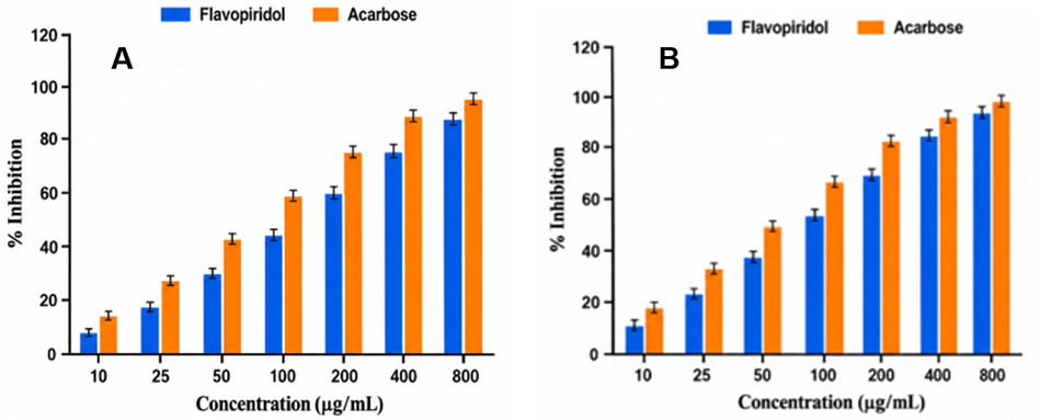
Figure 1: Percentage inhibition of α-amylase and α-glucosidase by flavopiridol and the standard drug acarbose at different concentrations. All experiments were performed in triplicate, and the results are expressed as mean ± SD (n = 3).
3.2 Effect of Flavopiridol on Fasting Blood Glucose
Figure 2 and Table 3show the fasting blood glucose values for all treatment groups. At baseline, diabetic animals had a significant elevation of fasting blood glucose (about 292–294 mg/dL), thereby confirming successful induction of experimental type 2 diabetes. During the period of treatment, fasting blood glucose was progressively increased in the diabetic control group, ultimately reaching a level of 405.7 13.2 mg/dL by the end of six weeks. Flavopiridol treatment induced significant (p < 0.05), dose-dependent reduction of fasting blood glucose relative to the diabetic control group. In the flavopiridol (1.25 mg/kg) group, fasting blood glucose was reduced from 293.2 7.8 mg/dL on Day 0 to 148.9 6.1 mg/dL after 42 days, indicating an overall decrease of approximately 49.2%. A significantly higher antihyperglycemic effect was observed in the flavopiridol (5 mg/kg) group, with a reduction in blood glucose from 294.1 8.3 mg/dL to 116.8 5.7 mg/dL, indicating an overall reduction of approximately 60.3%. The antihyperglycemic effect of flavopiridol (5 mg/kg) was comparable to that of the standard treatments of metformin (104.3 4.6 mg/dL) and pioglitazone (118.5 5.3 mg/dL). The reduction in fasting blood glucose levels became apparent beginning in the second week of the study and continued thereafter throughout the entire 42-day treatment period.
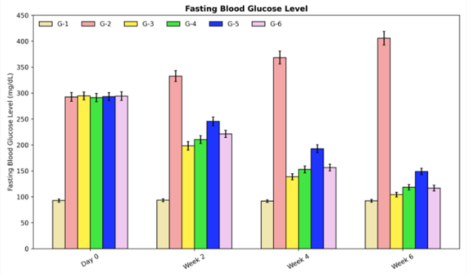
Figure 2: Effect of fasting blood glucose on diabetic wistar rats
G-1: Normal Control; G-2: Diabetic Control (HFD + STZ); G-3: Diabetic + Metformin (150–200 mg/kg/day, p.o.); G-4: Diabetic + Pioglitazone (10–20 mg/kg/day, p.o.); G-5: Diabetic + Flavopiridol (1.25 mg/kg/day, p.o.); G-6: Diabetic + Flavopiridol (5 mg/kg/day, p.o.).
3.2 Effect of Flavopiridol on Body Weight
Figure 3and Table 3 present the changes in body weight of rats throughout the experimental period. In normal control rats, body weight increased gradually from 198.6 4.2 g at the beginning to 223.4 5.3 g by the end of the experiment. However, untreated diabetic rats showed significant and progressive loss in body weight, decreasing from 201.8 5.1 g to 163.2 4.3 g at the end of the study, due to severely disrupted metabolism in this diabetic state. Treatment with flavopiridol markedly prevented this diabetes-associated weight loss. Rats receiving 1.25 mg/kg flavopiridol experienced a gradual rise in body weight to 208.7 4.9 g six weeks later. The increase was even more pronounced when rats were treated with 5 mg/kg flavopiridol (body weight 218.3 4.6 g), which reached levels similar to those achieved by treatment with metformin (216.8 4.7 g) or pioglitazone (220.1 4.8 g). The beneficial effect of flavopiridol on body weight could be observed from the second week and maintained itself during the course of treatment.
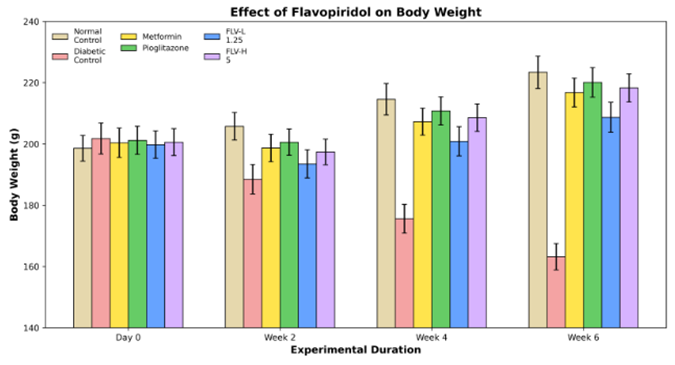
Figure 3: Effect of body weight on diabetic wistar rats
G-1: Normal Control; G-2: Diabetic Control (HFD + STZ); G-3: Diabetic + Metformin (150–200 mg/kg/day, p.o.); G-4: Diabetic + Pioglitazone (10–20 mg/kg/day, p.o.); G-5: Diabetic + Flavopiridol (1.25 mg/kg/day, p.o.); G-6: Diabetic + Flavopiridol (5 mg/kg/day, p.o.).
3.3 Effect of Flavopiridol on Serum Insulin Levels
The serum insulin concentrations measured at the end of the experimental period are shown in Figure 4 and Table 3. Induction of diabetes caused a marked reduction in circulating insulin levels, with the diabetic control group exhibiting a serum insulin concentration of 6.41 ± 0.52 µIU/mL, compared with 18.62 ± 0.84 µIU/mL in the normal control group. Treatment with flavopiridol significantly improved serum insulin concentrations in a dose-dependent manner. The low-dose flavopiridol group demonstrated an increase in serum insulin to 13.76 ± 0.61 µIU/mL, whereas the high-dose group showed a further increase to 16.28 ± 0.56 µIU/mL. The insulin-enhancing effect of high-dose flavopiridol was comparable to that observed with metformin (15.87 ± 0.63 µIU/mL) and pioglitazone (16.95 ± 0.58 µIU/mL). These findings suggest that flavopiridol effectively preserved pancreatic β-cell function and improved insulin secretion following prolonged treatment.
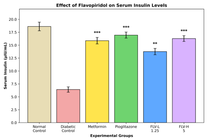
Figure 4: Effect of serum insulin levels on diabetic wistar rats
G-1: Normal Control; G-2: Diabetic Control (HFD + STZ); G-3: Diabetic + Metformin (150–200 mg/kg/day, p.o.); G-4: Diabetic + Pioglitazone (10–20 mg/kg/day, p.o.); G-5: Diabetic + Flavopiridol (1.25 mg/kg/day, p.o.); G-6: Diabetic + Flavopiridol (5 mg/kg/day, p.o.).
Table 3: Effect of the fasting blood glucose, body weight and serum insulin on diabetic wistar rats
|
Experimental Groups |
Fasting Blood Glucose (mg/dL)(Week 6) |
Body Weight (g)(Week 6) |
Serum Insulin (µIU/mL) |
|
Normal Control |
92.4 ± 2.7 |
223.4 ± 5.3 |
18.62 ± 0.84 |
|
Diabetic Control |
405.7 ± 13.2 |
163.2 ± 4.3 |
6.41 ± 0.52 |
|
Metformin |
104.3 ± 4.6*** |
216.8 ± 4.7*** |
15.87 ± 0.63*** |
|
Pioglitazone |
118.5 ± 5.3*** |
220.1 ± 4.8*** |
16.95 ± 0.58*** |
|
Flavopiridol |
148.9 ± 6.1*** |
208.7 ± 4.9** |
13.76 ± 0.61** |
|
Flavopiridol |
116.8 ± 5.7*** |
218.3 ± 4.6*** |
16.28 ± 0.56*** |
3.4 In vivo Antioxidant Activity
Effect of Flavopiridol on Reduced Glutathione (GSH)
Table 4 and Figure 5(A) indicate the levels of reduced GSH in the tissues. Normal control rats had highest level of reduced glutathione (63.24 ± 1.42 mol/g tissue) and diabetic control rats had significant reduction in GSH level (33.86 ± 1.58 mol/g tissue), indicating high degree of oxidative stress in diabetes. Administration of flavopiridol significantly normalized the level of GSH in the tissues. Fluvopiridol at 1.25 mg/kg level resulted in GSH level of 52.84 ± 1.41 mol/g tissue, which was increased to 59.36 ± 1.28 mol/g tissue when administered at 5 mg/kg dose level. Same trend was observed when these animals were treated with metformin (58.42 ± 1.36 mol/g tissue) and pioglitazone (60.18 ± 1.24 mol/g tissue). This suggests that flavopiridol enhanced the intra cellular antioxidants level in diabetic animals.
Effect of Flavopiridol on Glutathione Peroxidase (GPx)
The GPx activity in all the experimental groups has been summarized in Table 4 and Figure 5(B). Diabetic control rats had significantly decreased GPx activity (8.26 ± 0.38 U/mg protein) than normal control (18.14 ± 0.52 U/mg protein). The activity of GPx in the flavopiridol treated diabetic rats was restored to the levels close to the normal rats. Flavopiridol (low dose, 13.94 ± 0.46 U/mg protein; high dose, 16.31 ± 0.43 U/mg protein) as well as metformin (16.04 ± 0.44 U/mg protein) and pioglitazone (16.82 ± 0.48 U/mg protein) treatment group displayed a dose-dependent and significant increase in the activity of GPx.
Effect of Flavopiridol on Catalase (CAT)
Table 4 and Figure 5(C) shows the activity of CAT in the different treatment groups. As shown in Figure 3.7, diabetic control group has significantly low activity of CAT (8.34 0.49 U/mg protein) in comparison with normal control group (27.12 0.81 U/mg protein). Flavopiridol administration elevated the CAT activity in diabetic rats; low dose group shows the activity of 20.94 0.63 U/mg protein while the high dose shows the CAT activity of 24.42 0.67 U/mg protein. Flavopiridol with low and high dose restored the CAT activity almost to that obtained from animals treated with metformin (23.84 0.69 U/mg protein) and pioglitazone (25.16 0.74 U/mg protein) respectively, which means that the treatment effectively removes hydrogen peroxide in the diabetic tissues.
Effect of Flavopiridol on Superoxide Dismutase (SOD)
The activity of SOD in experiment rats is presented in Table 4 and Figure 5(D). In the induced diabetic control group, SOD activity was reduced significantly to 1.86 0.16 U/mg protein in compared to control 5.26 0.22 U/mg protein, oral administration of flavopiridol had recovery of SOD activity in dose dependent style; Low dose group showed SOD activity of 4.08 0.18 U/mg protein, High dose of flavopiridol treatment group. How SOD activity approached the control to 4.86 0.19 U/mg protein. As comparison, SOD activity in the pioglitazone (4.94 0.20 U/mg protein) and Metformin (4.72 0.19 U/mg protein) groups also had the tendency of restoring SODactivity.
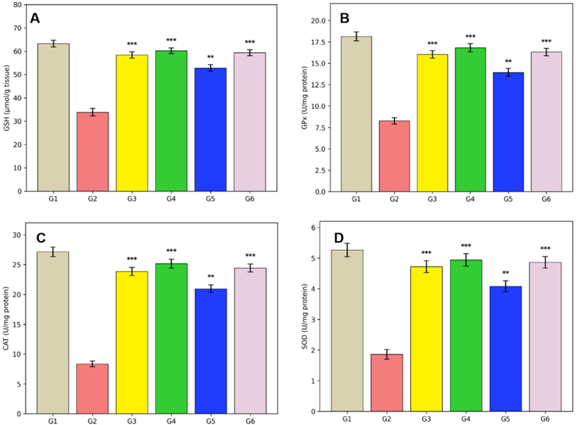
Figure 5: Effect of (A) GSH, (B) GPx, (C) CAT, (D) SOD on diabetic Wistar rats
Table 4: Effect of GSH, GPx, CAT and SOD on diabetic wistar rats
|
Groups |
GSH (µmol/g tissue) |
GPx (U/mg protein) |
CAT (U/mg protein) |
SOD (U/mg protein) |
|
Normal Control |
62.84 ± 1.35 |
17.92 ± 0.56 |
26.85 ± 0.84 |
5.12 ± 0.24 |
|
Diabetic Control |
34.28 ± 1.62 |
8.14 ± 0.42 |
8.42 ± 0.53 |
1.98 ± 0.17 |
|
Metformin |
57.46 ± 1.24*** |
15.92 ± 0.48*** |
23.48 ± 0.71*** |
4.62 ± 0.19*** |
|
Pioglitazone |
59.73 ± 1.18*** |
16.68 ± 0.51*** |
24.96 ± 0.76*** |
4.86 ± 0.21*** |
|
Flavopiridol (1.25 mg/kg) |
51.64 ± 1.37** |
13.72 ± 0.45** |
20.76 ± 0.64** |
3.98 ± 0.18** |
|
Flavopiridol (5 mg/kg) |
58.41 ± 1.26*** |
16.24 ± 0.47*** |
24.15 ± 0.69*** |
4.78 ± 0.20*** |
3.5 Histopathology of the Pancreas
HFD + low-dose STZ treatment caused remarkable histopathological changes in the pancreas, including degeneration of cells, shrinkage of islets, infiltration of inflammatory cells and deformation of normal pancreas tissue. These morphological alterations may cause impaired insulin secretion, leading to persistently raised blood glucose levels. Effects of flavopiridol treatment on pancreatic histopathology are presented in photomicrographs in Figure 6.
In control animals (normal group), pancreatic tissues were morphologically normal as revealed by normal architecture and well- distributed and well-defined pancreatic islets with complete and uniform populations of - cells, normal acinar tissue, and no evidence of inflammation (Fig.6A). Conversely, pancreatic tissue from the diabetic control group presented severe damage such as marked reduction of size and number of islets of Langerhans, massive degeneration of cells, cytoplasmic vacuolation, deformation of acinar tissue and significant inflammatory cell infiltration, suggesting that the experimental model of diabetes was successfully induced (Fig. 6B).
There was marked improvement in pancreatic morphology in the groups treated with metformin and pioglitazone, characterised by restoration of islets architecture, an increase in cell density, decrease in inflammation, and maintenance of normal acinar tissue (Fig. 6C, 6D). Pancreatic histopathology in rats treated with flavopiridol (1.25 mg/kg) revealed moderate regeneration of pancreatic islets and some degree of restoration of β-cell morphology and reduced degeneration in comparison to the diabetic control group (Fig. 6E). Histological improvement was more significant in rats that received a high dose of flavopiridol (5 mg/kg) in which the islets of Langerhans were larger, organized, rich in -cells, with minimal inflammation and almost normal pancreatic architecture comparable to that observed in standard treated groups (Fig. 6F). It can be concluded that flavopiridol produced a dose-dependent improvement in pancreatic histology. Preservation of islets morphology and cells integrity observed in the animals treated with a high dose of flavopiridol coincided well with the significant reduction in FBG levels and improvement of serum insulin levels, suggesting protection of the pancreatic tissues from diabetes-induced injury.
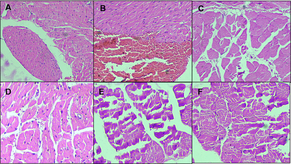
Figure 6: Histology of the pancreas (A- Normal control, B- Diabetic control, C- Metformin-treated, D- Pioglitazone-treated, E- Flavopiridol (1.25 mg/kg) and F- Flavopiridol (5 mg/kg)). Flavopiridol treatment produced dose-dependent restoration of islet architecture, increased β-cell density, and reduced inflammatory infiltration compared with the untreated diabetic group. This morphological improvement was most evident in the high-dose flavopiridol group, where pancreatic tissue closely resembled the normal histological appearance.
DISCUSSION:
Our current study revealed that flavopiridol exhibits promising antidiabetic activity via multiple mechanisms on the basis of in vitro inhibitory activity against α-amylase and α-glucosidase enzymes as well as in vivo antihyperglycemic, antioxidant, hepatoprotective and pancreatic protective activity in HFD-STZ-induced diabetic rats. Type 2 diabetes mellitus is accompanied by insulin resistance, inadequate insulin secretion, oxidative stress, and chronic inflammation, which may result in continuous hyperglycemia and progressive destruction of tissues. Hence, any therapy agent targeting several pathways can bring better control over the blood glucose level and may alleviate complications associated with diabetes. In vitro experiments confirmed that flavopiridol exhibited a significant concentration-dependent inhibitory effect on α-amylase and α-glucosidase. However, it was found that the inhibitory effect of acarbose against both enzymes was far better than flavopiridol, while still the treatment of flavopiridol provided notable inhibition to both carbohydrate-digesting enzymes, which implies that it may also hinder absorption of glucose in the intestine. To our surprise, the inhibition of α-glucosidase by flavopiridol was relatively much more compared to α-amylase, and in contrast to the latter, selective inhibition of α-glucosidase may not be associated with gastrointestinal adverse effects, which is often seen after inhibition of the potent α-amylase enzyme.
The present rat model (HFD-STZ induced diabetes) successfully mimicked various characteristic features of T2DM, such as sustained hyperglycemia, weight loss, depletion in insulin level in serum, presence of oxidative stress and injury in the pancreas. After repeated oral treatment of flavopiridol to diabetic rats the level of fasting blood glucose was found to be reduced significantly with an increase in the dose and remained significant throughout the duration of treatment. Moreover, at the highest dose (5 mg/kg), the reduction in blood glucose level was comparable to that of standard drugs i.e. Metformin and pioglitazone, which suggests that the glucose homeostasis is significantly improved in the animals treated with flavopiridol. Body Weight Body weight is a good index of metabolic status in diabetic animals. Diabetic rats without treatment lost weight gradually as a result of glucose malabsorption and increased catabolism of proteins. Flavopiridol produced a significant improvement in the body weight, which implied to bring the carbohydrate metabolism back towards normality and better absorption of nutrients.
Besides, a significantly high level of insulin in the serum of flavopiridol-treated rats indicated prevention or regeneration of pancreatic beta cells and good secretion of insulin. Oxidative Stress. The presence of free radicals may contribute to the development of diabetes complications. In the present study, the activities of endogenous antioxidant enzymes like SOD, CAT, GPx and GSH contents were reduced in diabetic control rats; on the contrary, the malondialdehyde level was found significantly increased in diabetic rats which indicated enhanced lipid peroxidation. Flavopiridol prevented depletion of endogenous antioxidant activities and reduced the level of lipid peroxide, thus suggesting attenuation of oxidative stress in treated animals. It indicated that prevention of oxidative stress may have contributed to the antihyperglycemic action of flavopiridol. Liver Function Biochemical estimations for liver damage indicated increased levels of AST and ALT in diabetic rats as an index of liver injury. Treatment with flavopiridol significantly brought back these values to the normal levels, which indicated protection against diabetes-induced liver damage. This finding is further supported by the histopathological observations where islets of Langerhans regenerated and the β-cell population increased in flavopiridol-treated pancreatic sections with diminished inflammatory infiltrations. Liver showed repair in normal architectural features and a significant reduction in cellular degeneration and vacuolations. Higher level of treatment with flavopiridol achieved the effect to a greater extent and even closer to the levels obtained by the standard antidiabetic drugs. Conclusion In conclusion, flavopiridol demonstrated significant antihyperglycemic action through inhibition of carbohydrate-digesting enzymes, augmentation of insulin secretion, abatement of oxidative stress and amelioration of liver damage along with the preservation of pancreatic architecture. A multifunctional approach may designate flavopiridol as a useful lead compound in developing potent agent against type 2 diabetes mellitus. Further mechanism-oriented studies and prolonged clinical studies would be necessary for definitive evaluation of this compound.
CONCLUSION :
In the current study, we observed that flavopiridol has potent antidiabetic activity via inhibiting both α-amylase and α-glucosidase, improving the control of blood glucose, rescuing the levels of serum insulin, strengthening the antioxidant defence mechanism, normalising liver enzymes, and preserving the architecture of pancreatic and liver tissues in HFD-STZ induced diabetic rats. These results indicate that flavopiridol can be used as an effective candidate drug to develop multi-target therapy for type 2 diabetes mellitus, and future research needs further mechanistic study and clinical trials to confirm the potential application.
BIBLIOGRAPHY:
1. Care D. (2024). 2. Diagnosis and classification of diabetes: Standards of care in diabetes—2024. Diabetes Care, 47(Suppl 1), S20-s42.
2. DeFronzo RA, Ferrannini E, Groop L, Henry RR, Herman WH, Holst JJ, Hu FB, Kahn CR, Raz I, Shulman GI, Simson DC, Testa MA, & Weiss R. (2015). Type 2 diabetes mellitus. Nature reviews Disease primers, 1(1), 15019.
3. Federation ID. (2021). IDF diabetes atlas 10th. 2021-12-06)[2024-11-18]. https://diabetesatlas. org/. html.
4. Galicia-Garcia U, Benito-Vicente A, Jebari S, Larrea-Sebal A, Siddiqi H, Uribe KB, Ostolaza H, & Martín C. (2020). Pathophysiology of type 2 diabetes mellitus. International journal of molecular sciences, 21(17), 6275.
5. Taylor R. (2013). Type 2 diabetes: etiology and reversibility. Diabetes care, 36(4), 1047-1055.
6. Chen X, Xie N, Feng L, Huang Y, Wu Y, Zhu H, Tang J, & Zhang Y. (2025). Oxidative stress in diabetes mellitus and its complications: From pathophysiology to therapeutic strategies. Chinese Medical Journal, 138(1), 15-27.
7. Senderowicz AM. (1999). Flavopiridol: the first cyclin-dependent kinase inhibitor in human clinical trials. Investigational new drugs, 17(3), 313-320.
8. Sedlacek HH (2001). Mechanisms of action of flavopiridol. Critical reviews in oncology/hematology, 38(2), 139-170.
9. Hydbring, P, Malumbres M, Sicinski P. (2016). Non-canonical functions of cell cycle cyclins and cyclin-dependent kinases. Nature Reviews Molecular Cell Biology, 17(5), 280–292.
10. Donath MY, & Shoelson SE. (2011). Type 2 diabetes as an inflammatory disease. Nature Reviews Immunology, 11(2), 98–107
11. Takada Y, Singh S, & Aggarwal BB. (2004). Identification of a p65 peptide that selectively inhibits NF-κ B activation induced by various inflammatory stimuli and its role in down-regulation of NF-κB-mediated gene expression and up-regulation of apoptosis. Journal of Biological Chemistry, 279(15), 15096-15104.
12. Malumbres M. (2014). Cyclin-dependent kinases. Genome biology, 15(6), 122.
13. Eusebio PS, Toledo JD, Mamauag REP, & Bernas MJG. (2004). Digestive enzyme activity in developing grouper (Epinephelus coioides) larvae. Advances in grouper aquaculture, CIAR Monograph.
14. Bernfeld P. (1955). [17] Amylases, α and β.
15. Kim YM, Jeong YK, Wang MH, Lee WY, & Rhee HI. (2005). Inhibitory effect of pine extract on α-glucosidase activity and postprandial hyperglycemia. Nutrition, 21(6), 756-761.
16. Apostolidis E, Kwon YI, & Shetty K. (2007). Inhibitory potential of herb, fruit, and fungal-enriched cheese against key enzymes linked to type 2 diabetes and hypertension. Innovative food science & emerging technologies, 8(1), 46-54.
17. Srinivasan K, Viswanad B, Asrat L, Kaul CL, & Ramarao PJPR.(2005). Combination of high-fat diet-fed and low-dose streptozotocin-treated rat: a model for type 2 diabetes and pharmacological screening. Pharmacological research, 52(4), 313-320.
18. Reed, MJ, Meszaros K, Entes LJ, Claypool MD, Pinkett JG, Gadbois TM, & Reaven GM. (2000). A new rat model of type 2 diabetes: the fat-fed, streptozotocin-treated rat. Metabolism-Clinical and Experimental, 49(11), 1390-1394.
19. Muthuraman P, Senthilkumar R, Srikumar K. Alterations in beta-islets of Langerhans in alloxan-induced diabetic rats by marine Spirulina platensis. J Enzyme Inhib Med Chem. 2009;24(6):1253–6.
20. Serrano-Rios M, Ramos F, Rodriguez-Minon JL, Vivanco F. Studies in prediabetes. Insulin response to oral glucose, intravenous tolbutamide and rapid intravenous glucose infusion in genetic prediabetics. Diabetologia. 1970;6(4):392–8.
21. Beutler E, Duron O, & Kelly BM. (1963). Improved method for determination of blood glutathione.
22. Paglia DE, & Valentine WN. (1967). Studies on the quantitative and qualitative characterization of erythrocyte glutathione peroxidase. The Journal of laboratory and clinical medicine, 70(1), 158-169.
23. Aebi H. (1984). [13] Catalase in vitro. In Methods in enzymology (Vol. 105, pp. 121-126). Academic press.
24. Misra HP, & Fridovich I. (1972). The role of superoxide anion in the autoxidation of epinephrine and a simple assay for superoxide dismutase. Journal of Biological chemistry, 247(10), 3170-3175.
25. Reitman S, & Frankel S. (1957). A colorimetric method for the determination of serum glutamic oxalacetic and glutamic pyruvic transaminases. American journal of clinical pathology, 28(1), 56-63.