Diagnostic Accuracy of Ultrasonography in the Diagnosis of Rotator Cuff tears, taking Magnetic Resonance Imaging as Gold Standard
- Najm ud Din , Postgraduate Resident Diagnostic Radiology, Department of Radiology, Mardan Medical Complex (MTI), Mardan, Pakistan
- Zubair Janan Orakzai , Associate Professor, Mardan Medical Complex (MTI), Mardan, Pakistan
- Taj Muhammad Khan , Postgraduate Resident Diagnostic Radiology, Department of Radiology, Mardan Medical Complex (MTI), Mardan, Pakistan
- Humayoun , Postgraduate Resident Diagnostic Radiology, Department of Radiology, Mardan Medical Complex (MTI), Mardan, Pakistan
- Muhammad Farah Adeed Khan , Postgraduate Resident Diagnostic Radiology, Department of Radiology, Mardan Medical Complex (MTI), Mardan, Pakistan
- Sumaira Noureen , Assistant Professor, Department of Radiology, Mardan MedicalComplex (MTI), Mardan, Pakistan
Article Information:
Abstract:
One of the most frequent causes of pain, weakness and limited range of motion in the shoulder is a tear in the rotator cuff. MRI is a very good tool for obtaining detailed information about rotator cuff pathology; however, it is somewhat costly and may not be available in all healthcare settings. A high resolution ultrasonography is a fast, cheap, and dynamic imaging tool, but there may be some differences in the diagnostic accuracy of the technique depending on the operator and the type of tears. The aim of this study was to assess the diagnostic value of ultrasonography in the diagnosis of rotator cuff tears compared to magnetic resonance imaging (MRI). Objective: To determine the diagnostic accuracy of ultrasonography for detecting rotator cuff tears, using magnetic resonance imaging as the reference standard. Methods: The cross-sectional validation study was carried out in the Department of Radiology, Mardan Medical Complex Mardan, from 23 November 2024 to 23 May 2025. One-hundred and thirty-seven patients (ages 25-70) with clinically diagnosed traumatic rotator cuff tear were recruited using non-probability consecutive sampling technique. All subjects received shoulder ultrasound (with an 8–15 MHz linear transducer) and then MRI (3-Tesla system). The two radiologists who read the two exams were not aware of each other's reports. The results of the ultrasonography were determined as true positive, false positive, false negative and true negative based on MRI as the reference standard. The following were calculated: sensitivity, specificity, positive predictive value, negative predictive value and overall diagnostic accuracy. Results: The mean age of the participants was 48.6 ± 11.7 years, and 82 (59.9%) were male. Ultrasonography identified rotator cuff tears in 70 (51.1%) patients, while MRI confirmed tears in 69 (50.4%). There were 61 true-positive, 59 true-negative, 9 false-positive and 8 false-negative results. Ultrasonography demonstrated a sensitivity of 88.4%, specificity of 86.8%, positive predictive value of 87.1%, negative predictive value of 88.1%, and overall diagnostic accuracy of 87.6%. Cohen’s kappa was 0.75, indicating substantial agreement between ultrasonography and MRI. Diagnostic performance was higher for full-thickness tears than for partial-thickness tears. Conclusion: High-resolution ultrasonography demonstrated good diagnostic accuracy and substantial agreement with MRI for detecting rotator cuff tears. It may be used as a cost-effective first-line imaging investigation, while MRI remains valuable for inconclusive findings, partial-thickness tears and preoperative assessment.
Keywords:
Article :
INTRODUCTION:
Rotator cuff tears are an important cause of shoulder pain, weakness and restricted movement, particularly among middle-aged and older adults. The rotator cuff is made up of the supraspinatus, infraspinatus, subscapularis and teres minor tendons which work together to stabilize the glenohumeral joint and facilitate shoulder motion. These tendons can suffer injury due to a sudden trauma, repetitive mechanical stresses, or from a combination of these. If left untreated, tears can gradually enlarge and can result in tendon retraction, muscle degeneration and long term functional disability [1-3].
Many patients presenting for a clinical consult regarding the shoulder have a rotator cuff disorder. Common clinical features are shoulder pain, limited and painful abduction, weakness, and night discomfort/daily activity impairments. But a diagnosis of a rotator cuff tear based solely on the clinical examination is not necessarily correct, as a diagnosis of subacromial bursitis, impingement, tendinopathy, adhesive capsulitis or labral pathology may also be considered. Imaging, therefore, is essential to confirm diagnosis, identify the tendon involved, differentiate partial from full-thickness tears and to check for any associated abnormalities [4-6].
MRI is considered to be a very accurate test to evaluate rotator cuff problems. It offers comprehensive imaging of the structure of tendons, the size of the tears, the thinning of the tendons, fatty infiltration, and the pullback of the tendon and the presence of other intra-articular abnormalities. However, MRI is costly, not easily available in resource-poor health care environments, and is more time-consuming. It also may not be appropriate for people who have some metal devices in their body or cardiac pacemakers or have severe claustrophobia. These restrictions may affect the time of diagnosis and the costs of the health services and the patients themselves [7, 8].
The use of high resolution ultrasonography in the investigations of rotator cuff tendons is a more accessible and economical imaging modality. It is portable, rapid, non-invasive and free from ionizing radiation. Ultrasound also can allow for dynamic study of the shoulder, evaluation during motion, and a comparison with the opposite side, which is done in real time. It can demonstrate tendon discontinuity, hypoechoic or anechoic defects, tendon retraction, bursal fluid, calcification and impingement. But it is dependent upon operator experience, scanning technique, the quality of the equipment, as well as the type or location of the tear [9-11].
Different estimates of the diagnostic value of ultrasonography have been reported in previous studies. Farooqi et al. found that the diagnostic performance of ultrasonography was similar to that of MRI, especially in cases of full-thickness and supraspinatus tears [4]. In a local study conducted by Shabana et al. the sensitivity was 81.82%, specificity was 77.27% and overall accuracy was 79.55% [12]. Nadeem et al reported a sensitivity of 91%, specificity of 87% and accuracy of 90.2% [13]. One study showed a reduction in sensitivity with partial-thickness tears and very high sensitivity with full-thickness tears [9]. These differences suggest that diagnostic accuracy could vary from patient to patient, examiner experience and imaging protocol.
In healthcare settings where MRI availability is limited, establishing the local diagnostic accuracy of ultrasonography is clinically important. Reliable ultrasound findings may allow early diagnosis, reduce unnecessary MRI examinations and support timely referral for conservative or surgical management. Therefore, this study was conducted to determine the sensitivity, specificity, positive predictive value, negative predictive value and overall diagnostic accuracy of ultrasonography for detecting rotator cuff tears, using MRI as the reference standard, among patients presenting to the Department of Radiology, Mardan Medical Complex, Mardan. The study protocol included standardized assessment of the rotator cuff tendons and blinded interpretation of ultrasonography and MRI examinations.
MATERIALS AND METHODS:
A cross-sectional validation study was conducted in the Department of Radiology, Mardan Medical Complex, Mardan, from 23 November 2024 to 23 May 2025. The study aimed to determine the diagnostic performance of ultrasonography for detecting rotator cuff tears, with magnetic resonance imaging used as the reference standard. Research approval was obtained from the College of Physicians and Surgeons Pakistan, Research Evaluation Unit, under reference number CPSP/REU/RAD-2021-028-3514, dated 23 November 2024. Permission was also obtained from the relevant institutional authorities before data collection. Written informed consent was obtained from every participant after explaining the study procedures, potential benefits, voluntary nature of participation and confidentiality of personal information.
The number of patients required was 137. It was determined by using a diagnostic sensitivity and specificity calculator at a 95% confidence level, with an expected prevalence of rotator cuff tears of 50.0%, an ultrasonographic sensitivity of 81.82%, a specificity of 77.27%, and absolute precision of 10%. A non-probability consecutive sampling technique was used to select the participants. Inclusion criteria were first, that all eligible patients who presented or were referred to the radiology department during the study period were included; and second, that the sample size was reached.
The inclusion criteria were being male and female, aged between 25 and 70 years, and having a clinically suspected traumatic rotator cuff injury. A suspected rotator cuff tear was defined as a history of shoulder trauma more than one month previously, a shoulder pain score of at least 3 on the visual analogue scale and clinically restricted shoulder movement. Patients who had metallic implants, shoulder prostheses, cardiac pacemakers, claustrophobia, previous shoulder surgery, shoulder fracture or dislocation, suspected tumour or malignancy, contraindications to MRI or who refused informed consent were excluded. A structured data collection form was used to capture basic information such as age, gender, duration of trauma, affected shoulder, rural-urban living and occupational category.
The shoulder ultrasonography was performed by a radiology resident, supervised by an experienced senior radiologist, with a high-frequency (8–15 MHz) linear transducer. The long head of the biceps tendon, subscapularis, supraspinatus, infraspinatus and teres minor tendons were scanned using a standardized scanning protocol. To minimize anisotropy, the ultrasound beam was held perpendicular to the direction of tendon fibres. Dynamic shoulder abduction was tested for impingement and compression of the rotator cuff was performed to help locate non-retracted tears. Whenever possible, a contralateral shoulder was compared to the symptomatic shoulder. Other abnormalities such as acromioclavicular joint changes, subacromial-subdeltoid bursal effusion, glenohumeral joint effusion, subcoracoid bursal effusion, biceps tendon sheath effusion, tendon calcification, muscle atrophy and labral abnormalities were also evaluated. A partial-thickness tear was diagnosed as a focal hypoechoic or anechoic defect in part of the tendon that did not involve the entire thickness of the tendon. A complete full-thickness tear was characterized as complete separation of the tendon fibres between the articular and bursal surface either with or without tendon retraction or non-visualization.
All participants subsequently underwent MRI of the affected shoulder using a 3-Tesla whole-body MRI system. Proton-density fat-saturated, T1-weighted and T2-weighted turbo spin-echo sequences were used for imaging in axial, sagittal and coronal planes. The MRI diagnosis of partial thickness tear was defined as the focal presence of fluid-equivalent hyperintense signal on the articular and/or bursal surface, or contained within the tendon substance. A hyperintense defect from the articular to the bursal side or as total avulsion of the tendon was considered as a full-thickness tear. The MRI was reported by the radiologist without the knowledge of the ultrasonography result and the ultrasonography examiner was not aware of the MRI result. This independent interpretation mitigated the observer and verification bias. MRI was considered the reference standard and the ultrasonography results were considered as true positive, false positive, false negative or true negative.
The data were collected and analyzed in IBM SPSS Statistics version 25.0. Continuous variables were tested for distribution using Shapiro–Wilk test. Variables that were normally distributed (e.g., age) were presented as mean and SD, while nonnormally distributed variables (e.g., duration since trauma) were presented as median and interquartile range. Categorical data such as gender, affected side, residence, occupation and imaging findings were reported as frequencies and percentages. Data was analyzed in terms of a 2 × 2 contingency table, which compared ultrasonography results to MRI results. The sensitivity, specificity, positive predictive value, negative predictive value and overall diagnostic accuracy were computed using standard formulas. The percentage agreement and Cohen's Kappa coefficient were used to determine the level of agreement between ultrasonography and MRI. Diagnostic performance was also broken down by age group, gender, duration since trauma, affected shoulder and residence and occupation. Comparisons among post-stratifications were carried out by the chi-square test or Fisher's exact test (as appropriate). A p-value ≤0.05 was considered statistically significant.
RESULTS:
There were 137 patients with clinically suspected traumatic rotator cuff tears included. The age range of the participants was 25–70 years, and the mean age was 48.6 ± 11.7 years. The average age of the respondents was 41-55 years. Males accounted for 82 (59.9%) patients, while 55 (40.1%) were females. The median time since trauma was 46 days (IQR: 31-72 days). In 79 patients (57.7%), the right shoulder was affected while 58 (42.3%) had left shoulder involvement. The total number of participants was 81 (59.1%) from rural communities and 56 (40.9%) from urban communities. The largest group of workers were field workers.
Table 1. Baseline and Clinical Characteristics of the Participants
|
Variable |
Frequency/Mean |
Percentage/SD |
|
Age, years |
48.6 |
±11.7 |
|
Age group |
||
|
25–40 years |
36 |
26.3% |
|
41–55 years |
58 |
42.3% |
|
56–70 years |
43 |
31.4% |
|
Gender |
||
|
Male |
82 |
59.9% |
|
Female |
55 |
40.1% |
|
Duration since trauma, days |
46 |
IQR: 31–72 |
|
Affected shoulder |
||
|
Right |
79 |
57.7% |
|
Left |
58 |
42.3% |
|
Residence |
||
|
Rural |
81 |
59.1% |
|
Urban |
56 |
40.9% |
|
Occupation |
||
|
Office-based |
33 |
24.1% |
|
Field-based |
62 |
45.3% |
|
Domestic work |
42 |
30.7% |
The 70 (51.1%) patients with ultrasonography findings of a rotator cuff tear had 69 (50.4%) confirmed by MRI. Of the 38 (27.7%) patients, ultrasonography identified partial-thickness tears and 40 (29.2%) patients, MRI identified partial-thickness tears. On ultrasonography, 32 (23.4%) had full-thickness tears, while MRI revealed 29 (21.2%) had full-thickness tears. Both imaging modalities involved the supraspinatus tendon as the most common tendon involved. Multiple tendon involvement was less common, and subacromial-subdeltoid bursal effusion was the most common associated abnormality.
Table 2. Comparison of Ultrasonography and MRI Findings
|
Imaging finding |
Ultrasonography, n (%) |
MRI, n (%) |
|
Any rotator cuff tear |
70 (51.1%) |
69 (50.4%) |
|
No rotator cuff tear |
67 (48.9%) |
68 (49.6%) |
|
Partial-thickness tear |
38 (27.7%) |
40 (29.2%) |
|
Full-thickness tear |
32 (23.4%) |
29 (21.2%) |
|
Supraspinatus involvement |
55 (40.1%) |
57 (41.6%) |
|
Infraspinatus involvement |
16 (11.7%) |
17 (12.4%) |
|
Subscapularis involvement |
12 (8.8%) |
13 (9.5%) |
|
Multiple-tendon involvement |
15 (10.9%) |
16 (11.7%) |
|
Tendon retraction |
19 (13.9%) |
21 (15.3%) |
|
Muscle atrophy |
11 (8.0%) |
13 (9.5%) |
|
Subacromial-subdeltoid bursal effusion |
34 (24.8%) |
36 (26.3%) |
|
Biceps tendon sheath effusion |
21 (15.3%) |
23 (16.8%) |
Of these, 61 patients were positive on both tests, and were considered as true-positive cases when compared with MRI. A total of 9 ultrasonography false-positive results were encountered, with a tear found on ultrasonography but not on MRI. 8 patients in the negative USG group were diagnosed with rotator cuff tears on MRI and were defined as false-negative cases. 59 patients were negative by ultrasonography and by MRI, which were true-negative results.
Table 3. Two-by-Two Comparison of Ultrasonography With MRI
|
Ultrasonography result |
MRI tear present |
MRI tear absent |
Total |
|
USG tear present |
61 true positive |
9 false positive |
70 |
|
USG tear absent |
8 false negative |
59 true negative |
67 |
|
Total |
69 |
68 |
137 |
When the MRI was used as the reference standard, ultrasonography had a sensitivity of 88.4% and a specificity of 86.8%. The positive predictive value was 87.1%, meaning that almost 9 of the patients who were positive on ultrasonography were positive on MRI. Negative predictive value was 88.1% and accuracy of the overall diagnosis was 87.6%. The agreement between ultrasonography and MRI was also 87.6%. The Cohen's kappa coefficient was 0.75, which means that there was a good level of agreement between the two imaging modalities.
Table 4. Diagnostic Accuracy of Ultrasonography for Rotator Cuff Tears
|
Diagnostic measure |
Value |
|
Sensitivity |
88.4% |
|
Specificity |
86.8% |
|
Positive predictive value |
87.1% |
|
Negative predictive value |
88.1% |
|
Overall diagnostic accuracy |
87.6% |
|
Percentage agreement |
87.6% |
|
Cohen’s kappa coefficient |
0.75 |
|
Interpretation of agreement |
Substantial |
Diagnostic performance of ultrasonography for FT tears was superior to that of PT tears. Sensitivity and specificity were 93.1% and 95.4%, respectively, and the overall diagnostic accuracy was 94.9% in the case of full-thickness tears. The sensitivity for partial-thickness tears was 80.0%, the specificity was 93.8%, and the overall accuracy was 89.8%. This indicates that ultrasonography is more likely to detect complete tears in the tendon than small or focal partial-thickness tears.
Table 5. Diagnostic Accuracy According to Tear Type
|
Type of tear |
Sensitivity |
Specificity |
PPV |
NPV |
Accuracy |
|
Partial-thickness tear |
80.0% |
93.8% |
84.2% |
91.9% |
89.8% |
|
Full-thickness tear |
93.1% |
95.4% |
84.4% |
97.1% |
94.9% |
|
Supraspinatus tear |
89.5% |
95.0% |
92.7% |
92.7% |
92.7% |
|
Multiple-tendon tear |
81.3% |
98.3% |
86.7% |
97.5% |
96.4% |
Stratified analysis demonstrated that the diagnostic accuracy was maintained at levels of >84% in all age and gender groups. The best accuracy was achieved in patients aged 25-40 years, and a slightly lower accuracy was obtained in patients aged 56-70 years. The diagnostic value of ultrasonography was similar for males and females. There was no significant difference in the diagnostic accuracy of the tests across age groups, gender, trauma duration, or affected shoulder (all p-values > 0.05).
Table 6. Stratified Diagnostic Accuracy of Ultrasonography
|
Variable |
Subgroup |
Sensitivity |
Specificity |
Accuracy |
p-value |
|
Age group |
25–40 years |
90.0% |
87.5% |
88.9% |
0.742 |
|
41–55 years |
88.6% |
87.0% |
87.9% |
||
|
56–70 years |
86.4% |
85.7% |
86.0% |
||
|
Gender |
Male |
89.2% |
86.7% |
87.8% |
0.814 |
|
Female |
87.5% |
86.8% |
87.3% |
||
|
Trauma duration |
≤45 days |
90.0% |
88.2% |
89.1% |
0.583 |
|
>45 days |
86.2% |
85.3% |
85.8% |
||
|
Affected shoulder |
Right |
89.7% |
87.5% |
88.6% |
0.671 |
|
Left |
86.7% |
85.7% |
86.2% |
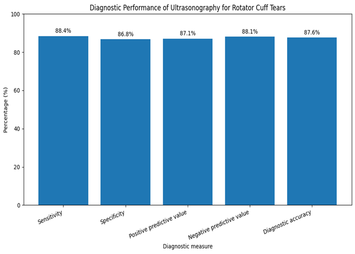
Figure 1. Diagnostic performance of ultrasonography for detecting rotator cuff tears using magnetic resonance imaging as the reference standard. Ultrasonography showed a sensitivity of 88.4%, specificity of 86.8%, positive predictive value of 87.1%, negative predictive value of 88.1%, and overall diagnostic accuracy of 87.6%.
DISCUSSION :
The present study evaluated the diagnostic performance of ultrasonography for detecting rotator cuff tears using MRI as the reference standard. Ultrasonography demonstrated a sensitivity of 88.4%, specificity of 86.8%, positive predictive value of 87.1%, negative predictive value of 88.1%, and overall diagnostic accuracy of 87.6%. Agreement between ultrasonography and MRI was substantial, with a Cohen’s kappa value of 0.75. These findings indicate that high-resolution ultrasonography can correctly classify most patients with suspected traumatic rotator cuff tears and may serve as an effective initial imaging investigation. The results are comparable to a 2024 study by Ganesh et al., which reported sensitivity of approximately 91%, specificity of 82%, and overall accuracy of nearly 89% for ultrasonography compared with MRI [14].
Stratified analysis revealed that for all age groups and gender, diagnostic accuracy was maintained at >84%. The highest accuracy was observed in patients within the age group 25–40 years and a slightly lower accuracy was recorded in patients aged 56–70 years. The sensitivity of the present study indicates that a large percentage of tears were identified by ultrasonography that were found by MRI. Farooqi et al. found similar results in their systematic review and meta-analysis, which reported a high sensitivity and specificity of ultrasonography for supraspinatus tears by expert operators, and its diagnostic accuracy was similar to MRI for both full- and partial-thickness tears [14]. Liang et al., also in this issue, concluded that ultrasonography was very efficient in diagnosis of rotator cuff tears and was suitable as a clinical diagnostic tool for patients with a suspected disease [15]. There is another study from Pakistan that got similar results with 89.8% sensitivity, 84.3% specificity, 92.9% positive predictive value, 78.2% negative predictive value, and 88.2% accuracy, which is very close to the results of the present analysis [16, 17].
The ultrasonography was shown to be more successful in evaluating full-thickness tears than partial-thickness tears. It demonstrated that the sensitivity and accuracy were 93.1% and 94.9% respectively in cases of full thickness tears, and 80.0% and 89.8% respectively in cases of partial-thickness tears. Full thickness tears typically show obvious discontinuity of the tendon fibres, fluid filled defects, non visualization or tendon retraction, which are easier to identify sonographically. Partial-thickness tears can be smaller and affecting only the articular or bursal surface, or within the tendon substance, which is often more difficult to diagnose. Previous studies also have demonstrated greater uniformity in ultrasonographic findings with full-thickness tears and greater variability with partial-thickness tears [14, 18].
The supraspinatus tendon was the most commonly injured tendon in the present study, as it is in many other studies, due to its anatomical position and susceptibility to mechanical impingement and repetitive stress. The diagnostic value of ultrasonography was high for supraspinatus tears, with a sensitivity of 89.5% and an accuracy of 92.7%. Ganesh et al. also found that the supraspinatus was the most commonly affected tendon and had good correlation between ultrasonography and MRI for the assessment of this tendon [19]. Further clinical benefits of ultrasound include the possibility of moving the shoulder and evaluating its movements, the ability to compare the normal and abnormal shoulder side by side and the ability to put abnormalities into the immediate context of the patient's pain. However, MRI offers more complete imaging of the dimensions of tears, retraction of the tendons, muscle atrophy, fatty infiltration, and abnormalities of the deep intra-articular structures, including the labrum [20].
The clinical significance of these results is of special relevance in medical environments where MRI availability is limited, due to cost or waiting time or contraindications. It is inexpensive, portable, readily available, and does not require ionizing radiation for use and is quite practical. It is therefore appropriate for use as a primary evaluation technique in patients who are suspected to have a rotator cuff tear, especially when used by experienced radiologists with a standardized scanning protocol. In addition to a high clinical index of suspicion, MRI should also be used for patients with negative or unclear ultrasound results, for suspected partial-thickness or complex tears and for those undergoing surgery or for those under consideration for surgery and intra-articular abnormalities. The decision between ultrasonography and MRI was highlighted in a recent 2024 review, taking into account diagnostic effectiveness, operator proficiency, access to imaging facilities, cost, and surgical planning requirements [20]
There are some limitations of this study. It was done at one centre and findings may not be generalizable to other hospitals and populations. Ultrasonography is operator dependent, and its results can be affected by the experience, training, and following of the scanning protocol by the operator. The reference standard used was MRI, which would mean that small tears not detected during MRI would not be confirmed during surgery. Patients with traumatic shoulder symptoms were included in this study; therefore, the results cannot be fully extrapolated to degenerative and asymptomatic tears of the rotator cuff. Moreover, interobserver and intraobserver reliability was not specifically assessed. Further multicentre studies are warranted with larger numbers of subjects, more blinded ultrasonographers, and separate analysis of the tears by tendon involved, tear size and tear surface, when clinically indicated, and arthroscopic confirmation of the tears.
CONCLUSION :
Ultrasonography showed high sensitivity, specificity, predictive values and overall diagnostic accuracy for detecting rotator cuff tears when compared with MRI. Its performance was particularly strong for full-thickness and supraspinatus tears, whereas partial-thickness tears remained comparatively more difficult to detect. The substantial agreement between the two imaging modalities supports the use of high-resolution ultrasonography as a reliable, accessible and cost-effective first-line investigation for patients with suspected rotator cuff tears. MRI remains important for inconclusive ultrasound examinations, complex or partial-thickness tears, associated intra-articular abnormalities and preoperative evaluation.
BIBLIOGRAPHY:
1. Liu, F., et al., Detecting rotator cuff tears: a network meta-analysis of 144 diagnostic studies. 2020. 8(2): p. 2325967119900356.
2. Pierce, J. and M.J.C.i.S.M. Anderson, Update on diagnostic imaging of the rotator cuff. 2023. 42(1): p. 25-52.
3. Zoga, A.C., et al., The evolving roles of MRI and ultrasound in first-line imaging of rotator cuff injuries. 2021. 217(6): p. 1390-1400.
4. Farooqi, A.S., et al., Diagnostic accuracy of ultrasonography for rotator cuff tears: a systematic review and meta-analysis. 2021. 9(10): p. 23259671211035106.
5. Gyftopoulos, S., et al., Postoperative imaging of the rotator cuff: a systematic review and meta-analysis. 2022. 219(5): p. 717-723.
6. Ahmed, A.A., et al., Comparative study between high resolution ultrasound and MRI in diagnosis of rotator cuff tears. 2023. 6(2): p. 639-651.
7. Vijayan, D., et al., Diagnostic accuracy of high-resolution ultrasonography in comparison with MRI for evaluation of rotator cuff pathologies. 2020. 5(2): p. 8-12.
8. Akhtar, M., et al., Diagnostic Accuracy and Agreement between Magnetic Resonance Imaging and Ultrasound Finding for the Diagnosis of Rotator Cuff Tears. 2023. 73(1): p. 231.
9. Gupta, A., et al., Diagnostic accuracy of ultrasound in rotator cuff and associated non-rotator cuff pathologies of shoulder joint compared to magnetic resonance imaging. 2022. 6(3).
10. Nasr, A.J., et al., Emerging role of quantitative ultrasound-based imaging techniques for characterizing rotator cuff tears: a scoping review. 2023. 13(12): p. 2011.
11. Kim, Y., et al., Ruling out rotator cuff tear in shoulder radiograph series using deep learning: redefining the role of conventional radiograph. 2020. 30(5): p. 2843-2852.
12. Shabana, et al., Diagnostic Accuracy and Agreement between Magnetic Resonance Imaging and Ultrasound Finding for the Diagnosis of Rotator Cuff Tears. Pakistan Armed Forces Medical Journal, 2023. 73(1): p. 231-34 DOI: 10.51253/pafmj.v73i1.5267.
13. Nadeem, R.D., et al., Accuracy of High Resolution Ultrasonography for Diagnosis of Rotator Cuff Tears Con-sidering Magnetic Resonance Imaging as Gold Standard. 2021. 27(2).
14. Farooqi, A.S., et al., Diagnostic Accuracy of Ultrasonography for Rotator Cuff Tears: A Systematic Review and Meta-analysis. Orthop J Sports Med, 2021. 9(10): p. 23259671211035106 DOI: 10.1177/23259671211035106.
15. Liang, W., et al., Diagnostic performance of ultrasound for rotator cuff tears: a systematic review and meta-analysis. Med Ultrason, 2020. 22(2): p. 197-202 DOI: 10.11152/mu-2352.
16. Toprak, U., et al., Diagnostic accuracy of ultrasound in subscapularis tendon abnormalities and the importance of operator experience. 2020. 54(4): p. 423.
17. Barad, H.V., et al., To determine the role of ultrasonography as a primary imaging modality as compared to MRI in patients with shoulder pain. 2022. 11(5): p. 2119-2122.
18. Okoroha, K.R., et al., Diagnostic accuracy of ultrasound for rotator cuff tears. Ultrasonography, 2019. 38(3): p. 215-220 DOI: 10.14366/usg.18058.
19. Ganesh, J., et al., Diagnostic Comparison of Ultrasound and Magnetic Resonance Imaging in Detecting Rotator Cuff Tears: A Study Conducted in the Population of Vijayapura. Cureus, 2024. 16(8): p. e68302 DOI: 10.7759/cureus.68302.
20. Toh, Y., Ultrasound Versus Magnetic Resonance Imaging as First-Line Imaging Strategies for Rotator Cuff Pathologies: A Comprehensive Analysis of Clinical Practices, Economic Efficiency, and Future Perspectives. Cureus, 2024. 16(4): p. e59231 DOI: 10.7759/cureus.59231.