Sonographic Gestational Age Estimation Discrepancies in Obese Mothers: A Cross-Sectional Study
- Laila Ameer , The University of lahore.
Article Information:
Abstract:
Maternal obesity poses significant challenges to accurate prenatal ultrasound assessment, particularly for gestational age (GA) estimation during the second and third trimesters. Accurate GA determination is essential for optimal obstetric management, yet the impact of maternal body mass index (BMI) on ultrasound-based GA estimation discrepancies remains incompletely understood. This study investigated whether maternal obesity is associated with significant discrepancies between GA estimated by last menstrual period (LMP) and ultrasound-based methods.Methods: A cross-sectional comparative study was conducted at Tehsil Headquarter Hospital Sadiq Abad, enrolling 230 pregnant women (116 normal BMI and 114 overweight/obese) during their second and third trimesters. Participants were categorized according to first-trimester BMI (normal: <24.9 kg/m²; overweight/obese: ≥25.0 kg/m²). GA was calculated using both LMP and ultrasound biometric parameters (biparietal diameter, femur length, abdominal circumference). Discrepancies were categorized as positive (ultrasound GA < LMP GA) or negative (ultrasound GA > LMP GA). Chi-square tests and independent samples t-tests were employed for statistical analysis.Results: The mean GA by LMP was 187.7 days (SD 45.3) for normal-weight and 209.6 days (SD 40.8) for obese participants. Chi-square analysis across five discrepancy categories revealed no significant difference between groups (p = 0.109). Similarly, when collapsed into positive, negative, and no-discrepancy categories, no significant difference was observed (p = 0.14). Independent samples t-test confirmed no significant difference in mean discrepancy between normal (mean 2.83 days, 95% CI 1.92–3.74) and obese (mean 2.00 days, 95% CI 0.94–3.05) groups (p = 0.23). Although a slight preponderance of negative discrepancies (suggesting fetal macrosomia) was noted among obese participants, this difference was not statistically significant.Conclusion: Maternal obesity does not significantly affect the discrepancy between LMP- and ultrasound-based GA estimation during the second and third trimesters. These findings suggest that ultrasound remains a reliable tool for GA assessment in overweight and obese pregnant women, despite the technical challenges associated with increased adipose tissue.
Keywords:
Article :
INTRODUCTION:
The global prevalence of obesity has increased substantially over recent decades, emerging as a significant public health concern across all populations, including women of reproductive age (1). The World Health Organization characterizes obesity as a pandemic, with higher prevalence observed in females than males (2). Consequently, a considerable proportion of pregnant women present with elevated body mass index (BMI), with estimates suggesting that at least one-third of pregnant women are overweight and 8% are classified as extremely obese (3).
Maternal obesity is associated with a spectrum of complications affecting both maternal and fetal health (4). These include gestational diabetes mellitus (GDM), pregnancy-induced hypertension (PIH), and delivery of large-for-gestational-age (LGA) neonates (5). Additionally, fetal growth abnormalities such as macrosomia may increase the likelihood of birth trauma, operative deliveries, and perinatal mortality (6,7). Such outcomes underscore the importance of accurate and timely prenatal assessments to mitigate risks and improve maternal and neonatal health outcomes (8).
Accurate determination of gestational age (GA) is a cornerstone of antenatal care, providing essential information for planning obstetric management—from timing diagnostic tests to assessing fetal growth and scheduling delivery (9). Traditionally, GA estimation has relied on the last menstrual period (LMP) as a reference point (10). However, this method is associated with limitations, including recall errors, irregular menstrual cycles, and variability in ovulation timing (11). These issues are particularly common among women with obesity, who may also experience hormonal imbalances affecting menstrual regularity (12).
Ultrasound has become the gold standard for GA estimation, particularly in the first trimester when fetal size exhibits minimal biological variability (13). During early pregnancy, measurements such as crown-rump length (CRL) offer high accuracy, often within 3–5 days of actual conception (14). However, with advancing pregnancy, sonographic estimation becomes increasingly challenging (15). Complex variations in fetal growth, maternal factors, and technical limitations contribute to reduced accuracy, especially during the second and third trimesters (16,17). These challenges are further aggravated in obese women, where excessive adipose tissue impairs fetal structure visualization, potentially leading to erroneous measurements of biometric parameters including biparietal diameter (BPD), femur length (FL), and abdominal circumference (AC) (18).
The rise in obesity among women of reproductive age has augmented the challenges associated with GA estimation (19). Studies suggest that maternal obesity can significantly impact sonographic measurements, particularly during the second and third trimesters when fetal growth shows greater variability (20). This suboptimal visualization due to large amounts of subcutaneous fat results in discrepancies of GA estimated by ultrasound when compared with that calculated using LMP (21). This confusion might potentially affect obstetric decision-making and hence pregnancy outcomes (22).
Despite advancements in ultrasound technology, including automated biometric calculations and artificial intelligence-assisted measurements (23), limited research has explored the extent of GA discrepancies in obese pregnant women during the second and third trimesters (24). Such discrepancies may have multiple implications for both maternal and fetal health, potentially resulting in unnecessary interventions or delayed delivery in cases of undiagnosed fetal distress (25). Moreover, the implications extend beyond individual cases to public health, as inaccurate GA estimation can distort population-level metrics, such as preterm birth rates and small-for-gestational-age (SGA) prevalence with effects on policy-making and planning at government level (26,27).
In settings with limited resources having constrained access to early prenatal care and first-trimester ultrasounds, the reliance on second- and third-trimester ultrasounds for GA estimation is certainly high (28). This dependence further highlights the importance of understanding and addressing the limitations of ultrasound-based GA assessment in obese populations (29). The present study aimed to compare discrepancies in sonographically determined GA between obese and non-obese mothers during the second and third trimesters of pregnancy.
METHODS:
Study Design
This was a cross-sectional comparative study.
Setting
The study was conducted at Tehsil Headquarter Hospital Sadiq Abad.
Study Duration
Data collection and analysis were completed over a 9-month period following synopsis approval.
Sample Size
Based on an assumed discrepancy between estimated date of delivery (EDD) by LMP and ultrasound of ≥14 days (30), with an anticipated odds ratio of 1.28, the sample size was calculated at 90% confidence level and 50% relative precision. The recommended sample size was 230 participants (116 normal BMI and 114 overweight/obese).
Sampling Technique
Convenient sampling was employed; all pregnant women fulfilling the inclusion criteria were enrolled.
Sample Selection
Inclusion Criteria
· Singleton pregnancy
· Certainty regarding LMP
· Availability of first antenatal record (10–12 post-menstrual weeks or earlier) for baseline maternal weight
Exclusion Criteria
· Fetal anomalies
· Multifetal pregnancy
· Hypertension or diabetes
Equipment
Ultrasound examinations were performed using a GE Logic P7 ultrasound machine equipped with a 5–7 MHz curvilinear probe.
Data Collection Procedure
Following ethical approval from the Research Ethical Committee of The University of Lahore, participants were recruited based on inclusion and exclusion criteria. Medical history, LMP, maternal weight, and BMI records were obtained. Participants were categorized into two groups according to BMI: normal (BMI < 24.9 kg/m²) and overweight/obese (BMI ≥ 25.0 kg/m²).
Scanning Technique
Transabdominal obstetric ultrasound was performed to assess ultrasound-based GA using standard parameters including BPD, FL, AC, head circumference (HC), and estimated fetal weight (EFW).
Data Analysis
Discrepancies were calculated as the difference in days between EDD by LMP and EDD by ultrasound. A negative discrepancy indicated that ultrasound EDD was later than LMP EDD. Discrepancies were categorized as:
· Category A1: Positive discrepancy ≤7 days
· Category A2: Positive discrepancy ≥8 days
· Category B1: Negative discrepancy ≤7 days
· Category B2: Negative discrepancy ≥8 days
· Category O: No discrepancy
Statistical analysis employed Chi-square tests and independent samples t-tests using SPSS software (version 26.0, IBM Corp., Armonk, NY, USA). A p-value <0.05 was considered statistically significant.
Ethical Considerations
Written informed consent was obtained from all participants. Confidentiality was maintained throughout the study, and participants were informed of their right to withdraw at any time without penalty. The study protocol was approved by the Institutional Review Board of The University of Lahore.
RESULTS:
Participant Demographics
A total of 230 pregnant women (116 normal, 114 overweight/obese) participated in the study. Descriptive statistics are presented in Table 1.
Table 1: Descriptive Statistics of Participants' Demographic Details
|
Variable |
Normal (n=116) |
Obese (n=114) |
|
GA LMP (days), mean (SD) |
187.7 (45.3) |
209.6 (40.8) |
|
Age (years), mean (SD) |
27.8 (5.1) |
30.5 (5.7) |
|
BMI (kg/m²), mean (SD) |
23.5 (1.1) |
27.4 (1.7) |
The mean age of normal-weight participants was 27.8 years (SD 5.1), while that of obese participants was 30.5 years (SD 5.7). The mean GA by LMP for normal-weight participants was 187.7 days (SD 45.3), and for obese participants was 209.6 days (SD 40.8).
Discrepancy Analysis
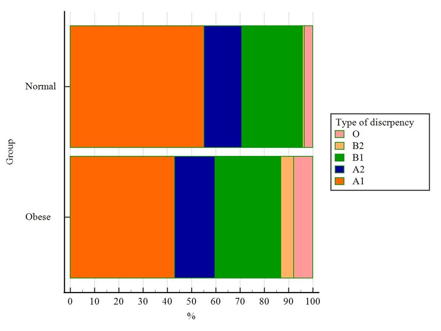
Table 2: Chi-Square Test for Comparison of Discrepancies Between Groups
|
Type of Discrepancy |
Normal (n=116) |
Obese (n=114) |
Total |
|
A1 (Positive ≤7 days) |
64 |
49 |
113 (49.1%) |
|
A2 (Positive ≥8 days) |
18 |
19 |
37 (16.1%) |
|
B1 (Negative ≤7 days) |
29 |
31 |
60 (26.1%) |
|
B2 (Negative ≥8 days) |
1 |
6 |
7 (3.0%) |
|
O (No discrepancy) |
4 |
9 |
13 (5.7%) |
Chi-squared test results: χ² = 7.563, df = 4, p = 0.109
Analysis across five discrepancy categories revealed no significant difference between groups (p = 0.109) (Figure 1). When categories were collapsed into positive, negative, and no-discrepancy groups (Table 3), the difference remained non-significant (p = 0.14).
Table 3: Collapsed Discrepancy Categories Between Groups
|
Discrepancy Type |
Normal (n=116) |
Obese (n=114) |
Total |
|
Positive |
82 (70.7%) |
68 (59.6%) |
150 (65.2%) |
|
Negative |
30 (25.9%) |
37 (32.5%) |
67 (29.1%) |
|
None |
4 (3.4%) |
9 (7.9%) |
13 (5.7%) |
Chi-squared test results: χ² = 3.94, df = 2, p = 0.14
Table 4: Independent Samples t-Test for Discrepancy Between Groups
|
Group |
n |
Mean Discrepancy (days) |
95% CI |
SD |
|
Normal |
116 |
2.83 |
1.92–3.74 |
4.93 |
|
Obese |
114 |
2.00 |
0.94–3.05 |
5.66 |
t = -1.19, df = 228, p = 0.23
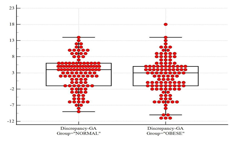
Independent samples t-test (Table 4) confirmed no significant difference in mean discrepancy between groups (p = 0.23). Although a slight preponderance of negative discrepancies was observed among obese participants (suggesting larger fetal size), this difference did not reach statistical significance. The Shapiro-Wilk test for normality (W = 0.9875, p = 0.0422) indicated slight deviation from normality, but the t-test remains robust to moderate violations.
DISCUSSION :
This study investigated whether maternal obesity is associated with significant discrepancies between LMP- and ultrasound-based GA estimation during the second and third trimesters. Our findings demonstrate that while slight differences exist, they are not statistically significant, suggesting that ultrasound remains a reliable tool for GA assessment in overweight and obese pregnant women.
The mean age of normal pregnant women (27.78 years) was lower than that of obese pregnant women (30.50 years) in our study, consistent with previous research suggesting higher BMI is associated with advanced maternal age (31). The mean GA LMP for obese participants (209.60 days, approximately 29.94 weeks) was higher than that for normal participants (187.70 days, approximately 26.81 weeks), reflecting the inclusion of participants across second and third trimesters (32).
The absence of significant discrepancy in our study aligns with some previous research but contradicts others. Bak et al. (33) reported that higher BMI was associated with delayed ultrasound EDD in a population-based cohort of 998 participants with 26–43 weeks' gestation. Similarly, Kullinger et al. (34) found that obese mothers exhibited the largest differences in EDD (±9 to ±20 days) in a population-based cross-sectional register study. Other significant factors that made a large difference included diabetes, young maternal age, history of gestational diabetes, multiple birth, and female fetus (34).
Gulersen and Chervenak (35) stated that higher BMI is significantly correlated with reduced sonographic fetal weight accuracy, although limited literature exists assessing the effect of such sonographic accuracy on neonatal and maternal consequences. Neel et al. (36) found that the sensitivity for detecting LGA was 61.0%, with a PPV of 54.8%, revealing the limited accuracy of third-trimester ultrasounds in identifying growth abnormalities in obese women.
Conversely, our findings are consistent with Field et al. (37), who assessed fetal weight in different BMI categories of pregnant females and reported no difference in sonographically assessed fetal weight and actual birth weight. They also included gestations of 26–43 weeks, similar to those used in our study. Morin et al. (38) found that determinants and consequences of discrepancies in menstrual and ultrasonographic GA estimates varied widely, further supporting the complexity of this relationship.
Several factors may explain divergent findings. First, the sample size in our study (230 participants) was smaller than reference studies (33,34), which may have limited statistical power to detect subtle differences. Second, our study included a larger proportion of overweight (BMI 25–29.9 kg/m²) rather than severely obese (BMI ≥30 kg/m²) participants, potentially attenuating the effect of obesity on GA estimation. This is consistent with Simic et al. (30) who noted that the most pronounced discrepancies occur in women with BMI >40 kg/m². Maxwell et al. (39) similarly demonstrated that obese women (BMI ≥30) had significantly lower success rates of routine anatomical ultrasounds (26% incomplete) compared to normal-weight controls (2.5% incomplete).
Dashe et al. (40) examined how maternal BMI impacts fetal anatomy visualization during second-trimester ultrasounds in a cohort of 10,112 women, finding that the ability to visualize fetal anatomy decreased as maternal BMI increased. This reduction in visibility could lead to errors in measuring fetal size and estimating GA. Paladini (41) reviewed the challenges faced by sonographers while performing ultrasound examinations of overweight and obese pregnant women, addressing technical issues associated with scanning obese mothers.
The slight preponderance of negative discrepancies (ultrasound GA > LMP GA) observed among obese participants in our study is noteworthy. Negative discrepancies suggest that fetuses of obese mothers appear larger for their GA—a finding consistent with established knowledge that maternal obesity is associated with fetal macrosomia (42). Gaudet et al. (43), in a meta-analysis, demonstrated that obesity in mothers correlates with fetal overgrowth, birth weight, and increased GA, likely mediated through mechanisms such as increased insulin resistance (even in nondiabetic women) and elevated fetal blood glucose and insulin levels.
Simic et al. (44) found that the risk of being SGA is significantly increased when GA according to ultrasonography is 7 days shorter than GA according to the menstrual cycle. They also noted that GA estimate according to menstrual cycle is longer than that according to ultrasound, but does not affect the infants or the incidence of morbidity (44). Our findings regarding the lack of significant discrepancy may reflect the inclusion criteria requiring certain LMP and first antenatal records, which potentially reduced variability.
Denison et al. (45) reported that maternal obesity affects length of gestation, risk of postdates pregnancy, and spontaneous onset of labour at term, further emphasizing the importance of accurate GA estimation in this population. Zhang et al. (46) conducted a long-term study with 468 obese and 2,334 non-obese pregnant women, showing that fetuses of obese mothers had consistently larger measurements across all key biometric parameters, including FL, humerus length, HC, and EFW, compared to those of non-obese mothers. By the 32nd week, the difference became especially noticeable.
Our findings have important clinical implications. Accurate GA estimation is crucial for appropriate timing of interventions, classification of fetal growth abnormalities, and prediction of preterm or post-term delivery risks (47). Although no significant discrepancies were found, clinicians should remain vigilant when assessing obese pregnant women, as technical challenges in fetal visualization may compromise measurement accuracy in individual cases (48). Xu et al. (49) demonstrated the importance of incorporating both second- and third-trimester ultrasound data and maternal factors in predicting fetal growth abnormalities.
The discrepancy in our findings compared to studies reporting significant associations may also relate to differences in population characteristics, ultrasound equipment, and operator expertise (50). The GE Logic P7 ultrasound machine employed in our study may provide different measurement accuracy compared to equipment used in other studies. Additionally, the use of convenient sampling may have introduced selection bias.
CONCLUSION :
This study concludes that maternal obesity does not significantly affect the discrepancy between LMP- and ultrasound-based GA estimation during the second and third trimesters. While a slight preponderance of negative discrepancies (suggesting larger fetal size) was observed among obese participants, this difference was not statistically significant. These findings suggest that ultrasound remains a reliable tool for GA assessment in overweight and obese pregnant women, despite the technical challenges associated with increased adipose tissue.
Recommendations
The information obtained from the present study can further be used to conduct other research and can provide foundation to future research by providing literature. Future studies should employ larger sample sizes with adequate representation of severely obese participants (BMI ≥30 kg/m²) to better evaluate the effect of maternal weight on GA measurement. Two groups consisting of adequate numbers of overweight as well as obese mothers are needed to evaluate the effect of increase in maternal weight on GA measurement on ultrasound. Additionally, prospective longitudinal studies incorporating early first-trimester ultrasound as a reference standard would provide more robust evidence. Clinicians should maintain awareness of potential technical limitations when scanning obese patients and consider appropriate adjustments as needed.
Limitations
The study's limitations include a relatively small sample size and the inclusion of a larger proportion of overweight rather than severely obese participants, which may have affected the results as suggested by previous studies (30). The cross-sectional design precludes assessment of temporal changes in discrepancy patterns. Additionally, the use of convenience sampling may limit generalizability of findings to broader populations. The Shapiro-Wilk test indicated slight deviation from normality, although the t-test remains robust to this violation.
REFERENCES:
1. Lim HJ, Xue H, Wang Y. Global trends in obesity. In: Handbook of Eating and Drinking: Interdisciplinary Perspectives. 2020. p. 1217-35.
2. World Health Organization. Obesity: preventing and managing the global epidemic. Report of a WHO consultation. World Health Organ Tech Rep Ser. 2000;894:i-xii, 1-253.
3. American College of Obstetricians and Gynecologists. ACOG Committee opinion no. 549: obesity in pregnancy. Obstet Gynecol. 2013;121(1):213-7.
4. Castro LC, Avina RL. Maternal obesity and pregnancy outcomes. Curr Opin Obstet Gynecol. 2002;14(6):601-6.
5. Ikedionwu CA, Dongarwar D, Yusuf KK, Ibrahimi S, Salinas-Miranda AA, Salihu HM. Pre-pregnancy maternal obesity, macrosomia, and risk of stillbirth: a population-based study. Eur J Obstet Gynecol Reprod Biol. 2020;252:1-6.
6. Yu YH, Bodnar LM, Himes KP, Brooks MM, Naimi AI. Association of overweight and obesity development between pregnancies with stillbirth and infant mortality in a cohort of multiparous women. Obstet Gynecol. 2020;135(3):634-43.
7. Dalbye R, Gunnes N, Blix E, Zhang J, Eggebø T, Nistov Tokheim L, et al. Maternal body mass index and risk of obstetric, maternal and neonatal outcomes: A cohort study of nulliparous women with spontaneous onset of labor. Acta Obstet Gynecol Scand. 2021;100(3):521-30.
8. Kramer MS. The epidemiology of adverse pregnancy outcomes: an overview. J Nutr. 2003;133(5):1592S-6S.
9. Kalish RB, Chervenak FA. Sonographic determination of gestational age. Ultrasound Rev Obstet Gynecol. 2005;5(4):254-8.
10. Tunon K, Eik-Nes SH, Grøttum P. A comparison between ultrasound and a reliable last menstrual period as predictors of the day of delivery in 15,000 examinations. Ultrasound Obstet Gynecol. 1996;8(3):178-85.
11. Chiazze L, Brayer FT, Macisco JJ, Parker MP, Duffy BJ. The length and variability of the human menstrual cycle. JAMA. 1968;203(6):377-80.
12. Deputy NP, Nguyen PH, Pham H, Nguyen S, Neufeld L, Martorell R, et al. Validity of gestational age estimates by last menstrual period and neonatal examination compared to ultrasound in Vietnam. BMC Pregnancy Childbirth. 2017;17:1-9.
13. Bisahnyui P, Nkfusai CN, Bede F, Kemjei M, Atuhaire C, Nchanji K, et al. Comparative study of clinical methods versus ultrasound methods for accurate gestational age determination in different trimesters of pregnancy, Ndop District Hospital, North West region, Cameroon. Pan Afr Med J. 2020;37(1).
14. Chervenak FA, McCullough LB, Chervenak JL. Prenatal informed consent for sonogram: an indication for obstetric ultrasonography. Am J Obstet Gynecol. 1989;161(4):857-60.
15. Mongelli M, Wilcox M, Gardosi J. Estimating the date of confinement: ultrasonographic biometry versus certain menstrual dates. Am J Obstet Gynecol. 2005;174(1 Pt 1):7-11.
16. Grisolia G, Milano K, Pilu G, Banzi C, David C, Gabrielli S, et al. Biometry of early pregnancy with transvaginal sonography. Ultrasound Obstet Gynecol. 1993;3(6):403-11.
17. Papageorghiou AT, Kemp B, Stones W, Ohuma EO, Kennedy SH, Purwar M, et al.; International Fetal and Newborn Growth Consortium for the 21st Century (INTERGROWTH-21st). Ultrasound-based gestational-age estimation in late pregnancy. Ultrasound Obstet Gynecol. 2016;48(6):719-26.
18. Caradeux J, Gratacós E, Figueras F. Normal and abnormal fetal growth. In: New Technologies and Perinatal Medicine. CRC Press; 2019. p. 18-22.
19. Weichert J, Hartge DR. Obstetrical sonography in obese women: a review. J Clin Ultrasound. 2011;39(4):209-16.
20. Beazley JM, Underhill RA. Fallacy of the fundal height. Br Med J. 1970;4(5732):404-6.
21. Dril L, Cacciaguerra G, Citossi A, Martina MD, Peressini L, Marchesoni D. Prepregnancy body mass index and adverse pregnancy outcomes. Arch Gynecol Obstet. 2008;278:23-6.
22. Galtier-Dereure F, Boegner C, Bringer J. Obesity and pregnancy: complications and cost. Am J Clin Nutr. 2000;71(5):1242S-8S.
23. Lee C, Willis A, Chen C, Sieniek M, Watters A, Stetson B, et al. Development of a machine learning model for sonographic assessment of gestational age. JAMA Netw Open. 2023;6(1):e2248685.
24. Campbell F, Johnson M, Messina J, Guillaume L, Goyder E. Behavioural interventions for weight management in pregnancy: a systematic review of quantitative and qualitative data. BMC Public Health. 2011;11:1-13.
25. Olesen AW, Thomsen SG. Prediction of delivery date by sonography in the first and second trimesters. Ultrasound Obstet Gynecol. 2006;28(3):292-7.
26. Bak GS, Sperling L, Källén K, Salvesen KÅ. Prospective population-based cohort study of maternal obesity as a source of error in gestational age estimation at 11–14 weeks. Acta Obstet Gynecol Scand. 2016;95(11):1281-7.
27. Li S, Zhao JH, Luben RN, Rodwell SA, Khaw KT, Ong KK, et al. Cumulative effects and predictive value of common obesity-susceptibility variants identified by genome-wide association studies. Am J Clin Nutr. 2010;91(1):184-90.
28. Erkamp JS, Voerman E, Steegers EA, Mulders AG, Reiss IK, Duijts L, et al. Second and third trimester fetal ultrasound population screening for risks of preterm birth and small-size and large-size for gestational age at birth: a population-based prospective cohort study. BMC Med. 2020;18:1-12.
29. Nelson L, Wharton B, Grobman WA. Prediction of large for gestational age birth weights in diabetic mothers based on early third-trimester sonography. J Ultrasound Med. 2011;30(12):1625-8.
30. Simic M, Wåhlin IA, Maršál K, Källén K. Maternal obesity is a potential source of error in mid-trimester ultrasound estimation of gestational age. Ultrasound Obstet Gynecol. 2010;35(1):48-53.
31. Salah Ali A, Fawzy Mohamed M, Soliman Taha W. The effect of maternal obesity on sonographic fetal weight estimation. Al-Azhar Med J. 2022;51(2):989-98.
32. Rekawek P, Liu L, Getrajdman C, Brooks C, Pan S, Overbey J, et al. Large-for-gestational age diagnosed during second-trimester anatomy ultrasound and association with gestational diabetes and large-for-gestational age at birth. Ultrasound Obstet Gynecol. 2020;56(6):901-5.
33. Bak GS, Sperling L, Källén K, Salvesen KÅ. Prospective population-based cohort study of maternal obesity as a source of error in gestational age estimation at 11–14 weeks. Acta Obstet Gynecol Scand. 2016;95(11):1281-7.
34. Kullinger M, Wesström J, Kieler H, Skalkidou A. Maternal and fetal characteristics affect discrepancies between pregnancy-dating methods: a population-based cross-sectional register study. Acta Obstet Gynecol Scand. 2017;96(1):86-95.
35. Gulersen M, Chervenak FA. Challenges of third-trimester screening in obese women. In: Obesity and Obstetrics. Elsevier; 2020. p. 69-73.
36. Neel A, Cunningham CE, Teale GR. A routine third trimester growth ultrasound in the obese pregnant woman does not reliably identify fetal growth abnormalities: A retrospective cohort study. Aust N Z J Obstet Gynaecol. 2021;61(1):116-22.
37. Field NT, Piper JM, Langer O. The effect of maternal obesity on the accuracy of fetal weight estimation. Obstet Gynecol. 1995;86(1):102-7.
38. Morin I, Morin L, Zhang X, Platt RW, Blondel B, Bréart G, et al. Determinants and consequences of discrepancies in menstrual and ultrasonographic gestational age estimates. BJOG. 2005;112(2):145-52.
39. Maxwell C, Dunn E, Tomlinson G, Glanc P. How does maternal obesity affect the routine fetal anatomic ultrasound? J Matern Fetal Neonatal Med. 2010;23(10):1187-92.
40. Dashe JS, McIntire DD, Twickler DM. Maternal obesity limits the ultrasound evaluation of fetal anatomy. J Ultrasound Med. 2009;28(8):1025-30.
41. Paladini D. Sonography in obese and overweight pregnant women: clinical, medicolegal and technical issues. Ultrasound Obstet Gynecol. 2009;33(6):720-9.
42. Glazer KB, Danilack VA, Field AE, Werner EF, Savitz DA. Term labor induction and cesarean delivery risk among obese women with and without comorbidities. Am J Perinatol. 2022;39(02):154-64.
43. Gaudet L, Ferraro ZM, Wen SW, Walker M. Maternal obesity and occurrence of fetal macrosomia: a systematic review and meta-analysis. Biomed Res Int. 2014;2014:640291.
44. Simic M, Amer-Wåhlin I, Lagercrantz H, Maršál K, Källén K. Survival and neonatal morbidity among extremely preterm born infants in relation to gestational age based on the last menstrual period or ultrasonographic examination. J Perinat Med. 2014;42(2):247-53.
45. Denison FC, Price J, Graham C, Wild S, Liston WA. Maternal obesity, length of gestation, risk of postdates pregnancy and spontaneous onset of labour at term. BJOG. 2008;115(6):720-5.
46. Zhang C, Hediger ML, Albert PS, Grewal J, Sciscione A, Grobman WA, et al. Association of maternal obesity with longitudinal ultrasonographic measures of fetal growth: findings from the NICHD Fetal Growth Studies–Singletons. JAMA Pediatr. 2018;172(1):24-31.
47. Papageorghiou AT, Kemp B, Stones W, Ohuma EO, Kennedy SH, Purwar M, et al. Ultrasound-based gestational-age estimation in late pregnancy. Ultrasound Obstet Gynecol. 2016;48(6):719-26.
48. Weichert J, Hartge DR. Obstetrical sonography in obese women: a review. J Clin Ultrasound. 2011;39(4):209-16.
49. Xu D, Shen X, Guan H, Zhu Y, Yan M, Wu X. Prediction of small-for-gestational-age neonates at 33–39 weeks' gestation in China: logistic regression modeling of the contributions of second-and third-trimester ultrasound data and maternal factors. BMC Pregnancy Childbirth. 2022;22(1):661.
50. Dashe JS, McIntire DD, Twickler DM. Maternal obesity limits the ultrasound evaluation of fetal anatomy. J Ultrasound Med. 2009;28(8):1025-30.