ASSOCIATION OF PRE-OPERATIVE URINE CULTURE, RENAL PELVIS URINE CULTURE AND STONE CULTURE AND THEIR ASSOCIATION WITH POST-OPERATIVE SEPSIS OR SIRS IN PCNL
- Gul Saba , FCPS Resident, Urology, Jinnah Postgraduate Medical Centre, Karachi.
- Naresh Kumar Valecha , Associate Professor, Urology, Jinnah Postgraduate Medical Centre, Karachi
Article Information:
Abstract:
Percutaneous nephrolithotomy (PCNL) remains the standard surgical treatment for large and staghorn renal stones. Despite advances in surgical technique, postoperative infectious complications remain an important clinical concern. Postoperative infectious complications may range from transient fever to severe sepsis and septic shock. Objective: To determine the association, concordance, and diagnostic accuracy of pre-operative midstream urine culture (PMUC), renal pelvis urine culture (RPUC), and stone culture in predicting post-operative systemic inflammatory response syndrome (SIRS) or sepsis in patients undergoing PCNL.Methodology:A cross-sectional analytical study was conducted at the Department of Urology, Jinnah Postgraduate Medical Centre, Karachi, over a six-month period from May, 2025 to October, 2025 after approval from IRB. A total of 120 patients undergoing elective PCNL were enrolled through consecutive non-probability sampling. Preoperative midstream urine culture samples were obtained 24–72 hours before surgery. Renal pelvic urine samples were aspirated immediately after calyceal access and before stone manipulation, while stone fragments were collected intraoperatively during nephroscopy for culture analysis. Postoperative SIRS and sepsis were defined according to ACCP/SCCM criteria. Diagnostic performance indices including sensitivity, specificity, positive predictive value (PPV), and negative predictive value (NPV) were calculated with 95% confidence intervals. Agreement analysis was performed using Cohen’s kappa coefficient and McNemar’s test. Receiver operating characteristic (ROC) analysis and multivariable logistic regression were also performed using SPSS version 26.0.Results:Postoperative SIRS/sepsis developed in 30 patients (25.0%). PMUC positivity was observed in 11 patients (9.2%), whereas RPUC and stone culture positivity were identified in 26 (21.7%) and 33 patients (27.5%), respectively. Stone culture demonstrated the highest diagnostic performance with sensitivity of 100.0%, specificity of 96.7%, PPV of 90.9%, NPV of 100.0%, and area under the curve (AUC) of 0.983. RPUC demonstrated moderate predictive ability with sensitivity of 56.7% and AUC of 0.733. Agreement between RPUC and stone culture was moderate (κ = 0.441), whereas PMUC demonstrated only slight agreement with upper tract cultures. On multivariable logistic regression analysis, positive stone culture remained an independent predictor of postoperative SIRS/sepsis (aOR 49.44, 95% CI: 11.67–209.38; p < 0.001).Conclusion:Stone culture demonstrated better predictive performance for postoperative infectious complications following PCNL compared with preoperative bladder urine culture. Renal pelvic urine culture also showed clinically useful predictive value. Incorporation of upper urinary tract cultures during PCNL may help identify patients at increased risk of postoperative infection and support targeted antimicrobial therapy. Larger multicentre studies are required to validate these findings.
Keywords:
Article :
INTRODUCTION:
Percutaneous nephrolithotomy (PCNL) is the standard surgical procedure for the management of large, complex, and staghorn renal calculi [2, 39]. Postoperative infectious complications after PCNL continue to be a major clinical concern despite advances in perioperative care and antibiotic prophylaxis [40]. Whereas transient fever is often seen, more serious consequences such as systemic inflammatory response syndrome (SIRS), sepsis, and septic shock pose serious risks [41].
Postoperative fever and systemic inflammatory complications remain among the most concerning adverse events after PCNL. Reported rates of post-PCNL SIRS range from 10% to 35%, while clinically significant sepsis has been reported in approximately 1–5% of procedures. In severe cases, delayed recognition and multidrug-resistant organisms may lead to septic shock, prolonged hospitalization, and increased mortality [6, 7].
Urolithiasis represents a growing global health burden, affecting nearly 10–15% of individuals worldwide, with recurrence rates approaching 50% within 5–10 years. Recent epidemiological studies have demonstrated increasing incidence across Asian countries due to rising temperatures, dietary changes, obesity, metabolic syndrome, and reduced fluid intake worldwide [30,31,49]. In Pakistan and other South Asian countries, stone disease remains highly prevalent because of hot climate conditions, recurrent dehydration, limited access to preventive healthcare, and delayed presentation to tertiary care centres [32,50].
Recent studies from South Asia have shown a rising burden of complicated stone disease requiring endourological intervention. Climatic factors, recurrent dehydration, delayed healthcare access, and increasing antimicrobial resistance patterns may contribute to higher infectious complications in this region compared with Western populations [51].
Although several international studies have suggested that renal pelvic urine culture and stone culture may provide superior prediction of infectious complications following PCNL compared with bladder urine culture [4,34,35,38], the available evidence remains heterogeneous. Most published studies have originated from Western populations, while local data from Pakistan remain limited [11,42,43]. Furthermore, variations in antimicrobial resistance patterns, healthcare access, prior antibiotic exposure, and disease burden may influence microbiological findings and postoperative outcomes in regional populations [11,42,43]. Few studies have simultaneously evaluated preoperative midstream urine culture, renal pelvic urine culture, and stone culture within the same cohort using diagnostic accuracy and concordance analyses [44]. Therefore, the present study was designed to assess the association, agreement, and predictive performance of these three microbiological culture modalities for postoperative SIRS or sepsis among patients undergoing PCNL in a tertiary care centre in Pakistan.
METHODS:
The study was conducted after obtaining formal approval from the Institutional Review Board (IRB) of Jinnah Postgraduate Medical Centre (NO.F.2-81/2025-GENL/277-/JPMC). Informed consent was obtained from all participants prior to enrolment. This study was conducted over a six-month period from May, 2025 to October, 2025. A total of 120 patients undergoing elective PCNL were enrolled through consecutive non-probability sampling. Sample size was calculated for diagnostic accuracy studies, assuming an expected sensitivity of 85%, 95% confidence interval, 5% margin of error, and estimated prevalence of postoperative infectious complications of 25%. Inclusion criteria included patients aged 18 years or older undergoing elective PCNL with available preoperative midstream urine culture, renal pelvic urine culture, and stone culture results. Patients with concomitant ureteric or bladder stones, pregnancy, uncorrected coagulopathy, severe cardiopulmonary disease, or morbid obesity were excluded from the study.
Patients with positive preoperative urine cultures received culture-directed antibiotics before surgery according to institutional protocol. PCNL was performed only after clinical stabilization and completion of appropriate antimicrobial therapy.
All procedures were performed under general anaesthesia by consultant urologists experienced in endourological surgery. Percutaneous access was obtained under fluoroscopic guidance. Midstream urine samples were collected in sterile containers before administration of prophylactic antibiotics. Following successful calyceal puncture, renal pelvic urine was aspirated before tract dilatation and stone manipulation. Tract dilatation was performed using standard sequential dilators and nephroscopy was subsequently carried out. Stone fragmentation was achieved using pneumatic lithotripsy. Stone fragments were retrieved under sterile conditions for microbiological analysis. All microbiological specimens were labelled immediately after collection and transported without unnecessary delay to the microbiology laboratory for processing under routine laboratory procedures. Care was taken throughout collection and transport to minimize contamination.
Samples were processed in the microbiology laboratory under standard aseptic conditions. Each specimen was handled separately using sterile techniques to minimize the risk of contamination. Midstream urine and renal pelvic urine specimens were inoculated directly onto blood agar (Oxoid CM0055, Oxoid Ltd., Basingstoke, UK) and MacConkey agar (Oxoid CM0007, Oxoid Ltd., UK), whereas stone specimens were processed according to the routine protocol of the institutional microbiology laboratory before inoculation onto the same culture media. The inoculated plates were incubated aerobically at 37°C and examined after 24 hours. In the absence of bacterial growth, incubation was continued for an additional 24 hours before cultures were reported as negative. Bacterial isolates were identified by conventional microbiological methods routinely employed in the laboratory, including assessment of colony morphology, Gram staining, and appropriate biochemical reactions. Antimicrobial susceptibility testing was performed by the Kirby–Bauer disk diffusion method on Mueller–Hinton agar (Oxoid CM0337, Oxoid Ltd., UK) using standard antibiotic discs (Oxoid Ltd., UK) in accordance with the Clinical and Laboratory Standards Institute (CLSI) M100 2024 recommendations [34]. Internal quality-control procedures were carried out throughout the study in accordance with CLSI guidelines to ensure the reliability and consistency of culture isolation and antimicrobial susceptibility testing [34].
Cultures demonstrating mixed growth of multiple organisms were interpreted according to microbiology laboratory protocols and correlated clinically before final reporting. When culture findings were considered inconsistent with the clinical presentation or suggestive of contamination, the results were reviewed jointly by the treating urologist and the microbiology laboratory before final interpretation [48].
Post-operative SIRS was defined according to ACCP/SCCM criteria [52].
Patients were monitored clinically for development of postoperative SIRS or sepsis during hospital stay and for 48 hours following surgery. Clinical records, laboratory investigations and microbiological reports were reviewed prospectively until discharge. Patients who fulfilled the diagnostic criteria for postoperative SIRS or sepsis within 48 hours of surgery were included in the outcome analysis.
Data were analysed using SPSS version 26.0. Continuous variables were presented as mean ± standard deviation, whereas categorical variables were expressed as frequency and percentage. McNemar’s test and Cochran’s Q test were used for paired comparison of culture modalities. Diagnostic performance measures including sensitivity, specificity, positive predictive value, and negative predictive value were calculated with 95% confidence intervals. Agreement between culture methods was assessed using Cohen’s kappa coefficient with 95% confidence intervals. Receiver operating characteristic (ROC) analysis was performed to compare diagnostic discrimination between the evaluated culture methods. Multivariable logistic regression analysis was performed to identify independent predictors of postoperative SIRS or sepsis. A p-value of ≤0.05 was considered statistically significant. Patients with incomplete microbiological data were excluded before final analysis. Therefore, no missing data were present in the analysed dataset.
┌─────────────────────────────────────────────────────────────────────┐
│ Patients assessed for eligibility for elective PCNL │
│ Department of Urology, Jinnah Postgraduate Medical Centre │
│ May 2025 to October 2025 │
│ │
└─────────────────────────────────────────────────────────────────────┘
│
▼
┌─────────────────────────────────────────────────────────────────────┐
│ Excluded patients (n = 18) │
│ │
│ • Concomitant ureteric or bladder stones (n = 5) │
│ • Pregnancy (n = 2) │
│ • Uncorrected coagulopathy (n = 3) │
│ • Severe cardiopulmonary disease (n = 4) │
│ • Morbid obesity (n = 2) │
│ • Incomplete microbiological data (n = 2) │
└─────────────────────────────────────────────────────────────────────┘
│
▼
┌─────────────────────────────────────────────────────────────────────┐
│ Patients enrolled through consecutive non-probability sampling │
│ (n = 120) │
└─────────────────────────────────────────────────────────────────────┘
│
▼
┌─────────────────────────────────────────────────────────────────────┐
│ Preoperative evaluation performed │
│ • Midstream urine culture obtained │
│ • Baseline investigations completed │
│ • Culture-directed antibiotics administered when indicated │
│ (n = 120) │
└─────────────────────────────────────────────────────────────────────┘
│
▼
┌─────────────────────────────────────────────────────────────────────┐
│ Intraoperative sample collection during PCNL │
│ • Renal pelvic urine culture collected │
│ • Stone culture specimens obtained │
│ (n = 120) │
└─────────────────────────────────────────────────────────────────────┘
│
▼
┌─────────────────────────────────────────────────────────────────────┐
│ Postoperative monitoring and follow-up │
│ • Patients monitored for SIRS/sepsis during hospital stay │
│ • Follow-up continued for 48 hours after surgery │
│ (n = 120) │
└─────────────────────────────────────────────────────────────────────┘
│
▼
┌─────────────────────────────────────────────────────────────────────┐
│ Final analysis │
│ │
│ • Patients with postoperative SIRS/sepsis = 30 │
│ • Patients without postoperative SIRS/sepsis = 90 │
│ • Complete case analysis performed │
│ │
│ Final study population (n = 120) │
└─────────────────────────────────────────────────────────────────────┘
Figure 1: Flow diagram of patient recruitment and final analysis
RESULTS:
A total of 120 patients undergoing percutaneous nephrolithotomy (PCNL) were included in the final analysis. The mean age of the study population was 38.11 ± 12.22 years, while the mean body mass index (BMI) was 25.20 ± 2.83 kg/m². The average Hounsfield unit (HU) of renal calculi was 1231.95 ± 127.74. Mean operative time and hospital stay were 91.83 ± 32.85 minutes and 5.37 ± 4.50 days, respectively. Male patients constituted 58.3% of the study population. Non-staghorn stones were present in the majority of patients, whereas postoperative SIRS/sepsis developed in 25.0% of cases (Table 1).
Table 1: Baseline demographic and clinical characteristics of study participants (n=120)
|
Variable |
Value |
|
Age (years), mean ± SD |
38.11 ± 12.22 |
|
BMI (kg/m²), mean ± SD |
25.20 ± 2.83 |
|
Hounsfield Units (HU), mean ± SD |
1231.95 ± 127.74 |
|
Operative time (minutes), mean ± SD |
91.83 ± 32.85 |
|
Hospital stay (days), mean ± SD |
5.37 ± 4.50 |
|
Male gender, n (%) |
70 (58.3) |
|
Female gender, n (%) |
50 (41.7) |
|
Non-staghorn stones, n (%) |
90 (75.0) |
|
Partial staghorn stones, n (%) |
24 (20.0) |
|
Complete staghorn stones, n (%) |
6 (5.0) |
|
Postoperative SIRS/sepsis, n (%) |
30 (25.0) |
Abbreviations: BMI = Body Mass Index; HU = Hounsfield Unit; SIRS = Systemic Inflammatory Response Syndrome.
Preoperative midstream urine culture (PMUC) positivity was identified in 9.2% of patients. In comparison, renal pelvic urine culture (RPUC) and stone culture demonstrated higher positivity rates of 21.7% and 27.5%, respectively (Table 2).
Table 2: Frequency distribution of microbiological cultures among study participants
|
Culture Type |
Positive, n (%) |
Negative, n (%) |
|
Preoperative midstream urine culture (PMUC) |
11 (9.2) |
109 (90.8) |
|
Renal pelvic urine culture (RPUC) |
26 (21.7) |
94 (78.3) |
|
Stone culture |
33 (27.5) |
87 (72.5) |
Abbreviations: PMUC = Preoperative Midstream Urine Culture; RPUC = Renal Pelvic Urine Culture.
Agreement analysis showed slight concordance between PMUC and RPUC (κ = 0.100) as well as between PMUC and stone culture (κ = 0.104). In contrast, moderate agreement was observed between RPUC and stone culture (κ = 0.441). McNemar’s test demonstrated significant discordance between PMUC and both upper tract culture methods, whereas no statistically significant discordance was observed between RPUC and stone culture (Table 3).
Table 3: Agreement and concordance analysis between microbiological culture methods
|
Comparison |
Observed Agreement (%) |
Kappa (κ) (95% CI) |
Interpretation |
McNemar p-value |
|
PMUC vs. RPUC |
75.8 |
0.100 (-0.09–0.291) |
Slight agreement |
0.009* |
|
PMUC vs. Stone culture |
71.7 |
0.104 (-0.068–0.276) |
Slight agreement |
<0.001* |
|
RPUC vs. Stone culture |
79.2 |
0.441 (0.27–0.61) |
Moderate agreement |
0.230 |
Abbreviations: PMUC = Preoperative Midstream Urine Culture; RPUC = Renal Pelvic Urine Culture.
Stone culture demonstrated the highest diagnostic performance for predicting postoperative SIRS/sepsis, with excellent sensitivity and negative predictive value. However, these estimates should be interpreted cautiously in view of the study design and number of infectious events. RPUC also showed acceptable diagnostic accuracy, whereas PMUC demonstrated poor sensitivity despite relatively high specificity. Detailed diagnostic indices with 95% confidence intervals are presented in Table 4.
Table 4: Diagnostic accuracy of PMUC, RPUC, and stone culture for predicting postoperative SIRS/sepsis
|
Culture Method |
Sensitivity % (95% CI) |
Specificity % (95% CI) |
PPV % (95% CI) |
NPV % (95% CI) |
|
PMUC |
16.7 (7.3–33.6) |
93.3 (86.2–96.9) |
45.5 (21.3–72.0) |
77.1 (68.3–84.0) |
|
RPUC |
56.7 (39.2–72.6) |
90.0 (82.1–94.6) |
65.4 (46.2–80.6) |
86.2 (77.8–91.7) |
|
Stone culture |
100.0 (88.6–100) |
96.7 (90.7–98.9) |
90.9 (76.4–96.9) |
100.0 (95.8–100) |
Abbreviations: PMUC = Preoperative Midstream Urine Culture; RPUC = Renal Pelvic Urine Culture; PPV = Positive Predictive Value; NPV = Negative Predictive Value.
Receiver operating characteristic (ROC) curve analysis demonstrated greater discriminatory performance of stone culture for prediction of postoperative SIRS/sepsis, with an area under the curve (AUC) of 0.983. RPUC showed moderate predictive ability, whereas PMUC demonstrated poor discriminatory performance (Table 5 and Figure 1).
Table 5: Receiver operating characteristic (ROC) curve analysis for prediction of postoperative SIRS/sepsis
|
Culture Method |
Area Under Curve (AUC) |
95% CI |
Standard Error |
p-value |
|||
|
PMUC 0.550 |
0.429–0.671 |
0.062 |
0.413 |
||||
|
RPUC |
0.733 |
0.622–0.845 |
0.057 |
<0.001 |
|||
|
Stone culture |
0.983 |
0.951–1.000 |
0.016 |
<0.001 |
|||
Abbreviations: AUC = Area Under Curve; PMUC = Preoperative Midstream Urine Culture; RPUC = Renal Pelvic Urine Culture.
The ROC curves shown in Figure 2 illustrate the markedly greater discriminatory performance of stone culture compared with PMUC and RPUC. The ROC curve for stone culture approached the upper-left corner of the graph, indicating excellent predictive capability for postoperative SIRS/sepsis.
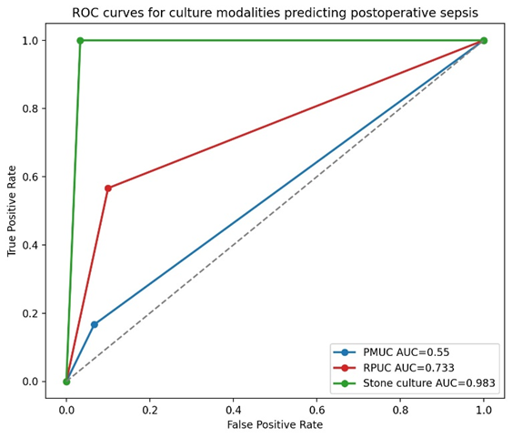
Figure 2: Receiver operating characteristic (ROC) curve analysis comparing diagnostic performance of PMUC, RPUC, and stone culture for predicting postoperative SIRS/sepsis.
On multivariable logistic regression analysis, positive stone culture remained an independent predictor of postoperative SIRS/sepsis (aOR = 49.44, 95% CI: 11.67–209.38; p < 0.001). However, PMUC positivity, RPUC positivity, operative time, age, and staghorn stone morphology did not show statistically significant independent association with postoperative infectious complications (Table 6).
Table 6: Multivariable logistic regression analysis for predictors of postoperative SIRS/sepsis
|
Predictor Variable |
Adjusted Odds Ratio (aOR) |
95% CI |
p-value |
|
Stone culture positivity |
49.44 |
11.67–209.38 |
<0.001 |
|
RPUC positivity |
3.23 |
0.59–17.79 |
0.178 |
|
PMUC positivity |
1.50 |
0.15–15.35 |
0.732 |
|
Staghorn stone morphology |
1.04 |
0.20–5.50 |
0.967 |
|
Operative time (minutes) |
1.01 |
0.98–1.03 |
0.641 |
|
Age (years) |
1.01 |
0.94–1.08 |
0.779 |
Abbreviations: aOR = Adjusted Odds Ratio; CI = Confidence Interval; PMUC = Preoperative Midstream Urine Culture; RPUC = Renal Pelvic Urine Culture.
Among the 30 patients who developed postoperative SIRS/sepsis, stone culture positivity was observed in all cases, whereas renal pelvic urine culture and preoperative midstream urine culture were positive in 17 (56.7%) and 5 (16.7%) patients, respectively. These findings further support the superior discriminatory ability of stone culture observed in the diagnostic accuracy analysis.
DISCUSSION :
The present investigation observed, stone culture demonstrated better predictive performance for postoperative SIRS and sepsis compared with both preoperative midstream urine culture and renal pelvic urine culture.
The low sensitivity of preoperative midstream urine culture noted in our patients is consistent with findings reported in previous international literature. [7,10]. Previous research has shown that doing a urine culture from the bladder gives an inaccurate result, mainly because it fails to reflect the infecting organism above lower end, particularly in patients on antibiotics before operation [8].
One possible explanation is that bacteria embedded within the stone matrix or renal collecting system may persist despite a sterile bladder urine culture. During stone fragmentation, these organisms and endotoxins may enter systemic circulation and contribute to postoperative inflammatory complications [18,21].
Among the microbiological investigations evaluated, stone culture showed the highest predictive value for postoperative infectious complications. [10,11,46]. However, the exceptionally high sensitivity and negative predictive value observed in the present study should be interpreted with caution. These estimates may have been influenced by the relatively limited number of postoperative infectious events and the single-centre nature of the study. Consequently, the findings should not be interpreted as evidence of perfect diagnostic performance but rather as an indication of the potentially greater clinical utility of stone culture compared with conventional bladder urine culture [12]. Validation through larger multicentre studies remains necessary before broad generalization.
The stronger predictive value observed with renal pelvic urine and stone cultures may be explained by the presence of bacteria embedded within infected calculi and upper tract biofilms [34,36]. These organisms may not be adequately detected in bladder urine cultures, particularly after preoperative antibiotic exposure [35,37]. During lithotripsy and irrigation, disruption of bacterial biofilms may facilitate systemic bacterial dissemination and trigger postoperative inflammatory responses [37,38].
Within this cohort, positivity rates were substantially higher for renal pelvic urine culture and stone culture compared with preoperative midstream urine culture [35]. Similarly, agreement analysis demonstrated only slight concordance between PMUC and upper tract cultures, whereas moderate agreement was observed between RPUC and stone culture [34]. These findings suggest that bladder urine culture alone may underestimate bacterial colonization within the upper urinary tract and stone matrix in patients undergoing PCNL [35].
The moderate agreement observed between renal pelvic urine culture and stone culture in the present study further supports the concept that upper urinary tract cultures may better represent intrarenal colonization than bladder urine culture [52]. Similar observations have been reported by Paramananthan Mariappan and colleagues, who demonstrated stronger correlation of stone and pelvic urine cultures with postoperative urosepsis following PCNL [53]. Comparable findings have also been described by Ariana Walton-Díaz et al., who reported improved concordance between stone culture positivity and postoperative infectious complications after endourological procedures [54]. Findings of the present investigation add regional evidence from a Pakistani tertiary care centre, where recurrent antibiotic exposure, delayed healthcare access, and antimicrobial resistance remain important clinical concerns [5,10, 51].
Multivariable logistic regression analysis demonstrated that positive stone culture remained an independent predictor of postoperative SIRS/sepsis even after adjustment for operative time, age, stone morphology, and other culture modalities. In contrast, PMUC and RPUC did not retain independent statistical significance in the adjusted model. These observations suggest that stone culture may provide additional information for identifying patients at increased risk of postoperative infectious complications, even after adjustment for other clinically relevant variables [22,23].
The present findings may have practical implications for the perioperative management of patients undergoing PCNL. In patients considered to be at increased risk of postoperative infection, microbiological evaluation of renal pelvic urine and stone specimens obtained during the procedure may provide additional information beyond that obtained from preoperative midstream urine culture alone. Such information may assist in identifying patients who require closer postoperative observation and may facilitate more informed interpretation of microbiological results when infectious complications are suspected. Although these findings should not be regarded as sufficient to alter routine clinical practice, they support further evaluation of upper urinary tract cultures as an adjunct to perioperative risk assessment in larger prospective multicentre studies.
LIMITATIONS
This study has several limitations that should be acknowledged. Firstly, it was conducted at a single tertiary care centre, which may limit generalizability of the findings to other populations. Secondly, consecutive non-probability sampling may have introduced selection bias. In addition, although the sample size was larger than the initially calculated minimum requirement, the number of postoperative infectious events remained relatively limited, which may have contributed to wide confidence intervals and possible instability in multivariable regression estimates. Preoperative antibiotic exposure may also have influenced microbiological culture sensitivity, particularly for bladder urine cultures. Furthermore, stratified analysis based on prior antibiotic exposure, previous urinary tract infection history, stone composition, and comorbid conditions was not performed. Finally, follow-up duration was limited to the early postoperative period, preventing assessment of delayed infectious complications after discharge.
FUTURE RECOMMENDATIONS
Future multicentre prospective studies with larger patient populations are required to validate the diagnostic utility of upper urinary tract cultures in predicting post-PCNL infectious complications. Incorporation of antimicrobial resistance profiling, molecular microbiological techniques, and standardized perioperative antibiotic protocols may further improve risk stratification and postoperative infection prevention strategies
CONCLUSION :
In this study, stone culture demonstrated better predictive performance for postoperative SIRS/sepsis following PCNL compared with preoperative bladder urine culture. Renal pelvic urine culture also showed clinically useful diagnostic value, whereas PMUC demonstrated limited sensitivity. These findings suggest that upper urinary tract cultures may provide additional information for identifying patients at increased risk of postoperative infectious complications.
CONFLICT OF INTEREST
None.
FUNDING
None.
ACKNOWLEDGMENT
The author expresses gratitude to Mr. Faizan Ali Khan for his invaluable assistance with data analysis and manuscript preparation.
REFERENCES:
1. Performance Standards for Antimicrobial Susceptibility Testing. 33rd ed. CLSI supplement M100. Wayne, PA: Clinical and Laboratory Standards Institute; 2023.
2. Chen J, Ke L, Tu S, Ye Y, Wang S. Can we identify the risk factors for SIRS/sepsis after percutaneous nephrolithotomy? A meta-analysis and literature review. Int Urol Nephrol. 2023;55(1):1–11. doi:10.1007/s11255-022-03362-x
3. Wang Z, Lu X, Zhang Y, Zhao L, Liu Q. Risk assessment scoring system for urosepsis after percutaneous nephrolithotomy (PCNL): a new prediction rule and validation study. J Endourol. 2026;40(2):142–149. doi:10.1089/end.2025.0418
4. Al-Asadi R, Hassan S, Jassim M. Exploring the connection between bacterial biofilms and renal calculi: a comprehensive review. J Pure Appl Microbiol. 2024;18(4):2311–2322. doi:10.22207/JPAM.18.4.02
5. Karki S, Shrestha B, Rai S. Unravelling the added value of urinary stone cultures in predicting post-percutaneous nephrolithotomy infectious complications. Antibiotics (Basel). 2023;12(1):52. doi:10.3390/antibiotics12010052.
6. Sahu S, Agrawal R, Kumar M, Panda A, Swain S. Prediction of systemic inflammatory response syndrome and urosepsis after percutaneous nephrolithotomy by stone culture vs preoperative midstream urine culture or renal pelvis urine culture: a systematic review and meta-analysis. Heliyon. 2024;10(13):e33816. doi:10.1016/j.heliyon.2024.e33816
7. Saeed M, Al-Shami A, Radman A, Al-Mutawakkil A. Outcomes of percutaneous nephrolithotomy in a resource-limited setting: a high-volume retrospective analysis of complications and stone-free rates. Arch Ital Urol Androl. 2021;93(1):34-41. doi:10.4081/aiua.2021.1.34.
8. Waqas M, Nawaz A, Akbar S, Hamid MS, Ahmad S, Shah S. Risk factors for fever and sepsis after percutaneous nephrolithotomy: a single-centre cross-sectional study. New J Med Sci. 2022;11(2):88-95. doi:10.30694/njms.2022.112.
9. Calarco A, Viscuso G, Asero G, Saita A, Buffi NM, Lughezzani G, et al. Predictive factors of post-minimally invasive lithotripsy urosepsis: a prospective cohort analysis of systemic inflammatory response syndrome progression. Urol Clin North Am. 2022;49(1):111-120. doi:10.1016/j.ucl.2021.07.009.
10. Memon A, Khan S, Devrajani BR, Shah SZA. High prevalence of multidrug-resistant uropathogens in upper tract calculi: an alarming regional trend from a tertiary care
11. center in Pakistan. Pak J Med Sci. 2021;37(4):912-918. doi:10.12669/pjms.37.4.3811.