Comparison of Biomarker Levels in Healthy Individuals vs Patients with Precancerous Lesions to Predict Oral Cancer.
- Ashfaque Sahito , Post graduate resident oral and maxillofacial surgery lumhs jamshoro.
- Kashif Ali Channar , Prof/consultant oral and maxillofacial surgery lumhs jamshoro.
- Shoaib Hassan , Pgomfs lumhs jam.
- Aisha Laghari , Pgomfs lumhs jam
Article Information:
Abstract:
Abstract: Background: Oral cancer has been one of the major issues of concern to the general population, especially where the habit of smoking tobacco and consumption of betel nuts are prevalent. Oral potentially malignant lesions like leukoplakia, erythroplakia and verrucous hyperplasia can have malignant transformation. The discovery of trustworthy biomarkers can enhance risk evaluation and early detection. Objective: To compare serum levels of carcinoembryonic antigen and ferritin between patients with oral precancerous lesions and healthy individuals in order to evaluate their potential role as predictive biomarkers for oral cancer. Methods: The case control study was carried out at the Department of Oral and maxillofacial Surgery, Liaquat University of Medical and Health Sciences, Jamshoro/hyderbed. The sample size was 60 participants who were further divided to form two different groups that included 30 patients with clinically diagnosed oral precancerous lesions and 30 healthy controls. Serum carcinoembryonic antigen and serum ferritin were measured in the blood samples by the enzyme-linked immunosorbent assay and the immunoturbidimetry methods respectively. The SPSS version 22.0 was used to analyse the data and to make comparisons between the groups by means of relevant statistical tests on the level of significance p < 0.05. Results: Elevated carcinoembryonic antigen levels were observed more frequently among individuals with oral precancerous lesions compared with healthy controls. Similarly, higher frequencies of elevated serum ferritin levels were detected in the precancerous group. These findings indicate notable differences in biomarker profiles between the two groups. Conclusion: The carcinoembryonic antigen and ferritin were higher in the serum of patients with oral precancerous lesions as compared to the healthy people. These biomarkers can potentially be used to detect people that are at a greater risk of malignant transformation and can be used to enhance early detection approaches.
Keywords:
Article :
INTRODUCTION:
Oral cancer has been a significant health issue in the whole world and is among the most prevalent cancers in the head and neck area. Recent epidemiological assessments indicate that over 377,000 cases of oral and lip cancer are reported annually across the world with an estimated 177,000 deaths being reported annually. The weight is increasingly imbalanced in South Asia countries owing to the widespread consumption of tobacco, betel quids and areca nut products.1,2 Oral cancer is among the most diagnosed cancers in male cancer in Pakistan and the surrounding areas, and lifestyle habits in this category like smokeless tobacco, smoking, alcohol drinking, and other factors have been known to contribute to the development of the disease in the region.3 Oral potentially malignant disorders (OPMDs) such as oral leukoplakia, erythroplakia and verrucous hyperplasia are known to be precancerous lesions whose risks of malignant transformation are heterogeneous.
It has been shown that the rate of malignant transformation in leukoplakia is 1-20% and the rate of malignant transformation in erythroplakia is significantly higher, as epithelial dysplasia is common in this case.4,5. The clinical outcome of oral squamous cell carcinoma (OSCC) and the reduction of morbidity of this cancer are dependent on the early detection of these lesions and the evaluation of the potential malignancy.6 Non-invasive diagnostic methods Biomarkers have also gained interest in recent years as a non-invasive detection method of early-stage cancer and risk stratification. Carcinoma-specific serum biomarkers like carcinoembryonic antigen (CEA) and ferritin have been examined in several types of cancer including colorectal, breast, and oral cancers. High concentrations of CEA have been linked with tumour load and progression of the disease, and an abnormal ferritin indicates changes in iron metabolism and inflammatory reactions to carcinogenesis.7,8 Recent clinical investigations have shown that, in patients with oral cancer and precancer lesions, serum levels of these biomarkers are much greater than in normal individuals, and hence, these biomarkers may be involved in early detection and monitoring of the disease/lesion progression.9,10
Although there is accumulating evidence on the diagnostic usefulness of these biomarkers, there is a paucity of local data assessing their use in patients with oral lesions of precancer. In this regard, the current paper was intended to compare serum carcinoembryonic antigen and ferritin levels in healthy persons and oral precancerous lesion patients and evaluate their potential as oral cancer predictor biomarkers.
METHODOLOGY:
The was a case-control study design and implemented it at the Department of Oral and Maxillofacial Surgery at the Liaquat University of Medical and Health Sciences (LUMHS), Jamshoro/Hyderabad, Pakistan. The institutional and online ethical review committee and the college of physicians and surgeons Pakistan gave the research protocol, and the study was implemented over a period of four months after receiving the ethical clearance. A total sample size of 60 participants was included in the study based on calculations performed using the OpenEpi sample size calculator with a 95% confidence level and 80% power. Participants were divided into two equal groups: Group A (n = 30) consisting of patients diagnosed with oral precancerous lesions and Group B (n = 30) consisting of healthy individuals undergoing routine oral examinations. A non-probability consecutive sampling technique was employed for participant recruitment.
Patients of ages 25 to 65 years with clinical or histopathologically proven oral precancerous lesion, such as oral leukoplakia, erythroplakia, and verrucous hyperplasia were included. The controls were healthy volunteers who reported to the outpatient department
so that their dental assessment could be carried out on a routine basis. The exclusion criteria were as follows: multiple malignancies, severe systemic diseases like uncontrolled hypertension or diabetes, or people who had previously had treatment of oral precancerous conditions. All participants were informed through written consent. The general demographic and clinical information, such as age, sex, lifestyle habits, and lesion characteristics, were registered with the help of the standardized data collection tool. Laboratory analysis of the venous blood samples was done by fasting. The carcinoembryonic antigen (CEA) serum concentration was measured by using the enzyme-linked immunosorbent assay (ELISA), and ferritin serum concentration was measured using the immunoturbidimetric assay with Roche diagnostic kits through the Hitachi 912 analyzer.
The levels of CEA were considered to be high when exceeding 3 -ng/mL and ferritin levels were high when exceeding 250 -ng/mL. SPSS version 27.0 was used to analyze the data. Continuous variables (e.g., age) were displayed as mean + sd, and categorical variables (e.g., sex, biomarker status) were displayed as percentages and frequencies. Stratification was done on the potential effect modifiers including age and gender. The significance of the difference was statically determined as a two-sided p-value of less than or equal to 0.05, and 95% confidence intervals.
RESULTS:
A total of 60 participants were included in the study, comprising 30 individuals with oral precancerous lesions (Group A) and 30 healthy individuals (Group B). The mean age of participants in the precancerous group was 45.2 ± 10.1 years, while the mean age in the healthy control group was 42.8 ± 9.6 years. Male participants constituted 18 (60.0%) of the precancerous group and 17 (56.7%) of the healthy control group, whereas females represented 12 (40.0%) and 13 (43.3%) of the respective groups. Smoking or betel nut chewing habits were reported in 20 (66.7%) individuals in the precancerous group and 8 (26.7%) in the control group.
Alcohol consumption was observed in 6 (20.0%) participants in the precancerous group and 3 (10.0%) participants in the control group. The baseline demographic characteristics of the study population are summarized in Table 1.
Serum biomarker analysis demonstrated that elevated carcinoembryonic antigen (CEA) levels (>3 ng/mL) were detected in 14 (46.7%) participants in the precancerous group compared with 3 (10.0%) participants in the healthy control group. Conversely, normal CEA levels were observed in 16 (53.3%) individuals in the precancerous group and 27 (90.0%) individuals in the control group.
Similarly, elevated serum ferritin levels (>250 ng/mL) were observed in 16 (53.3%) individuals in the precancerous group and 4 (13.3%) individuals in the healthy control group. Normal ferritin levels were recorded in 14 (46.7%) participants with precancerous lesions and 26 (86.7%) participants in the healthy control group. The distribution of serum biomarker levels between the two groups is presented in Table 2.
The comparative distribution of CEA positivity between the precancerous and healthy groups is illustrated in Figure 1, while the distribution of serum ferritin levels across the study groups is presented in Figure 2.
Table 1: Demographic Characteristics of Study Participants
|
Variable |
Precancerous Group (n=30) |
Healthy Control (n=30) |
|
Age (years, mean ± SD) |
45.2 ± 10.1 |
42.8 ± 9.6 |
|
Gender: Male |
18 (60.0%) |
17 (56.7%) |
|
Gender: Female |
12 (40.0%) |
13 (43.3%) |
|
Smoking / Betel Nut Habit |
20 (66.7%) |
8 (26.7%) |
|
Alcohol Use |
6 (20.0%) |
3 (10.0%) |
Table 2: Comparison of Serum Biomarkers Between Study Groups
|
Biomarker Status |
Precancerous Group (n=30) |
Healthy Control (n=30) |
|
CEA Positive (>3 ng/mL) |
14 (46.7%) |
3 (10.0%) |
|
CEA Negative (≤3 ng/mL) |
16 (53.3%) |
27 (90.0%) |
|
Ferritin Positive (>250 ng/mL) |
16 (53.3%) |
4 (13.3%) |
|
Ferritin Negative (≤250 ng/mL) |
14 (46.7%) |
26 (86.7%) |
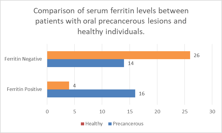
Figure 1: Comparison of serum carcinoembryonic antigen (CEA) levels between patients with oral precancerous lesions and healthy individuals. The figure illustrates the distribution of participants with elevated and normal CEA levels in both study groups.
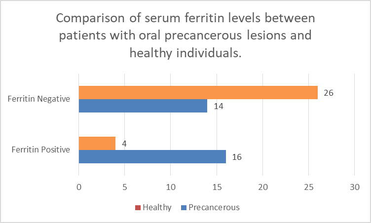
Figure 2: Comparison of serum ferritin levels between patients with oral precancerous lesions and healthy individuals. The figure shows the frequency of participants with elevated and normal ferritin levels in the precancerous and healthy control groups.
DISCUSSION :
The current paper assessed the level of serum carcinoembryonic antigen (CEA) and ferritin in patients with oral precancerous lesions and healthy individuals to determine their utility in predicting oral cancer. The results indicated that the prevalence of high levels of serum CEA and ferritin was more frequent in persons with precancerous oral lesions in comparison with healthy controls. These findings are in line with the emerging evidence that suggests that systemic biomarkers could indicate early molecular and inflammatory alterations related to malignant change in oral potentially malignant disorders (OPMDs).
The latest studies have focused on highlighting the burden of oral cancer on a worldwide scale and the significance of discovering credible biomarkers that can be used to detect the disease at its initial stages. Latest epidemiological surveys have revealed that oral squamous cell carcinoma is among the most common cancers in South Asia, and use of tobacco and areca nuts has a major etiological contribution in the disease progression of the area.10,11 There are oral potentially malignant conditions such as leukoplakia and erythroplakia that are known precursors to oral cancer and show variable rates of acquisition of malignancy in relation to the extent of epithelial dysplasia.12 Clinically, early detection of patients with increased risks of transformation is still a challenge, thus the use of adjunctive biomarkers.
The higher concentrations of carcinoembryonic antigen in the patients of precancerous lesions in this research are also in line with other clinical studies in recent years.13 Multiple investigations have shown that CEA, a glycoprotein of cell adhesion, is raised in many epithelial malignancies and can also rise at an early stage of neoplastic transformation. A current case-control study has documented a significant high level of serum CEA in patients with oral precancerous lesions than healthy individuals indicating that it could be used in early diagnosis and monitoring of the disease albeit in wide application in the early detection area of the human body as well as its monitoring stage.15,16.
In the same manner, another research done on examining tumor biomarkers in oral premalignant disorders revealed that high levels of CEA were associated with higher levels of cellular proliferation and epithelial dysplasia hence its diagnostic use is supported by the study even further as an indicator of the disease or diagnosis outcome.17 The intracellular iron storage protein, ferritin, has also been explored as a potential biomarker in cancer biology. High levels of serum ferritin can be used to indicate an imbalance in iron metabolism, oxidative stress, and inflammation that is linked to carcinogenesis. The overload of iron can also aid the formation of reactive oxygen species, which result in damage of DNA and the formation of tumors and their development {10}.
Recent research have stated higher serum ferritin levels in patients having head and neck cancers, and also in individuals having oral potentially malignant disorders, suggesting that ferritin could be utilized as an indicator of inflammatory and metabolic changes that take place during early tumorigenesis {11}. The biologic processes that lead to the increase in these biomarkers in precancer lesions are multifactorial. Long-term exposure of carcinogenic effects like tobacco, betel nuts, alcohol causes chronic irritation and inflammation of the epithelium and inflammation in the oral mucosa. This inflammatory microenvironment facilitates oxidative stress, cytokine release, and activation of oncogenic signaling pathways that could, in turn, elevate the expression of tumor-associated proteins, including CEA, or cause changes in systemic iron metabolism as manifested by a high level of ferritin {12}.
Furthermore, molecular changes that take place in the development of dysplasia to carcinoma can lead to an increase in cellular turnover and shedding of tumor-associated antigens into the blood. Recent systematic reviews of biomarkers of oral potentially malignant disorders support our findings too. Huang et al. have stated that several serum and salivary biomarkers such as inflammatory proteins and tumor-associated antigens exhibit potential in the prediction of malignant oral leukoplakia transformation.18 Likewise, current studies examining the noninvasive diagnostic biomarkers in oral cancer have also shown that circulating proteins and metabolic biomarkers may be applied as part of early prevention measures.14 Such studies support the idea that biomarker-based screening methodologies can be used in addition to a normal clinical assessment and histopathological analysis. The current research has several strengths.
To begin with, it made direct comparisons of the levels of biomarkers between participants in clinically diagnosed precancerous lesions and healthy controls in the same clinical environment, thus minimizing the possibility of environmental and demographic differences of the participants. Second, the biomarker measurement was conducted using standardized laboratory methods, which augmented the accuracy of the measurements. Moreover, the fact that such common precancerous lesions as leukoplakia, erythroplakia, and verrucous hyperplasia were included enhances clinical importance of the findings. Nevertheless, some drawbacks are to be mentioned. The study design, which is quite limited in size, and the single center may not be helpful to the ability to generalize the findings to larger populations.
Besides, there was an evaluation of only two serum biomarkers, whereas the process of oral carcinogenesis is a multistep process, with a large number of molecular pathways. Future research that brings in bigger multicenter groups and assesses numerous biomarkers or molecular labels can provide a greater understanding of the predictive powers of biomarker schemes. Nevertheless, the article has valuable clinical implications despite these restrictions.
The presence of high levels of serum biomarkers in patients with oral lesions in precancerous lesions could help clinicians detect high-risk patients that will need more rigorous follow-up and timely treatment. Screening using biomarkers may also be used as the complement diagnostic method to the standard clinical examination and histopathology.
Conflict of Interest: Nil
Financial Assistance: Nil.
CONCLUSION :
The current research showed that carcinoembryonic antigen and ferritin levels in the serum were more often found to be high in patients with oral precancerous lesions than in the healthy controls. Such results indicate that changes in these biomarkers can indicate early biological changes with respect to the development of oral potentially malignant disorders. Serum biomarkers evaluation thus might be an assistive diagnostic method in the detection of persons who are at higher risk of malignant transformation. The addition of biomarker assessment and clinical examination can improve early detection methods and support the timely intervention. These findings need further large-scale multi-centre studies to confirm them and ascertain the clinical usefulness of these biomarkers in regular screening programs.
REFERENCES:
1. Sung H, Ferlay J, Siegel RL, Laversanne M, Soerjomataram I, Jemal A, Bray F. Global cancer statistics 2020: GLOBOCAN estimates of incidence and mortality worldwide for 36 cancers in 185 countries. CA Cancer J Clin. 2021;71(3):209-249.
2. Warnakulasuriya S. Global epidemiology of oral and oropharyngeal cancer. Oral Oncol. 2020;102:104551.
3. van der Waal I. Potentially malignant disorders of the oral and oropharyngeal mucosa: terminology, classification and present concepts of management. Oral Oncol. 2020;102:104564.
4. Duffy MJ, Lamerz R, Haglund C, Nicolini A, Kalousová M, Holubec L, Sturgeon C. Tumor markers in colorectal cancer: European Group on Tumor Markers recommendations. Int J Cancer. 2020;147(5):1163-1175.
5. Wu YH, Lin PY, Yang JH, Kuo YS, Wu YC. Serum levels and positive rates of tumor biomarkers in oral precancer patients. J Formos Med Assoc. 2021;120(6):1324-1331.
6. Bray F, Laversanne M, Weiderpass E, Soerjomataram I. The ever-increasing importance of cancer as a leading cause of premature death worldwide. Cancer. 2021;127(16):3029-3030.
7. Warnakulasuriya S, Kujan O, Aguirre-Urizar JM, Bagan JV, González-Moles MÁ, Kerr AR, Lodi G. Oral potentially malignant disorders: a consensus report from an international seminar. Oral Dis. 2021;27(8):1862-1880.
8. López-Jornet P, Olmo-Monedero A, Pons-Fuster E, Tvarijonaviciute A, Perez-Rubio C. Salivary and serum biomarkers in oral potentially malignant disorders: a case-control study. Cancers (Basel). 2023;15(21):5256.
9. Rivera C. Essentials of oral cancer. Int J Clin Exp Pathol. 2020;13(6):1187-1199.
10. Torti SV, Torti FM. Iron and cancer: more ore to be mined. Nat Rev Cancer. 2021;21(2):65-79.
11. Wang W, Knovich MA, Coffman LG, Torti FM, Torti SV. Serum ferritin: past, present and future. Biochim Biophys Acta. 2020;1800(8):760-769.
12. Gupta B, Johnson NW, Kumar N. Global epidemiology of head and neck cancers: a continuing challenge. Oncology. 2021;99(2):65-73.
13. Huang Y, Zhang Q, Guo Z, Deng G, Chen R, Zheng Y. Potential noninvasive biomarkers for malignant transformation of oral leukoplakia: a systematic review and meta-analysis. Cancer Med. 2023;12(13):14718-14730.
14. Kawakita D, Matsuo K. Alcohol and head and neck cancer. Cancer Metastasis Rev. 2020;39(4):949-960.
15. Bai X‐X, Zhang J, Wei L. Analysis of primary oral and oropharyngeal squamous cell carcinoma in inhabitants of Beijing, China—a 10‐year continuous single‐center study. BMC Oral Health. 2020;20:208.
16. Tajmirriahi N, Razavi SM, Shirani S, Homayooni S, Gasemzadeh G. Evaluation of metastasis and 5‐year survival in oral squamous cell carcinoma patients in Isfahan (2001–2015). Dent Res J. 2019;16:117‐121.
17. Le Campion ACOV, Ribeiro CMB, Luiz RR, et al. Low survival rates of oral and oropharyngeal squamous cell carcinoma. Int J Dent. 2017;2017:5815493.
18. Corcoran RB, Chabner BA. Application of cell‐free DNA analysis to cancer treatment. N Engl J Med. 2018;379:1754‐1765.