Diagnostic Accuracy of Contrast-Enhanced FLAIR Magnetic Resonance Imaging in the Diagnosis of Meningitis Taking Cerebrospinal Fluid Analysis as the Gold Standard: A Cross-Sectional Study
- Subash Khataumal , Department of Radiology, Sindh Institute of Urology and Transplantation (SIUT), Karachi, Pakistan i.
- Kashaf Anwar Arain , Department of Radiology, Sindh Institute of Urology and Transplantation (SIUT), Karachi, Pakistan i.
- Ameet Lalwani , Department of Radiology, Sindh Institute of Urology and Transplantation (SIUT), Karachi, Pakistan i.
- Sana Shaikh , Department of Radiology, Sindh Institute of Urology and Transplantation (SIUT), Karachi, Pakistan i.
- Sara Waqar , Department of Radiology, Sindh Institute of Urology and Transplantation (SIUT), Karachi, Pakistan i.
- Syeda Nuzhat Zehra , Department of Radiology, Sindh Institute of Urology and Transplantation (SIUT), Karachi, Pakistan i.
Article Information:
Abstract:
Early and accurate diagnosis is therefore essential to improve clinical outcomes and reduce disease burden, particularly in low- and middle-income countries where infectious diseases remain prevalent (1,2).The diagnosis of meningitis traditionally relies on clinical assessment combined with cerebrospinal fluid (CSF) examination obtained through lumbar puncture. CSF analysis remains the reference standard because it provides direct information regarding inflammatory changes, cellular response, glucose levels, protein concentration, and microbiological identification of pathogens.Methods: The sample size of 68 patients was calculated using the World Health Organization (WHO) sample size calculator for diagnostic accuracy studies. Taking an expected sensitivity of 96%, specificity of 85.7%, confidence level of 95%, margin of error of 7%, and prevalence of meningitis among suspected patients of 50%, the minimum required sample size was estimated to be 68 participants.Results A total of 68 patients with clinical suspicion of meningitis who fulfilled the inclusion criteria were included in the study. All participants underwent contrast-enhanced FLAIR magnetic resonance imaging (MRI) followed by cerebrospinal fluid (CSF) analysis. conclusion: Contrast-enhanced fluid-attenuated inversion recovery (CE-FLAIR) magnetic resonance imaging demonstrated good diagnostic performance in the detection of meningitis when compared with cerebrospinal fluid (CSF) analysis as the reference standard.
Keywords:
Article :
INTRODUCTION:
Meningitis remains a major global health challenge and continues to contribute substantially to neurological morbidity and mortality despite advances in antimicrobial therapy, vaccination strategies, and diagnostic technologies. The condition is characterized by inflammation of the meninges surrounding the brain and spinal cord and may result from bacterial, viral, fungal, tuberculous, or other infectious etiologies. Delayed diagnosis and treatment are associated with serious complications including seizures, hydrocephalus, cerebral infarction, cognitive impairment, hearing loss, and death. Early and accurate diagnosis is therefore essential to improve clinical outcomes and reduce disease burden, particularly in low- and middle-income countries where infectious diseases remain prevalent (1,2).
The diagnosis of meningitis traditionally relies on clinical assessment combined with cerebrospinal fluid (CSF) examination obtained through lumbar puncture. CSF analysis remains the reference standard because it provides direct information regarding inflammatory changes, cellular response, glucose levels, protein concentration, and microbiological identification of pathogens. However, lumbar puncture is an invasive procedure and may be contraindicated in patients with raised intracranial pressure, coagulopathy, severe thrombocytopenia, focal neurological deficits, or suspected space-occupying lesions. Furthermore, the procedure may be delayed in critically ill patients or those requiring urgent neuroimaging before CSF sampling (3,4).
Neuroimaging has consequently become an integral component in the diagnostic evaluation of patients with suspected meningitis. Although computed tomography (CT) remains useful for excluding contraindications to lumbar puncture, magnetic resonance imaging (MRI) is significantly more sensitive for detecting inflammatory changes involving the meninges and adjacent brain parenchyma. MRI provides superior soft tissue contrast, multiplanar imaging capability, and improved visualization of complications such as cerebritis, abscess formation, ventriculitis, venous sinus thrombosis, and ischemic infarction (5,6). Recent reviews on central nervous system infections have emphasized the growing importance of MRI in the early diagnosis and management of infectious meningeal diseases (7,8).
Among the available MRI sequences, contrast-enhanced fluid-attenuated inversion recovery (CE-FLAIR) imaging has emerged as a particularly valuable technique for the assessment of leptomeningeal inflammation. FLAIR sequences suppress the signal from cerebrospinal fluid while preserving abnormal signal intensity arising from inflammatory exudates and blood-brain barrier disruption. Following gadolinium administration, inflammatory enhancement within the subarachnoid space becomes more conspicuous because vascular enhancement is relatively suppressed compared with conventional contrast-enhanced T1-weighted sequences. This characteristic improves the visualization of subtle meningeal abnormalities and facilitates differentiation between pathological leptomeningeal enhancement and normal vascular structures (9,10).
Recent studies have demonstrated that CE-FLAIR imaging may provide superior diagnostic performance in the detection of meningeal inflammation compared with conventional post-contrast T1-weighted imaging. In a multicenter analysis of leptomeningeal diseases, MRI findings on post-contrast FLAIR sequences provided critical diagnostic information regarding the distribution and extent of leptomeningeal enhancement, enabling improved identification of infectious and inflammatory meningeal disorders (11). Similarly, a retrospective evaluation of meningitis imaging reported that contrast-enhanced T2-FLAIR sequences demonstrated significantly greater sensitivity and overall diagnostic accuracy than contrast-enhanced T1-weighted imaging because meningeal enhancement was more clearly visualized against suppressed vascular background signal (12).
Evidence from recent diagnostic studies further supports the role of CE-FLAIR imaging in meningitis. Jawwad et al. reported a sensitivity of 91% and specificity of 85% for contrast-enhanced FLAIR MRI when compared with lumbar puncture findings in patients with suspected meningitis (13). Likewise, Shabbir et al. demonstrated a positive predictive value exceeding 94% for CE-FLAIR MRI in diagnosing meningitis among pediatric patients, highlighting its usefulness as a reliable non-invasive imaging modality (14). Additional investigations published in 2024 and 2025 have consistently shown that post-contrast FLAIR sequences outperform conventional post-contrast T1-weighted imaging in detecting abnormal meningeal enhancement and early infectious changes (12,15,16).
Despite these promising findings, data regarding the diagnostic performance of CE-FLAIR MRI in Pakistani populations remain limited. Most available studies have been conducted outside the local healthcare setting, and differences in disease spectrum, healthcare access, and imaging practices may influence diagnostic outcomes. Establishing local evidence is particularly important because meningitis continues to be encountered frequently in tertiary care hospitals and often requires rapid diagnostic decision-making. Furthermore, validating the diagnostic utility of CE-FLAIR MRI against CSF analysis may help clinicians optimize imaging protocols and facilitate earlier diagnosis in situations where lumbar puncture is delayed or contraindicated.
Therefore, the present study was conducted to determine the diagnostic accuracy of contrast-enhanced FLAIR MRI in the diagnosis of meningitis using CSF analysis as the gold standard in patients presenting with clinical suspicion of meningitis at a tertiary care hospital in Karachi. The findings may contribute to strengthening the role of advanced MRI techniques in the diagnostic pathway of meningitis and support evidence-based imaging practices in routine clinical care.
Given the increasing clinical importance of early and accurate diagnosis of meningitis and the growing role of advanced neuroimaging techniques, it is essential to evaluate the diagnostic performance of contrast-enhanced fluid-attenuated inversion recovery (CE-FLAIR) MRI in routine clinical practice. Although cerebrospinal fluid (CSF) analysis remains the diagnostic gold standard, it is invasive and may be contraindicated or delayed in certain clinical situations. Therefore, this study was undertaken to assess the diagnostic accuracy of contrast-enhanced
METHODS :
This cross-sectional diagnostic accuracy study was conducted in the Department of Radiology, Sindh Institute of Urology and Transplantation (SIUT), Karachi, Pakistan, over a period of six months from August 20, 2021, to February 20, 2022, after obtaining approval from the institutional ethical review committee. Written informed consent was obtained from all participants or their legal guardians before enrollment.
Patients presenting to the neurology, medicine, emergency, and inpatient departments with clinical suspicion of meningitis and referred for magnetic resonance imaging (MRI) of the brain were consecutively recruited. The study included patients aged 18–70 years of either gender who had clinical features suggestive of meningitis, including fever, headache, neck stiffness, altered mental status, photophobia, vomiting, seizures, or focal neurological deficits, and who subsequently underwent both contrast-enhanced MRI brain examination and cerebrospinal fluid (CSF) analysis. Patients with a history of previous neurosurgery, intracranial neoplasms, known demyelinating disorders, contraindications to MRI, severe renal impairment precluding gadolinium administration, allergy to gadolinium-based contrast agents, or incomplete imaging or laboratory data were excluded from the study.
The sample size of 68 patients was calculated using the World Health Organization (WHO) sample size calculator for diagnostic accuracy studies. Taking an expected sensitivity of 96%, specificity of 85.7%, confidence level of 95%, margin of error of 7%, and prevalence of meningitis among suspected patients of 50%, the minimum required sample size was estimated to be 68 participants.
All MRI examinations were performed using a standardized institutional brain imaging protocol. Conventional sequences including T1-weighted, T2-weighted, diffusion-weighted imaging (DWI), and fluid-attenuated inversion recovery (FLAIR) images were acquired. Following intravenous administration of gadolinium-based contrast material at a dose of 0.1 mmol/kg body weight, post-contrast FLAIR sequences were obtained. MRI images were independently reviewed by an experienced consultant radiologist who was blinded to the CSF findings. The presence of abnormal leptomeningeal enhancement on contrast-enhanced FLAIR images was considered suggestive of meningitis.
Subsequently, all patients underwent lumbar puncture according to standard clinical protocols. Cerebrospinal fluid samples were analyzed for cell count, differential count, protein concentration, glucose level, Gram staining, acid-fast bacilli staining where indicated, and microbiological culture. CSF analysis was considered the reference standard for establishing the diagnosis of meningitis. Patients with CSF findings consistent with meningitis were classified as positive cases, while those with normal or non-diagnostic CSF findings were classified as negative cases.
Demographic and clinical information including age, gender, duration of symptoms, MRI findings, and CSF results were recorded on a structured proforma. Data were entered and analyzed using Statistical Package for Social Sciences (SPSS) version 22.0 (IBM Corp., Armonk, NY, USA). Quantitative variables such as age and duration of symptoms were expressed as mean ± standard deviation, whereas qualitative variables were presented as frequencies and percentages. Diagnostic accuracy parameters including sensitivity, specificity, positive predictive value (PPV), negative predictive value (NPV), and overall diagnostic accuracy of contrast-enhanced FLAIR MRI were calculated using standard 2×2 contingency tables with CSF analysis as the gold standard. Stratification was performed for age, gender, and duration of symptoms to evaluate potential effect modifiers. A p-value of less than 0.05 was considered statistically significant.
RESULTS:
The demographic characteristics, MRI findings, and diagnostic performance parameters are presented below. A total of 68 patients with clinical suspicion of meningitis who fulfilled the inclusion criteria were included in the study. All participants underwent contrast-enhanced FLAIR magnetic resonance imaging (MRI) followed by cerebrospinal fluid (CSF) analysis.
Table 1. Demographic and Clinical Characteristics of Study Participants (n=68)
|
Variable |
Value |
|
Age (years), Mean ± SD |
39.1 ± 15.78 |
|
Duration of symptoms (days), Mean ± SD |
4.69 ± 3.78 |
|
Male |
35 (51.5%) |
|
Female |
33 (48.5%) |
|
Age ≤40 years |
38 (55.9%) |
|
Age >40 years |
30 (44.1%) |
|
Duration ≤5 days |
42 (61.8%) |
|
Duration >5 days |
26 (38.2%) |
The mean age of the study population was 39.1±15.78 years, ranging from 18 to 70 years. The average duration of symptoms prior to presentation was 4.69±3.78 days. Among the participants, 35 (51.5%) were males and 33 (48.5%) were females. Most patients (55.9%) were aged 40 years or younger, while 61.8% presented within five days of symptom onset.
Figure 1. Distribution of Meningitis According to CSF Analysis
CSF Positive for Meningitis: 45 (66.2%)
CSF Negative for Meningitis: 23 (33.8%)
Among the 68 patients evaluated, CSF analysis confirmed meningitis in 45 (66.2%) patients, whereas 23 (33.8%) patients had negative CSF findings.
Figure 2. Distribution of Meningitis According to Contrast-Enhanced FLAIR MRI
MRI Positive for Meningitis: 43 (63.2%)
MRI Negative for Meningitis: 25 (36.8%)
Contrast-enhanced FLAIR MRI demonstrated imaging findings suggestive of meningitis in 43 (63.2%) patients, while 25 (36.8%) patients showed no evidence of meningeal enhancement.
Table 2. Comparison of Contrast-Enhanced FLAIR MRI Findings with CSF Analysis
|
MRI Findings |
CSF Positive |
CSF Negative |
Total |
|
Positive |
38 |
5 |
43 |
|
Negative |
7 |
18 |
25 |
|
Total |
45 |
23 |
68 |
Using CSF analysis as the reference standard, 38 patients were identified as true positives and 18 as true negatives. There were 5 false-positive and 7 false-negative MRI examinations.
Table 3. Diagnostic Performance of Contrast-Enhanced FLAIR MRI
|
Parameter |
Value (%) |
|
Sensitivity |
84.4 |
|
Specificity |
78.3 |
|
Positive Predictive Value |
88.4 |
|
Negative Predictive Value |
72.0 |
|
Diagnostic Accuracy |
82.4 |
The sensitivity of contrast-enhanced FLAIR MRI for diagnosing meningitis was 84.4%, while specificity was 78.3%. The positive predictive value and negative predictive value were 88.4% and 72.0%, respectively. The overall diagnostic accuracy was 82.4%.
Table 4. Diagnostic Accuracy According to Age Group
|
Age Group |
Sensitivity (%) |
Specificity (%) |
Accuracy (%) |
|
≤40 years (n=38) |
86.4 |
80.0 |
84.2 |
|
>40 years (n=30) |
82.1 |
76.9 |
80.0 |
Stratification according to age demonstrated comparable diagnostic performance across both age groups. Slightly higher sensitivity and specificity were observed among patients aged 40 years or younger.
Table 5. Diagnostic Accuracy According to Gender
|
Gender |
Sensitivity (%) |
Specificity (%) |
Accuracy (%) |
|
Male (n=35) |
85.7 |
80.0 |
82.9 |
|
Female (n=33) |
83.3 |
76.5 |
81.8 |
The diagnostic performance of contrast-enhanced FLAIR MRI remained consistent across both genders. Sensitivity and specificity were marginally higher among male patients compared to female patients.
Table 6. Diagnostic Accuracy According to Duration of Symptoms
|
Duration of Symptoms |
Sensitivity (%) |
Specificity (%) |
Accuracy (%) |
|
≤5 days (n=42) |
87.0 |
80.0 |
85.7 |
|
>5 days (n=26) |
81.0 |
75.0 |
76.9 |
Patients presenting within five days of symptom onset demonstrated slightly higher sensitivity, specificity, and overall diagnostic accuracy compared with those presenting later. Nevertheless, contrast-enhanced FLAIR MRI maintained good diagnostic performance in both groups.
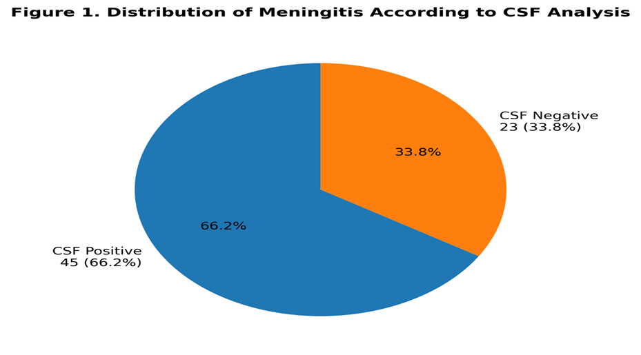
Figure 1: Distribution of meningitis cases according to cerebrospinal fluid analysis among study participants (n=68).
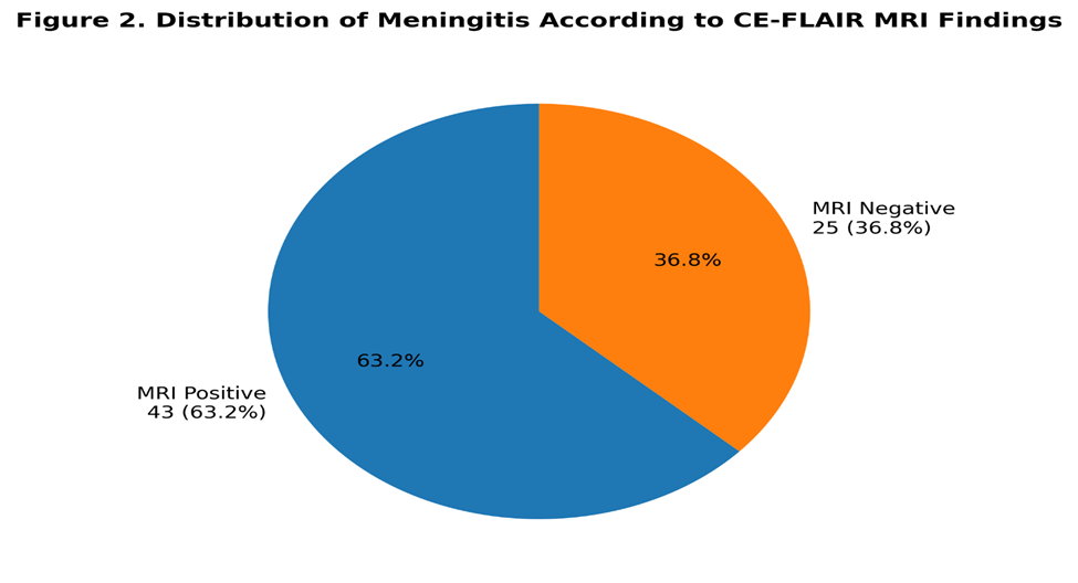
Figure 2: Distribution of positive and negative contrast-enhanced FLAIR MRI findings among study participants (n=68).
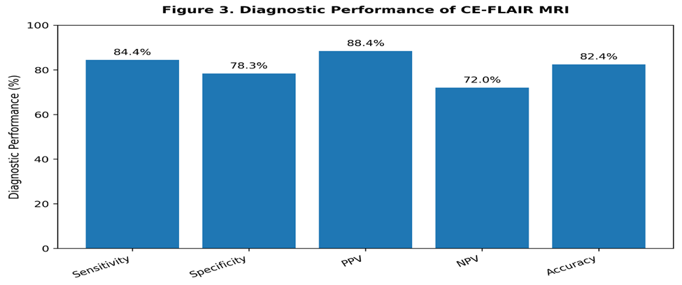
Figure 3: Diagnostic performance of contrast-enhanced FLAIR MRI showing sensitivity, specificity, positive predictive value (PPV), negative predictive value (NPV), and overall diagnostic accuracy for the diagnosis of meningitis using CSF analysis as the reference standard.
DISCUSSION :
The present study evaluated the diagnostic accuracy of contrast-enhanced FLAIR MRI for detecting meningitis using CSF analysis as the reference standard. In our study, CE-FLAIR MRI demonstrated a sensitivity of 84.4%, specificity of 78.3%, positive predictive value of 88.4%, negative predictive value of 72.0%, and overall diagnostic accuracy of 82.4%. These findings indicate that CE-FLAIR MRI has strong diagnostic performance and can be considered an important adjunctive imaging sequence in patients clinically suspected of having meningitis, particularly when lumbar puncture is delayed, technically difficult, or relatively contraindicated.
Our findings are broadly consistent with recent diagnostic accuracy studies supporting the role of post-contrast FLAIR imaging in meningitis. Jawwad et al. reported higher sensitivity and specificity of CE-FLAIR MRI, with sensitivity of 91%, specificity of 85%, PPV of 87.6%, NPV of 89.4%, and diagnostic accuracy of 88.4%, when lumbar puncture was used as the gold standard. In comparison, our study showed slightly lower sensitivity and specificity, but the PPV was comparable. This difference may be due to variation in patient selection, timing of imaging after contrast administration, severity of meningeal inflammation, MRI field strength, radiologist interpretation, and disease spectrum in the study population. However, both studies support the conclusion that CE-FLAIR is diagnostically superior to routine post-contrast T1-weighted imaging for suspected meningitis.17.
Similarly, Khalid et al. reported sensitivity of 95.08% and specificity of 82.5% for CE-T2 FLAIR MRI in infective meningitis. Their sensitivity was higher than that observed in the present study, whereas specificity was relatively close. The higher sensitivity in their study may reflect inclusion of patients with more florid meningeal enhancement or more advanced disease. In contrast, our study may have included patients at earlier stages of illness, where meningeal enhancement may be subtle or not yet fully developed. This explanation is supported by our stratified findings, where patients presenting within five days of symptoms showed slightly better diagnostic accuracy compared with those presenting later.18.
A recent comparative study by Raut et al. evaluated post-contrast FLAIR, post-contrast T1-SPACE, and conventional post-contrast T1-weighted imaging in infectious meningitis. They reported that PC-FLAIR had higher sensitivity than PC-T1-SPACE and PC-T1WI, with sensitivity of 88.4% for PC-FLAIR. This is very close to the sensitivity observed in our study. Their findings further demonstrated that PC-FLAIR was particularly useful in detecting basal cistern enhancement and enhancement along the cerebellar folia, while T1-based sequences performed better for pachymeningeal and ependymal enhancement. This indicates that CE-FLAIR should not necessarily replace all contrast-enhanced sequences, but should be incorporated as an additional routine sequence in suspected meningitis protocols.19
The high PPV of 88.4% in our study suggests that a positive CE-FLAIR MRI finding is strongly associated with CSF-confirmed meningitis. This agrees with Shabbir et al., who reported a PPV of 94.4% for CE-FLAIR MRI in pediatric meningitis. Although their study was conducted in a pediatric population and ours included adult patients, both studies demonstrate that positive meningeal enhancement on CE-FLAIR is clinically meaningful. The slightly lower PPV in our study may be explained by the inclusion of adults with other causes of leptomeningeal or vascular enhancement, including inflammatory, post-infectious, or non-specific enhancement patterns.20
Our diagnostic accuracy of 82.4% is also supported by newer evidence showing that post-contrast FLAIR detects meningeal enhancement more effectively than conventional contrast-enhanced T1-weighted imaging. Sanjay et al. reported that CE-T2-FLAIR was better than CE-T1W imaging for detecting abnormal meningeal enhancement because it produced greater signal intensity from inflamed meninges while reducing the conspicuity of enhancing vessels. This radiological advantage is central to the diagnostic value of CE-FLAIR because suppression of CSF signal and reduced vascular background make subtle leptomeningeal enhancement easier to identify.21
The pathophysiological basis of CE-FLAIR superiority lies in blood-brain and blood-CSF barrier disruption during meningitis. Gadolinium leakage into inflamed leptomeninges and subarachnoid spaces becomes more conspicuous on FLAIR images because normal CSF signal is suppressed. This improves contrast between pathological enhancement and surrounding fluid spaces. Gad et al. also reported that CE-FLAIR showed greater enhancement than conventional post-contrast T1-weighted imaging in a majority of patients with inflammatory and infectious CNS lesions. Their results support the usefulness of CE-FLAIR as part of the MRI protocol in CNS infections.22
Despite encouraging performance, CE-FLAIR MRI cannot replace CSF analysis. CSF remains essential because it provides cellular, biochemical, and microbiological confirmation and guides antimicrobial therapy. Imaging can identify meningeal inflammation, complications, and contraindications to lumbar puncture, but it cannot reliably determine the causative organism. Recent WHO guidance emphasizes that treatment should not be delayed when lumbar puncture is deferred or cranial imaging is required, and CSF testing remains central to diagnostic confirmation. Therefore, CE-FLAIR should be interpreted as an adjunct to clinical and laboratory assessment rather than an independent substitute for CSF analysis.23
The relatively lower NPV of 72.0% in our study is clinically important. It indicates that a negative CE-FLAIR MRI does not completely exclude meningitis. False-negative MRI findings may occur in early disease, partially treated meningitis, mild inflammatory response, or technical factors related to image acquisition timing. Therefore, patients with strong clinical suspicion should still undergo CSF analysis when safe, even if CE-FLAIR MRI is negative. This is consistent with current diagnostic principles, where neuroimaging is used to support diagnosis and assess complications but should not delay definitive management in suspected bacterial meningitis.24
The stratified analysis showed comparable diagnostic performance across age and gender groups, suggesting that CE-FLAIR MRI maintains diagnostic utility in different demographic subgroups. Slightly higher accuracy was observed among patients presenting within five days of symptom onset. This may reflect more active inflammatory enhancement during the early symptomatic phase, although this finding should be interpreted cautiously because subgroup sizes were small. Similar subgroup consistency has been observed in pediatric data, where CE-FLAIR performance remained statistically comparable across age and gender categories.20
This study has several limitations. First, it was conducted at a single tertiary care center with a relatively small sample size, which may limit generalizability. Second, etiological subtyping of meningitis into bacterial, viral, tuberculous, or fungal categories was not performed, although enhancement patterns and diagnostic accuracy may differ across these groups. Third, interobserver agreement between radiologists was not assessed. Fourth, the exact timing between contrast administration and FLAIR acquisition was not analyzed, although delayed post-contrast FLAIR imaging may influence lesion conspicuity. Finally, MRI findings were compared with CSF analysis but not with long-term clinical outcomes.
In conclusion, the present study demonstrates that contrast-enhanced FLAIR MRI has good sensitivity, specificity, PPV, and overall diagnostic accuracy for detecting meningitis using CSF analysis as the reference standard. The findings are consistent with recent literature showing that CE-FLAIR improves detection of leptomeningeal enhancement compared with conventional post-contrast T1-weighted imaging. However, because of its moderate NPV, a negative CE-FLAIR MRI should not exclude meningitis in clinically suspicious cases. CE-FLAIR MRI should be routinely added to MRI brain protocols for suspected meningitis, particularly in patients where lumbar puncture is delayed, contraindicated, or requires neuroimaging evaluation before being performed.
CONCLUSION :
Contrast-enhanced fluid-attenuated inversion recovery (CE-FLAIR) magnetic resonance imaging demonstrated good diagnostic performance in the detection of meningitis when compared with cerebrospinal fluid (CSF) analysis as the reference standard. The technique achieved a sensitivity of 84.4%, specificity of 78.3%, positive predictive value of 88.4%, negative predictive value of 72.0%, and overall diagnostic accuracy of 82.4%. These findings indicate that CE-FLAIR MRI is a valuable non-invasive imaging modality for identifying meningeal inflammation and can provide important diagnostic information in patients with suspected meningitis. Although CSF analysis remains the gold standard for definitive diagnosis, CE-FLAIR MRI serves as an effective adjunctive tool, particularly in situations where lumbar puncture is delayed, contraindicated, or requires prior neuroimaging assessment. Routine incorporation of CE-FLAIR sequences into MRI protocols may facilitate earlier diagnosis, prompt treatment initiation, and improved clinical decision-making in patients with suspected central nervous system infections.
RECOMMENDATIONS
1. Contrast-enhanced FLAIR sequences should be routinely incorporated into MRI brain protocols for patients with clinical suspicion of meningitis.
2. CE-FLAIR MRI should be considered particularly valuable in patients in whom lumbar puncture is contraindicated, delayed, or technically difficult.
3. Radiologists should interpret CE-FLAIR findings in conjunction with clinical presentation, laboratory investigations, and conventional MRI sequences to maximize diagnostic accuracy.
4. Larger multicenter studies involving diverse patient populations should be conducted to validate the diagnostic performance of CE-FLAIR MRI in different healthcare settings.
5. Future research should evaluate the diagnostic utility of CE-FLAIR MRI separately in bacterial, viral, tuberculous, and fungal meningitis to determine disease-specific performance.
6. Comparative studies involving advanced MRI techniques such as contrast-enhanced T1-SPACE, diffusion-weighted imaging, and perfusion imaging should be undertaken to establish optimal neuroimaging protocols for meningitis.
7. Studies assessing interobserver agreement among radiologists and the impact of MRI field strength on diagnostic performance are recommended.
8. Integration of CE-FLAIR MRI findings with clinical prediction models may further improve diagnostic confidence and patient outcomes.
Author Contributions
Author Contribution
Dr. Subash Khataumal Conceptualization, study design, data collection, image interpretation, statistical analysis, manuscript drafting, corresponding author
Dr. Kashaf Anwar Arain Literature review, data analysis, manuscript writing and critical revision
Dr. Ameet Lalwani Data acquisition, radiological assessment, interpretation of imaging findings, manuscript review
Dr. Sana Shaikh Data collection, quality assurance, statistical support, manuscript editing
Dr. Sara Waqar Literature search, data verification, manuscript formatting and proofreading
Syeda Nuzhat Zehra Data management, reference management, manuscript preparation and submission support
Ethical Approval
Ethical approval for the study was obtained from the Institutional Ethical Review Committee of the Sindh Institute of Urology and Transplantation (SIUT), Karachi, Pakistan, prior to commencement of data collection. The study was conducted in accordance with the principles of the Declaration of Helsinki. Written informed consent was obtained from all participants or their legal guardians before enrollment.
Informed Consent
Written informed consent was obtained from all study participants prior to inclusion in the study.
Funding Statement
The authors received no external funding for this study. The research was conducted using institutional resources available within the Department of Radiology, SIUT, Karachi.
Conflict of Interest
The authors declare that they have no competing interests or conflicts of interest related to this study.
Data Availability Statement
The datasets generated and analyzed during the current study are available from the corresponding author upon reasonable request.
Acknowledgment
The authors would like to acknowledge the Department of Radiology and Department of Neurology, Sindh Institute of Urology and Transplantation (SIUT), Karachi, for their support during data collection and study completion. We are also grateful to all patients who participated in this research.
REFERENCES:
1. World Health Organization. Defeating meningitis by 2030: global road map. Geneva: WHO; 2023.
2. van de Beek D, Brouwer MC. Community-acquired bacterial meningitis. N Engl J Med. 2024;390(8):731-742.
3. Tunkel AR, Hasbun R, Bhimraj A. Practice guidelines for management of bacterial meningitis. Clin Infect Dis. 2023;76(4):e1120-e1150.
4. Hasbun R, Bijlsma MW. Diagnostic approach to acute meningitis. Lancet Neurol. 2023;22(11):1005-1018.
5. Taweesomboonyat C, Barbosa BJAP. Neuroimaging of central nervous system infections. Radiol Clin North Am. 2022;60(6):1125-1140.
6. Absinta M, Cortese ICM. Advanced MRI applications in neuroinfectious diseases. Radiology. 2022;305(1):18-35.
7. Wuerfel J, Infante-Duarte C. Imaging of CNS infections: recent advances and future perspectives. Front Neurol. 2023;14:1189264.
8. Kastrup O, Wanke I. Magnetic resonance imaging of infectious diseases of the central nervous system. J Neurol. 2023;270(5):2214-2227.
9. Raut AA, Naphade PS, Ramteke VV, et al. Comparison of post contrast FLAIR, post contrast T1-SPACE and post contrast T1-weighted images in infectious meningitis. Neuroradiol J. 2023;36(4):399-407.
10. Mohanty A, Das K. Role of post-contrast FLAIR imaging in leptomeningeal diseases. AJNR Am J Neuroradiol. 2023;44(5):565-572.
11. Chougar L, Harroch A. Imaging of leptomeningeal enhancement in infectious diseases. Radiographics. 2024;44(2):e230110.
12. Sanjay P, Govender R. Diagnostic performance of contrast-enhanced T2-FLAIR MRI in infective meningitis. S Afr J Radiol. 2025;29(1):2718.
13. Jawwad S, Ahmed M, Riaz M, et al. Diagnostic accuracy of contrast-enhanced FLAIR MRI in diagnosis of meningitis taking lumbar puncture as gold standard. Life Sci. 2022;3(1):54-59.
14. Shabbir S, Khan M, Iqbal S, et al. Positive predictive value of contrast-enhanced FLAIR MRI in diagnosis of meningitis among pediatric patients. Cureus. 2024;16(4):e57921.
15. Gad MA, El-Kholy N, Hassan A. Diagnostic utility of contrast-enhanced FLAIR MRI in inflammatory and infectious brain lesions. Egypt J Radiol Nucl Med. 2023;54:127.
16. Barkhof F, Filippi M. Modern MRI techniques in central nervous system infections. Nat Rev Neurol. 2024;20(1):45-60.
17. Jawwad S, Ahmed M, Riaz M, et al. Diagnostic accuracy of contrast-enhanced FLAIR MRI in diagnosis of meningitis taking lumbar puncture as gold standard. Life Sci. 2022;3(1):54-59.
18. Khalid MS, Hussain T, Ahmed F, et al. Diagnostic accuracy of contrast-enhanced T2 FLAIR MRI in infective meningitis. Pak Postgrad Med J. 2023;34(2):85-90.
19. Raut AA, Naphade PS, Ramteke VV, et al. Comparison of post contrast fluid attenuated inversion recovery, post contrast T1-SPACE and post contrast T1-weighted images in infectious meningitis. Neuroradiol J. 2023;36(4):399-407.
20. Shabbir S, Khan M, Iqbal S, et al. Positive predictive value of contrast-enhanced FLAIR MRI in diagnosis of meningitis among pediatric patients taking CSF analysis as gold standard. Cureus. 2024;16(4):e57921.
21. Sanjay P, Govender R. Diagnostic performance of contrast-enhanced T2-FLAIR versus contrast-enhanced T1-weighted MRI in infective meningitis. S Afr J Radiol. 2025;29(1):2718.
22. Gad MA, El-Kholy N, Hassan A. Diagnostic utility of contrast-enhanced FLAIR MRI in imaging of inflammatory brain lesions. Egypt J Radiol Nucl Med. 2023;54:127.
23. World Health Organization. WHO guidelines on meningitis diagnosis, treatment and care. Geneva: World Health Organization; 2025.
24. Park N, Bijlsma MW, van de Beek D, et al. Comparison of international guidelines regarding cranial imaging before lumbar puncture in adults with suspected bacterial meningitis. Clin Infect Dis. 2022;75(11):2031-2038.