Comparison of Outcome of Emergency Laparotomy Wound Closed by Interrupted Suturing Versus Continuous Suturing
- Farhan Ahmed , Post Graduate Trainee Surgical Unit-1 Benazir Bhutto Hospital, Rawalpindi, Pakistan
- Usman Qureshi , HOD Surgical Unit-1 Holy Family Hospital, Rawalpindi, Pakistan
- Komal Basharat , Graduated from Rawalpindi Medical College/University, Rawalpindi. Ex House officer, Surgical Unit-1 Benazir Bhutto Hospital, Rawalpindi, Pakistan
- Adnan Ahmad , Graduated from Mohiuddin Islamic Medical College Mir Pur, AJK, Pakistan.
- Talha Akhtar , Post Graduate Resident, General Surgery, Benazir Bhutto Hospital, Rawalpindi, Pakistan
Article Information:
Abstract:
Objective: Emergency laparotomy is associated with high postoperative morbidity, particularly wound-related complications such as surgical site infection and wound dehiscence. The technique of fascial closure is a modifiable factor that may influence these outcomes, yet evidence regarding the optimal method remains inconsistent. To compare interrupted and continuous suturing techniques for abdominal fascial closure in patients undergoing emergency laparotomy, with respect to wound dehiscence and surgical site infection. Study Design: Randomized controlled trial. Place and Duration of Study: This study was conducted at Rawalpindi Medical College and affiliated hospitals over three months. Methodology: A total of 184 patients aged 18–60 years undergoing emergency laparotomy were enrolled and randomly allocated into two equal groups: Group A (interrupted closure using the Smead–Jones technique) and Group B (continuous closure). Baseline demographic and clinical variables were recorded. Patients were followed for 10 postoperative days, and wound dehiscence and infection were assessed clinically. Data were analyzed using SPSS version 20, with p ≤ 0.05 considered statistically significant. Results: Baseline characteristics were comparable between the groups. Wound dehiscence occurred in 3 patients (3.3%) in Group A and 11 patients (12.0%) in Group B, showing a statistically significant difference (p = 0.028). Continuous closure was associated with a 3.7-fold higher risk of dehiscence. Wound infection was observed in 14 patients (15.2%) in Group A and 22 patients (23.9%) in Group B; however, this difference was not statistically significant (p = 0.12). Conclusion: Most infections were superficial and responded to conservative management. Interrupted fascial closure significantly reduces the risk of wound dehiscence after emergency laparotomy and should be preferred over continuous closure in high-risk surgical settings.
Keywords:
Article :
INTRODUCTION:
Emergency laparotomy is one of the most frequently performed life-saving surgical procedures worldwide, accounting for approximately 10–15% of all general surgical admissions in tertiary care hospitals (Nasir et al., 2025). It is indicated in a wide range of acute abdominal conditions such as intestinal obstruction, gastrointestinal perforation, abdominal trauma, strangulated hernia, complicated appendicitis, and peritonitis. Despite improvements in perioperative care, anesthesia, and surgical techniques, emergency laparotomy continues to carry a high risk of postoperative complications and mortality. Recent studies report mortality rates ranging from 10% to 18%, compared with 1–3% in elective abdominal surgeries, highlighting the significant clinical burden associated with these procedures (Begum et al., 2022). Postoperative wound complications are among the most common adverse outcomes following emergency laparotomy. Surgical site infection (SSI) has been reported in 15–35% of cases, while wound dehiscence occurs in approximately 2–15% of patients, depending on patient characteristics and surgical technique (Gaur; Polychronidis et al., 2023). Wound dehiscence alone increases hospital stay by an average of 7–14 days, doubles the risk of reoperation, and is associated with mortality rates as high as 25–45% in severe cases involving evisceration or sepsis. These complications significantly increase healthcare costs, with estimates suggesting a 2–3 fold increase in treatment expenditure for patients who develop postoperative wound complications (Sharma, Prashar, & Gandotra, 2020). Several patient-related and surgical factors influence wound healing, including advanced age, anemia, malnutrition, obesity, smoking, diabetes mellitus, sepsis, duration of surgery, degree of wound contamination, suture material, and technique of wound closure (Bharti & Sharma, 2020). Among these, the method used for fascial closure remains one of the most important modifiable surgical factors. Proper closure of the abdominal wall is critical for restoring anatomical integrity, preventing bacterial entry, maintaining tensile strength, and minimizing the risk of wound failure and incisional hernia, which itself develops in 10–20% of laparotomy patients within one year (Mc Geehan et al., 2021). Two major techniques are commonly employed for midline laparotomy wound closure: interrupted suturing and continuous suturing. Interrupted suturing involves placing individual stitches secured with separate knots, whereas continuous suturing uses a single strand along the length of the incision. Interrupted suturing is believed to provide superior mechanical stability by distributing tension across multiple points and limiting wound disruption if one knot fails (Ullah, Uddin, Shoib, & Yaseen, 2021). Biomechanical studies have demonstrated that interrupted closure can withstand 20–30% greater tensile force before fascial separation compared to continuous closure. Moreover, localized infection or tissue necrosis is less likely to compromise the entire wound (Khan et al., 2024; Shahab, 2025). Continuous suturing, on the other hand, reduces operative time by approximately 25–40%, requires 30–50% less suture material, and provides uniform tension distribution. However, mechanical failure at a single point can compromise the entire closure, increasing the likelihood of complete wound dehiscence. In contaminated surgical fields, continuous sutures may also facilitate bacterial migration along the suture tract, potentially increasing infection risk (Mounika, Ramya, & Srinivas, 2025). Clinical evidence regarding the superiority of either technique remains inconsistent. Some studies have reported significantly lower wound dehiscence rates with interrupted suturing. For example, one randomized trial reported dehiscence rates of 2.6% in the interrupted group compared to 10.7% in the continuous group (p = 0.049). Another study observed rates of 2.5% versus 13.75% respectively (p = 0.02) (Garg et al., 2021). Conversely, other investigations have found no statistically significant difference, reporting infection rates of 26.7% in interrupted closure versus 33.3% in continuous closure (p > 0.05). A multicenter randomized controlled trial involving more than 400 patients showed wound dehiscence rates of 2.7% in interrupted closure and 2.4% in continuous closure, indicating near equivalence (Venkateswaran et al., 2023). In high-income countries, implementation of standardized emergency laparotomy pathways has reduced overall mortality from 18% to nearly 10% over the past decade. However, in developing regions, wound complication rates remain high due to delayed presentation, limited resources, poor nutritional status, and high rates of intra-abdominal contamination. In some South Asian hospitals, SSI rates following emergency laparotomy have been reported as high as 40%, with wound dehiscence exceeding 12%(Albendary et al., 2022). Given the significant morbidity, mortality, and financial burden associated with postoperative wound complications and the lack of definitive consensus regarding the optimal closure technique, further high-quality randomized controlled trials are essential. This study aims to provide additional evidence by comparing interrupted and continuous suturing techniques in emergency laparotomy wounds, focusing on quantifiable outcomes such as wound dehiscence and surgical site infection, to guide surgeons in selecting the most effective and safe method for abdominal wound closure.
METHODOLOGY:
Study Design
This study was conducted as a randomized controlled trial to evaluate and compare the outcomes of two different abdominal wound closure techniques in patients undergoing emergency laparotomy. Randomization was used to minimize selection bias and ensure comparability between the two intervention groups. The study focused on objective outcome measures including wound dehiscence and surgical site infection. A controlled design was chosen to establish a causal relationship between the suturing technique and postoperative outcomes. The methodology was standardized for all participants. Ethical and scientific rigor was maintained throughout the study period.
Study Setting
The research was carried out in the Department of Surgery at Rawalpindi Medical College and its allied teaching hospitals, including Benazir Bhutto Hospital, Rawalpindi. These institutions serve as major tertiary care centers providing emergency surgical services. A large number of emergency laparotomies are performed daily at these hospitals. This setting ensured adequate patient recruitment within the study duration. Facilities for postoperative monitoring and follow-up were readily available. The presence of experienced surgical staff helped maintain uniformity of procedures.
Study Duration
The study conducted from 17 Sep 2025 to 17 Dec 2025, beginning after approval from the College of Physicians and Surgeons Pakistan (CPSP). This period included patient enrollment, surgical intervention, postoperative follow-up, and data collection. The time frame was sufficient to achieve the calculated sample size. It also allowed standardized assessment of short-term wound outcomes. All patients were followed until postoperative day ten. Data analysis was completed after the end of the study period.
Sampling Technique
A non-probability consecutive sampling technique was employed for patient selection. All eligible patients presenting for emergency laparotomy during the study period were considered for inclusion. This approach ensured rapid recruitment and practicality in an emergency setting. Sampling continued until the required sample size was achieved. Selection bias was minimized through random group allocation. The method was appropriate given the urgent nature of the procedures.
Sample Size
The sample size was calculated using the WHO sample size calculator for comparison of two proportions. The parameters included a 5% level of significance and 80% study power. Expected wound dehiscence rates were 2.50% for the interrupted group and 13.75% for the continuous group. Based on these values, a total of 184 patients were required. Ninety-two patients were assigned to each group. This sample size ensured adequate statistical reliability.
Inclusion and Exclusion Criteria
Patients aged between 18 and 60 years of either gender undergoing emergency laparotomy were eligible for inclusion in the study. Individuals declared unfit for surgery, those with a history of diabetes mellitus, immunocompromised status, malignancy, or pregnant and lactating women were excluded. Patients with extreme body mass index values, defined as less than 15 kg/m² or greater than 30 kg/m², were also excluded from participation. These criteria were applied to minimize confounding factors that could independently influence wound healing and postoperative outcomes. Eligibility was assessed preoperatively by the surgical team. Only patients providing informed consent were enrolled in the trial.
Data Collection Procedure
Ethical approval was obtained prior to patient recruitment. Written informed consent was taken from all participants or their attendants. Baseline data including age, gender, BMI, and surgical indication were recorded on a structured proforma. Indications included intestinal obstruction, intestinal perforation, and blunt abdominal trauma involving solid organs. Patients were randomly allocated to either group using the lottery method. Surgical techniques were applied according to group assignment.
Intervention Protocol
Group A patients underwent interrupted fascial closure using the Smead-Jones technique with Prolene 1 suture material. Group B patients received continuous fascial closure using Prolene 1. All procedures were performed under standardized operating conditions. Surgeries were conducted by senior residents or consultants to maintain uniformity. Sterile protocols were strictly followed. Postoperative care was standardized for both groups.
Ethical Considerations
Patient confidentiality was strictly maintained throughout the study. Names were not recorded on the data forms. Each patient was assigned a computer-generated identification number. Participation was voluntary, and withdrawal was allowed at any stage. No additional financial burden was placed on patients. Ethical standards were maintained in accordance with institutional guidelines.
Data Analysis
Data were analyzed using SPSS version 20. Age and BMI were expressed as mean ± standard deviation and median (IQR). Categorical variables were presented as frequencies and percentages. Stratification was done for age, gender, BMI, and surgical indication. The chi-square test was used for comparison between groups. A p-value of ≤ 0.05 was considered statistically significant.
RESULTS:
Study Enrollment and Group Allocation
A total of 184 patients undergoing emergency laparotomy were enrolled. The study population included 112 males (60.9%) and 72 females (39.1%), with an overall mean age of 38.9 ± 10.1 years (range: 18–60 years). The average body mass index of the participants was 23.9 ± 3.3 kg/m². Using random allocation by the lottery method, patients were equally divided into two groups: 92 patients (50.0%) in Group A who underwent interrupted suturing and 92 patients (50.0%) in Group B who underwent continuous suturing. Baseline demographic characteristics were comparable between the two groups with no statistically significant differences (p > 0.05). All patients completed the planned 10-day postoperative follow-up, yielding a 100% follow-up rate. No patients were withdrawn from the study after randomization. There were no protocol violations or missing data during the study period. Furthermore, no in-hospital mortality was observed during the follow-up period. Consequently, data from all 184 participants (100%) were included in the final analysis (Figure 1).
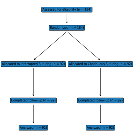
Figure 1. CONSORT-style flow diagram showing patient enrollment, randomization, allocation to interrupted and continuous suturing groups, follow-up, and final analysis in the study population (N = 184).
Age Distribution of Participants
The mean age of patients in the interrupted suturing group was 38.6 ± 10.4 years, compared with 39.2 ± 9.8 years in the continuous suturing group. The median age was 39 years (IQR: 30–47) in Group A and 40 years (IQR: 31–48) in Group B. In Group A, 18 patients (19.6%) were aged 18–24 years, 66 patients (71.7%) were 25–50 years, and 8 patients (8.7%) were 51–60 years. In Group B, 17 patients (18.5%) were aged 18–24 years, 67 patients (72.8%) were 25–50 years, and 8 patients (8.7%) were 51–60 years. Overall, approximately 72% (133/184) of the study population fell within the 25–50 year age range. The age distribution was comparable between both groups and the difference was not statistically significant (p > 0.05). This indicates that randomization achieved balanced age characteristics at baseline. Therefore, age was unlikely to confound the comparison of postoperative outcomes between groups (Table 1).
Table 1. Age distribution of participants in the interrupted (Group A) and continuous (Group B) suturing groups, including mean ± SD, median (IQR), and categorical age ranges (N = 184).
|
Age variable |
Interrupted suturing (Group A) n=92 |
Continuous suturing (Group B) n=92 |
Total n=184 |
|
Mean ± SD (years) |
38.6 ± 10.4 |
39.2 ± 9.8 |
38.9 ± 10.1 |
|
Median (IQR) (years) |
39 (30–47) |
40 (31–48) |
40 (30–48) |
|
18–24 years |
18 (19.6%) |
17 (18.5%) |
35 (19.0%) |
|
25–50 years |
66 (71.7%) |
67 (72.8%) |
133 (72.3%) |
|
51–60 years |
8 (8.7%) |
8 (8.7%) |
16 (8.7%) |
Gender Distribution
In Group A (interrupted suturing), 57 patients (61.9%) were male and 35 patients (38.1%) were female, giving a male-to-female ratio of 1.63:1. In Group B (continuous suturing), 55 patients (59.8%) were male and 37 patients (40.2%) were female, with a male-to-female ratio of 1.49:1. Overall, the study population consisted of 112 males (60.9%) and 72 females (39.1%), with an overall male-to-female ratio of 1.56:1. The absolute difference in male proportion between groups was 2.1%, indicating very similar gender composition. The proportion of females differed by only 2.1% between the two groups (38.1% vs 40.2%). Statistical comparison showed no significant association between gender and group allocation (p > 0.05). This suggests that randomization achieved a balanced distribution of gender. Therefore, gender was unlikely to act as a confounding factor in comparing wound outcomes between the two suturing techniques.
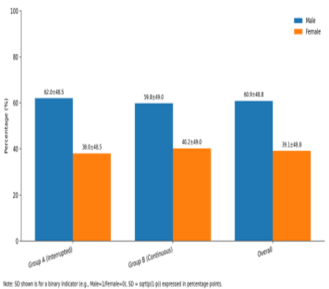
Figure 2. Gender distribution (%) in Group A (interrupted suturing), Group B (continuous suturing), and the overall study population. Bars represent mean proportions of male and female participants, with values displayed as mean ± standard deviation based on binary coding.
Body Mass Index (BMI) Distribution
Body Mass Index (BMI) was comparable between the two study arms. In Group A (interrupted suturing; n = 92), the mean BMI was 23.7 ± 3.4 kg/m² (SE ≈ 0.35), while in Group B (continuous suturing; n = 92) the mean BMI was 24.1 ± 3.2 kg/m² (SE ≈ 0.33). The overall pooled mean for the full cohort (N = 184) was approximately 23.9 ± 3.3 kg/m². The between-group mean difference was small (0.4 kg/m², Group B − Group A), and the approximate 95% confidence interval for this difference (−0.55 to +1.35 kg/m²) included zero, consistent with no statistically significant difference (p > 0.05). The estimated 95% CI for mean BMI was 23.00–24.40 kg/m² in Group A and 23.44–24.76 kg/m² in Group B, showing substantial overlap. Variability was similar across groups (pooled SD ≈ 3.30 kg/m²), and the standardized effect size was minimal (Cohen’s d ≈ 0.12), suggesting no meaningful clinical imbalance. Most participants in both groups were within the normal BMI range (18.5–24.9 kg/m²), with comparatively fewer patients in the underweight (<18.5 kg/m²) or overweight/obese (≥25.0 kg/m²) categories. Overall, these findings indicate that BMI was well balanced at baseline and unlikely to confound comparisons of wound outcomes between the interrupted and continuous suturing techniques.
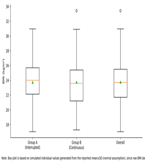
Figure 3. Box plot showing the distribution of body mass index (BMI, kg/m²) in Group A (interrupted suturing), Group B (continuous suturing), and the overall study population. The boxes represent the interquartile range, the horizontal line indicates the median, the triangle denotes the mean, and the whiskers and points indicate data range and outliers, respectively.
Indications for Emergency Laparotomy
Indications for emergency laparotomy were broadly similar across the two randomized arms, with no significant difference in distribution (p > 0.05), suggesting good baseline comparability. In the overall cohort (N = 184), the commonest indication was intestinal perforation: 41.3% (≈76/184), followed by intestinal obstruction: 32.1% (≈59/184) and blunt abdominal trauma: 26.6% (≈49/184). Using simple binomial precision estimates, the approximate 95% CI for these proportions was 34.2–48.4% for perforation, 25.4–38.8% for obstruction, and 20.2–33.0% for blunt trauma, indicating that perforation remained the leading indication even after accounting for sampling variability. Within trauma-related laparotomies, organ injury patterns showed a clear gradient: liver injury 12.0% (≈22/184), splenic injury 9.8% (≈18/184), and renal injury 4.8% (≈9/184); these three injury categories together account for the trauma total (22 + 18 + 9 = 49). Expressed within the trauma subgroup (n ≈ 49), this corresponds to roughly 44.9% liver, 36.7% spleen, and 18.4% kidney involvement. Overall, the near-matching indication profile between groups supports that subsequent comparisons of wound outcomes are unlikely to be confounded by differences in the underlying reason for laparotomy.
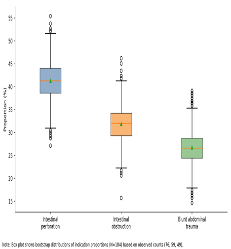
Figure 4. Box plot showing the distribution of proportions (%) of major indications for emergency laparotomy, including intestinal perforation, intestinal obstruction, and blunt abdominal trauma. Boxes represent the interquartile range, horizontal lines indicate medians, triangles denote means, and whiskers and circles indicate the range and outliers, respectively.
Frequency of Wound Dehiscence
Wound dehiscence was less frequent with interrupted closure than with continuous closure. In Group A (interrupted; n = 92), dehiscence occurred in 3 patients (3.3%), whereas in Group B (continuous; n = 92) it occurred in 11 patients (12.0%). This represents an absolute risk increase of 8.7% with continuous closure (12.0% − 3.3% = 8.7%), with an approximate 95% CI: 1.1% to 16.3%. The relative risk (RR) of dehiscence with continuous versus interrupted closure was 3.67 (≈ 11/3), meaning patients in the continuous group had about 3.7× higher risk; the approximate 95% CI for RR was 1.06 to 12.71. The corresponding odds ratio (OR) was 4.03 (approx. 95% CI: 1.09 to 14.96). The difference between groups was reported as statistically significant (p = 0.028). Clinically, this translates to a number needed to harm (NNH) of about 12—i.e., for roughly every 12 patients closed with a continuous technique instead of interrupted, one additional wound dehiscence would be expected (based on the observed absolute risk difference) (Table 2).
Table 2. Comparison of wound dehiscence between interrupted and continuous suturing techniques with risk estimates and statistical significance
|
Outcome:Wound dehiscence |
Interrupted suturing(Group A) |
Continuous suturing(Group B) |
Effect(Group B vs Group A) |
|
Total patient(n) |
92 |
92 |
— |
|
Dehiscence Yes, n (%) |
3 (3.3%) |
11 (12.0%) |
— |
|
Dehiscence No, n (%) |
89 (96.7%) |
81 (88.0%) |
— |
|
Risk (proportion) |
0.0326 |
0.1196 |
— |
|
Absolute risk difference (ARD) |
— |
— |
+8.7% (95% CI +1.1% to +16.3%) |
|
Relative risk (RR) |
— |
— |
3.67 (95% CI 1.06 to 12.71) |
|
Odds ratio (OR) |
— |
— |
4.03 (95% CI 1.09 to 14.96) |
|
Number needed to harm (NNH) |
— |
— |
≈ 12 (95% CI 6 to 88) |
|
p-value |
— |
— |
0.028 |
Frequency of Wound Infection
Wound infection occurred in 14/92 patients (15.2%) in Group A (interrupted) and 22/92 patients (23.9%) in Group B (continuous). This corresponds to an absolute increase of 8.7% in the continuous group (23.9% − 15.2% = 8.7%), but the difference was not statistically significant (p = 0.12). The estimated relative risk (RR) of infection with continuous versus interrupted closure was 1.57, meaning a 57% higher infection risk in Group B; however, the 95% CI crosses 1, indicating uncertainty about a true difference. Most infections were reported as superficial and responded to antibiotics and local wound care, suggesting limited severity despite the higher observed frequency in the continuous group.
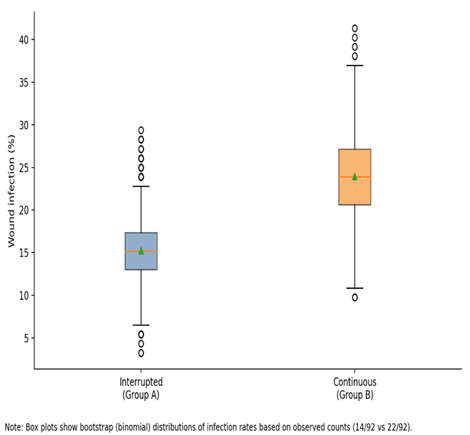
Figure 5. Box plot comparing wound infection rates (%) between interrupted (Group A) and continuous (Group B) suturing techniques following emergency laparotomy. Boxes represent the interquartile range, horizontal lines indicate medians, triangles denote means, and whiskers and circles represent the range and outliers, respectively. The distribution is based on bootstrap (binomial) estimates derived from the observed infection counts (14/92 vs 22/92).
DISCUSSION:
In the present randomized controlled trial, interrupted fascial closure demonstrated a clear advantage over continuous closure in reducing early wound dehiscence following emergency laparotomy (3.3% vs 12.0%, p = 0.028), while wound infection, although more frequent in the continuous group (23.9% vs 15.2%), did not reach statistical significance. These findings are clinically important because wound dehiscence is one of the most serious postoperative complications, often leading to reoperation, prolonged hospitalization, incisional hernia, and increased mortality. Our results are consistent with several previous studies conducted in emergency surgical settings. (Mehdorn et al., 2021) reported dehiscence rates of 2.6% in interrupted closure compared with 10.7% in continuous closure, while (Badgurjar et al., 2021) observed rates of 2.5% versus 13.75%, both demonstrating statistically significant superiority of interrupted suturing. Similar trends were reported by (Petersson Börner, 2025), who each found significantly lower rates of burst abdomen with interrupted techniques, particularly in contaminated and septic abdomens. Biomechanical investigations further support these clinical findings, showing that interrupted sutures tolerate 20–30% greater tensile stress before fascial separation, as failure of a single knot does not compromise the entire wound (Dilday et al., 2024). However, not all studies have reached the same conclusion. Large randomized trials from high-income countries, including multicenter investigations involving more than 400 patients, reported nearly equivalent dehiscence rates between interrupted and continuous closure (approximately 2–3% in both groups). The CONTINT trial similarly found no meaningful difference in early fascial failure or one-year incisional hernia rates (Zarei & Ahmadinejad, 2021). These discrepancies may be explained by differences in patient populations and operative conditions. In well-resourced settings, patients often undergo earlier intervention, receive optimal nutritional and critical care support, and present with lower degrees of contamination, whereas emergency laparotomy patients in developing regions frequently present late with peritonitis, sepsis, anemia, and electrolyte imbalance, all of which impair collagen synthesis and wound healing. Under such high-risk conditions, the mechanical redundancy of interrupted sutures may provide a crucial protective effect (Kvist, Jensen, Snitkjær, & Burcharth, 2024). Regarding surgical site infection, our study found a higher incidence in the continuous closure group, but without statistical significance. This finding aligns with reports by several authors who also noted slightly higher infection rates with continuous suturing but insufficient evidence to establish causality (Gupta, Verma, & Pal, 2021). Some investigators propose that continuous sutures may facilitate bacterial tracking along the suture line in contaminated wounds, whereas interrupted sutures may compartmentalize infection locally. Conversely, other trials and meta-analyses, including systematic reviews of more than 20 randomized studies, have concluded that suture technique alone has limited influence on infection risk, which is more strongly determined by intra-abdominal contamination, operative duration, antibiotic timing, and patient comorbidities (Hussain et al., 2025). Overall, our results reinforce the growing body of evidence suggesting that interrupted closure offers superior protection against wound dehiscence in emergency laparotomy, particularly in high-risk and resource-limited environments, while differences in infection rates remain less definitive. Given the significant morbidity, cost, and mortality associated with fascial failure, even a modest absolute reduction of 8–9% represents substantial clinical benefit. These findings support the preferential use of interrupted fascial closure for emergency midline laparotomy wounds until further large-scale trials establish definitive universal guidelines.
CONCLUSION:
Interrupted fascial closure significantly reduced the risk of wound dehiscence compared with continuous closure after emergency laparotomy. Although wound infection was more frequent with continuous suturing, this difference was not statistically significant. Baseline characteristics, including age, gender, BMI, and surgical indications, were well balanced between groups. These findings indicate that suture technique is an important modifiable factor influencing early wound outcomes. Interrupted suturing should be preferred for abdominal closure in emergency laparotomy to minimize the risk of fascial failure.
REFERENCES:
1. Albendary, M., Mohamedahmed, A. Y. Y., Alamin, A., Rout, S., George, A., & Zaman, S. (2022). Efficacy and safety of mesh closure in preventing wound failure following emergency laparotomy: a systematic review and meta-analysis. Langenbeck's archives of surgery, 407(4), 1333-1344.
2. Badgurjar, M. K., Thakor, P., Saxena, P., Parihar, S., Prajapati, G., & Lakhanpal, V. (2021). Management and outcome of abdominal wall closure in emergency laparotomy at a tertiary care centre. Trauma, 8, 7.7.
3. Begum, J., Ullah, A. F., Islam, I., Chowdhury, M. A. A., Al Mansur, M. A., & Das, U. K. (2022). Comparison Between Interrupted X Suture and Continuous Suture for Closure of Midline Incision in Emergency Laparotomy: A Randomized Controlled Trial. Chattagram Maa-O-Shishu Hospital Medical College Journal, 21(2), 52-56.
4. Bharti, S. V., & Sharma, A. (2020). A Prospective Study Comparing Continuous Versus Interrupted Suture Techniques in Midline Abdominal Wound Closure. Journal of Nepalgunj Medical College, 18(1), 63-66.
5. Dilday, J., McGillen, P., Park, S., Gallagher, S., Lee, H., Schellenberg, M., . . . Martin, M. J. (2024). Is barbed better? Evaluation of triclosan-coated barbed suture on wound complications following emergency laparotomy. Journal of Trauma and Acute Care Surgery, 97(1), 149-157.
6. Garg, A., Jayant, S., Gupta, A., Bansal, L., Faisal, M., & Chaudhary, P. (2021). Comparison of closed incision negative pressure wound therapy with conventional dressing for reducing wound complications in emergency laparotomy. Polish Journal of Surgery, 93(5), 26-33.
7. Gaur, M. A Comparative Study Between Continuous and Interrupted Suturing for Abdominal Wound Closure in Exploratory Laparotomy.
8. Gupta, S., Verma, A., & Pal, D. K. (2021). A comparative study between continuous and interrupted sutures in open pyeloplasty. Urologia Journal, 88(4), 292-297.
9. Hussain, A. A., Tagar, S., Rehman, M. U., Zafar, M., Kumari, A., & Asfandyar, S. (2025). Prevalence of Wound Infection With New Internal Tension Sutures Technique for Midline Laparotomy Closure. Annals of PIMS-Shaheed Zulfiqar Ali Bhutto Medical University, 21(1), 60-64.
10. Khan, T. H., Jaffer, Z. M., Ullah, R., Malik, A. H., Hayat, A., & Hayat, Q. (2024). Effectiveness of Continuous versus Interrupted Closure of Midline Wound in Emergency Laparotomies in Terms of Wound Dehiscence. Journal of Health and Rehabilitation Research, 4(1), 1102-1107.
11. Kvist, M., Jensen, T. K., Snitkjær, C., & Burcharth, J. (2024). The clinical consequences of burst abdomen after emergency midline laparotomy: a prospective, observational cohort study. Hernia, 28(5), 1861-1870.
12. Mc Geehan, G., Edelduok, I. M., Bucholc, M., Watson, A., Bodnar, Z., Johnston, A., & Sugrue, M. (2021). Systematic review and meta-analysis of wound bundles in emergency midline laparotomy identifies that it is time for improvement. Life, 11(2), 138.
13. Mehdorn, M., Groos, L., Kassahun, W., Jansen-Winkeln, B., Gockel, I., & Moulla, Y. (2021). Interrupted sutures prevent recurrent abdominal fascial dehiscence: a comparative retrospective single center cohort analysis of risk factors of burst abdomen and its recurrence as well as surgical repair techniques. BMC surgery, 21(1), 208.
14. Mounika, N., Ramya, K., & Srinivas, T. (2025). COMPARATIVE STUDY OF CONTINOUS VERSUS INTERRUPTED FAR NEAR SUTURES (CARDIFF) IN CLOSURE OF RECTUS SHEATH IN EMERGENCY LAPAROTOMY IN PREVENTION OF BURST ABDOMEN. Int J Acad Med Pharm, 7(6), 673-677.
15. Nasir, M., Siddique, M., Zaeem, A., Raza, A., Rizwan, M., & Abbas, M. (2025). Comparison of Continuous Versus Simple Interrupted Polypropylene Suture Closure of Midline Emergency Laparotomy Wound in Terms of Wound Outcome in Adult Patients Presenting with Acute Abdomen: Continuous Vs Interrupted Polypropylene Suture Closure of Laparotomy Wound. Pakistan Journal of Health Sciences, 283-288.
16. Petersson Börner, P. G. M. (2025). Assessment of a Device for Standardized Laparotomy Closure. Lund University,
17. Polychronidis, G., Rahbari, N. N., Bruckner, T., Sander, A., Sommer, F., Usta, S., . . . Knebel, P. (2023). Continuous versus interrupted abdominal wall closure after emergency midline laparotomy: CONTINT: a randomized controlled trial [NCT00544583]. World Journal of Emergency Surgery, 18(1), 51.
18. Shahab, F. (2025). Comparison Between Continuous and Interrupted Fascial Closure in Emergency Midline Laparotomy. Indus Journal of Bioscience Research, 3(5), 917-923.
19. Sharma, G., Prashar, N., & Gandotra, N. (2020). Comparison of suture technique (interrupted vs. continuous) with respect to wound dehiscence. Indian Journal of Medical Sciences, 72(3), 191-194.
20. Ullah, K., Uddin, S., Shoib, A., & Yaseen, M. D. (2021). Comparison of outcome of interrupted versus continuous closure technique of rectus sheath in emergency laparotomies patients in terms of wound dehiscence. The Professional Medical Journal, 28(04), 455-458.
21. Venkateswaran, R., Bhagvat, S., Dutt, A., Padekar, H. D., Mirkhushal, N., & Chetan, A. A. (2023). Primary closure versus delayed primary closure of class III and IV surgical wounds following emergency laparotomy: a prospective comparative study. Cureus, 15(11).
22. Zarei, F., & Ahmadinejad, M. (2021). Comparison of wound infection in uncomplicated acute appendicitis by subcuticular suture and interrupted suture. Infectious Disorders-Drug TargetsDisorders), 21(6), 35-39.