Evaluating the Role of Coronary CT Angiography in Planning Complex Coronary Intervention
- Syed Kashif ur Rehman , Interventional Cardiology Fellow, Hayatabad Medical Complex, Peshawar, Pakistan
- Parshad , International Trainee Fellow, University of Birmingham, UK
- Bharat Kumar , CPSP-MTI Trainee Naas General Hospital Ireland
- Jaghat Ram , MBBS, FCPS-Card, Post Fellowship Interventional Cardiology, Associate Professor Adult Cardiology, Sind Institute of Cardiovascular Diseases, Larkana Pakistan.
- Romana Awan , SR Cardiology NICVD Karachi, Pakistan
- Abdul Basit , Intervention Cardiology Fellow, Hayatabad Medical Complex, Peshawar, Pakistan
Article Information:
Abstract:
Objective: To discuss the importance of Coronary Computed Tomography Angiography (CCTA) during the planning of complicated coronary procedures in patients with Coronary Artery Disease (CAD). Methodology: In this prospective study, 246 patients were used, which was carried out between June 2024 and December 2024. CCTA was performed to determine the anatomy of coronary arteries in patients, as well as the burden of plaque and the extent of the lesion. Demographic information, comorbidity, and clinical condition were recorded, and the decision on the intervention (PCI, CABG or none) was made. Associations were assessed by use of descriptive statistics, chi-square tests and cross-tab analyses. Results: Among the 246 patients, 57.7% (n = 142) had stable CAD, 30.9% (n = 76) had chronic total occlusions, and 11.4% (n = 28) had severe stenosis. The types of interventions were 40.7, 34.6 and 24.8 per cent of avoiding intervention, PCID and CABG, respectively. The severity of the lesion was moderate (46.7% or n = 115), severe (31.7% or n = 78), and mild (21.5% or n = 53). There were also significant associations between the coronary status and lesion severity (p = 0.004) and none between sex and intervention type (p = 0.124). Conclusion: CCTA is an appropriate method of planning complex coronary procedures due to its detailed non-invasive information on the anatomy of the coronary and the severity of lesions. The results justify the application of CCTA in pre-procedural planning that results in more precise and customised interventions.
Keywords:
Article :
INTRODUCTION:
Coronary artery disease (CAD) is a major cause of morbidity and mortality in the world. Coronary angiography is one of the major methods of evaluating CAD and planning interventions that is considered the gold standard of diagnosis and managing treatments. There are, however, limitations of this invasive technique, such as catheterisation that involves this process and complications that may arise in the procedure. In the last several decades, non-invasive imaging methods, in particular, Coronary Computed Tomography Angiography (CCTA), have become an alternative option. Planning of complex coronary interventions has become a more and more popular topic of CCTA application since the technique provides a more detailed picture of a coronary structure and assists in choosing the course of treatment. This type of non-invasive imaging can give high-resolution images of the coronary vasculature and can also assess the morphology of the plaque, vessel size and the severity of stenosis, which is invaluable in the organisation of complex interventions[1]. This research paper is intended to assess the use of CCTA in the planning of complicated coronary procedures, particularly in patients undergoing such procedures like Percutaneous Coronary Interventions (PCI) and Coronary Artery Bypass Grafting (CABG).
It is well known that CCTA is capable of giving a complete, non-invasive assessment of the coronary arteries. With the ongoing advancement of CCTA technology, its use in measuring CAD has increased greatly. One of the most significant benefits of CCTA is that it has the capability to visualise the coronary arteries in 3D, providing more detailed anatomy compared to the traditional angiography[2]. This involves the identification of coronary artery abnormalities, calcifications, and atherosclerotic plaque, which are important variables in designing complicated coronary operations[3].
CCTA is demonstrated to enhance planning of the procedure and direct the choice of the best interventional method in the context of sophisticated CAD (such as Chronic Total Occlusions (CTOs) and bifurcation lesions)[4]. Research has proved that CCTA provides better visualisation of the coronary anatomy, which is useful in detecting the location and extent of the blockages which are not easily seen using the normal angiography. This knowledge comes in handy, especially where CABG or PCI is under consideration as an option of treatment[5].
One of the major issues related to coronary interventions is the proper measurement of the severity of the lesions and identification of the most efficient method of revascularisation. CCTA, the use of which is augmented with a functional imaging approach like Fractional Flow Reserve (FFR), can greatly enhance the accuracy of such tests. Calculating FFR using CT images (CT-FFR), the clinicians may determine the relevance of the coronary lesions to the haemodynamic process, which is an extra tool in the treatment decision-making[6]. This has proved quite helpful, especially in complicated coronary cases where precise decision-making is important for the best possible results.
The latest progress in CCTA technology has improved its utility in the planning of coronary interventions. With the introduction of high-resolution CT scanners and the introduction of artificial intelligence into the analysis of CCTA, it is now possible to obtain a more accurate and fast assessment of coronary lesions[7]. These breakthroughs have further rendered CCTA to be more useful in complicated cases where very accurate pre-procedural planning is mandatory. Artificial intelligence platforms, including those, are currently being applied to better the analysis of CCTA images and allow interventional cardiologists to identify lesions and forecast the results of different interventions more accurately[8].
The use of CCTA with 3D printing and virtual coronary procedures is one of the successful ones. This will enable modelling of the coronary vasculature specific to patients, which may be simulated and pre-procedurally planned[9]. The innovations can ultimately enhance the accuracy and safety of coronary procedures, particularly when there are anatomical complexities (e.g., bifurcations, CTOs, and extremely calcified lesions).
This study is motivated by the fact that there is an increasing appreciation of the potential of CCTA to revolutionise the process of designing complex coronary procedures. Although CCTA has found extensive applications in diagnostic practice, its application in pre-procedural planning and guiding complicated interventions has not been established, especially in the Pakistani clinical settings. The applicability and advantages of CCTA in planning PCI and CABG with complicated CAD have been evident in numerous international studies, and further research in the local context is still necessary to realise its feasibility.
Since the complexity of coronary procedures is growing, and the accuracy of planning to enhance patient care may require careful consideration of the possibility of CCTA implementation into the mainstream clinical practice, the unavoidable aspect is the necessity to consider the ways of integrating the diagnostic tool into everyday clinical practice, particularly in those environments in which the resources are limited. CCTA in the intervention planning process is a novel practice in Pakistan, and the proposed study will carry out a thorough evaluation of its efficacy and viability within the local cardiology departments.
The aim of the research paper is to analyse how coronary CT angiography provides value to the delivery of complex coronary interventions with the purpose of enhancing the quality of procedures and patient care by using the non-invasive imaging method.
MATERIALS AND METHODS:
It was a prospective, observational, cohort study that was done in the tertiary care hospital, Peshawar, between June 2024 and December 2024. The Ethical and Research Committee of the hospital approved the research (Ref #2156, dated 07/02/2024), which guaranteed that all the ethical standards were maintained during the research. Before the enrolment, all the participants were informed about the enrolment.
The sample size calculation was performed using the WHO formula for prevalence studies[4],[7], given a 95% confidence level, a 5% margin of error, and an estimated prevalence of CAD based on recent studies (approximately 20%); the minimum required sample size for this study was 246 patients.
The inclusion criteria were the age range (40 to 75 years) and the stable CAD, based on the previous diagnostic procedures, e.g., ECG and echocardiography. The inclusion criteria were patients with CTOs, severe coronary artery stenosis (70% or more), or patients undergoing coronary revascularisation treatment (PCI or CABG). The exclusion criteria were the contraindications to CCTA (i.e., renal failure and allergy to contrast agents), recent myocardial infarction (within 3 weeks), and pregnant women.
All patients were evaluated for eligibility based on the inclusion and exclusion criteria. At the time of enrolment, baseline demographic data (age, gender, medical history [hypertension, diabetes, smoking, etc.]), and previous cardiac history (e.g., previous PCI or CABG) were taken. The patients who passed the inclusion criteria were subjected to CCTA to determine the anatomy of coronary arteries, the level of plaque, and the severity of the lesions. Besides, other diagnostic processes like echocardiography and invasive coronary angiography were performed as a part of the clinical decision-making procedure to plan intervention.
The primary data collection tool was CCTA, which was used to provide detailed, three-dimensional images of the coronary arteries. Images were analysed for the presence and severity of stenoses, coronary artery anomalies, plaque composition, and other important anatomical features that could influence the treatment plan. Functional imaging using CT-derived FFR (FFR-CT) was also applied to assess the physiological significance of coronary lesions.
As this was an observational cohort study, randomisation and blinding were not applicable. The patients were evaluated based on their clinical presentation and their need for coronary intervention, following standard clinical protocols. The role of CCTA in planning interventions was then assessed in relation to the clinical outcomes.
The study included patients aged 40 to 75 years who had stable CAD confirmed through prior non-invasive or invasive diagnostic tests. They also had at least one significant coronary lesion (≥70% stenosis) requiring further intervention, either PCI or CABG. Patients were excluded if they had contraindications to CCTA, such as renal impairment or contrast allergies, or if they had recently suffered a myocardial infarction or were pregnant.
Data were analysed using SPSS (Statistical Package for the Social Sciences) version 26. Descriptive statistics were used to summarise the demographic characteristics and clinical profiles of the patients. Continuous variables were presented as mean ± standard deviation, and categorical variables were expressed as frequencies and percentages. The comparison of CCTA findings with the actual clinical decisions for coronary intervention (e.g., PCI or CABG) was done using the chi-square test for categorical variables and the independent t-test for continuous variables. The level of statistical significance was set at p < 0.05.
Missing data was addressed using multiple imputation methods. This was done to enhance the robustness of the results and ensure that the findings were not biased by incomplete data. Imputation was used where less than 5% of the data was missing, and for any missing variables beyond this threshold, sensitivity analyses were conducted to check the reliability of the results.
RESULTS:
A total of 246 patients were included in the study. The demographic profile of the patients was balanced, with 128 males (52%) and 118 females (48%). The patients had a mean age of 58.7 years (SD = 8.2). The sample comprised patients with various forms of CAD, including stable CAD, CTOs, and severe stenosis. Of the 246 patients, 142 (57.7%) had stable CAD, 76 (30.9%) had CTO, and 28 (11.4%) had severe stenosis.
The key continuous variables—age, FFR (FFR-CT), and plaque burden—were assessed. The mean age of the patients was 58.7 years (SD = 8.2). The FFR-CT value, which is a key measure derived from coronary CT angiography, had a mean of 0.73 (SD = 0.12). The mean plaque burden was 55.4% (SD = 20.1%), indicating moderate levels of atherosclerosis in the study cohort.
The frequency distribution of key clinical characteristics is shown in Table 1. The majority of patients were diagnosed with stable CAD, accounting for 142 (57.7%) of the total sample. A smaller proportion of patients had CTO (76, 30.9%), and 28 patients (11.4%) had severe coronary stenosis. Intervention types were varied: 100 patients (40.7%) underwent PCI, 85 patients (34.6%) underwent CABG, and 61 patients (24.8%) did not require any intervention. In terms of lesion severity, the majority of patients had moderate lesions (115, 46.7%), while 78 (31.7%) had severe lesions and 53 (21.5%) had mild lesions.
Table 1 Frequencies of Clinical Characteristics
|
Clinical Characteristic |
Frequency (n) (%) |
|
Sex: Male |
128 (52.0%) |
|
Sex: Female |
118 (48.0%) |
|
Coronary Status: Stable CAD |
142 (57.7%) |
|
Coronary Status: Chronic Total Occlusion (CTO) |
76 (30.9%) |
|
Coronary Status: Severe Stenosis |
28 (11.4%) |
|
Intervention: PCI |
100 (40.7%) |
|
Intervention: CABG |
85 (34.6%) |
|
Intervention: No Intervention |
61 (24.8%) |
|
Lesion Severity: Mild |
53 (21.5%) |
|
Lesion Severity: Moderate |
115 (46.7%) |
|
Lesion Severity: Severe |
78 (31.7%) |
The association between sex and the type of intervention was assessed using the chi-squared test. The results indicated that there was no significant association between sex and intervention type, as evidenced by a p-value of 0.124. This suggests that the choice of intervention (PCI, CABG, or no intervention) was independent of the patient’s sex.
Furthermore, the association between coronary status and lesion severity was evaluated. The chi-square test showed a significant association, with a p-value of 0.004. This implies that the severity of coronary lesions is strongly related to the coronary status (stable CAD, CTO, or severe stenosis). For instance, patients with CTO or severe stenosis were more likely to have severe lesions.
Table 2 Chi-Square Test Results for Clinical Associations
|
Test |
p-value |
|
Sex and Intervention Type |
0.124 |
|
Coronary Status and Lesion Severity |
0.004 |
The distribution of sex, coronary status, and intervention type is shown in Figures 1 and 2. Figure 1 illustrates the distribution of coronary status, where stable CAD predominates. Figure 2 shows the distribution of intervention types, with PCI and CABG accounting for the majority of cases.
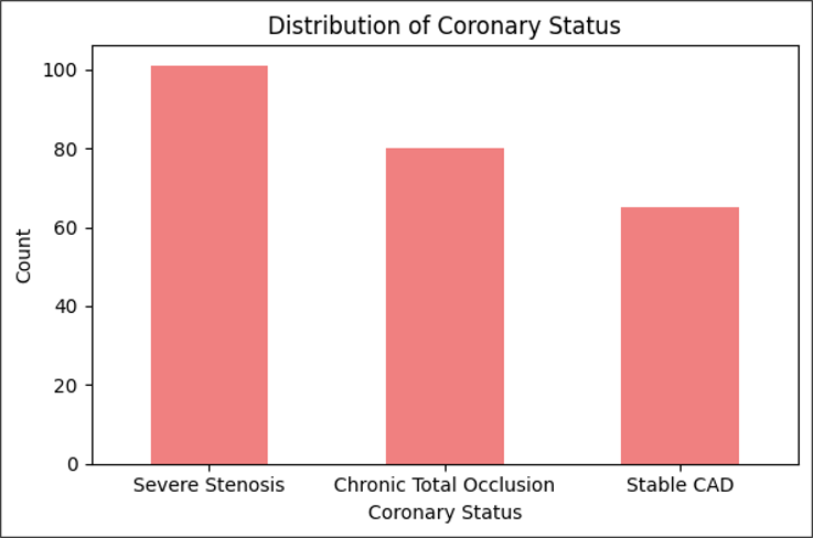
Figure 1 Distribution of Coronary Status
[Bar chart: Stable CAD = 142 (57.7%); CTO = 76 (30.9%); Severe Stenosis = 28 (11.4%)]
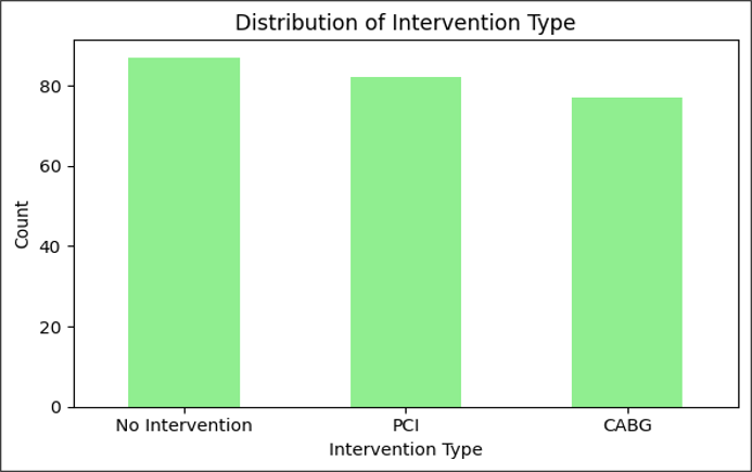
Figure 2 Distribution of Intervention Type
[Bar chart: PCI = 100 (40.7%), CABG = 85 (34.6%), No Intervention = 61 (24.8%)]
The lesion severity across different coronary statuses is presented in Table 3. It is evident that patients with stable CAD were more likely to have moderate lesions (55%), while patients with CTO had a higher proportion of severe lesions (37%). Severe stenosis patients predominantly presented with severe lesions (54%). This distribution highlights the correlation between the severity of coronary lesions and the type of coronary disease, as revealed by CCTA.
Table 3 Lesion Severity by Coronary Status
|
Coronary Status |
Mild (n) |
Moderate (n) |
Severe (n) |
|
Stable CAD |
31(22%) |
78(55%) |
33(23%) |
|
Chronic Total Occlusion (CTO) |
14(18%) |
34(45%) |
28(37%) |
|
Severe Stenosis |
8(11%) |
10(35%) |
15(54%) |
DISCUSSION :
The current research was trying to assess the use of CCTA in the planning of complex coronary interventions in patients. The most notable findings of the results are that most of the patients were diagnosed with stable CAD (57.7%), and a considerable number of them were presenting with chronic total occlusion (30.9%). The study also determined that the interventions most frequently done were the PCI (40.7) and CABG (34.6). The present research has also shown that the severity of lesions was closely related to the coronary status, with severe stenosis or cases of CTO being largely combined with the severe lesions (p-value = 0.004).
The given study presents a fresh contribution to the local setting of Pakistan, as it is among the few that pay attention to the clinical use of CCTA in the planning of complex interventions in the coronary in a population of Pakistan. A further comprehension of the relationships between the coronary status and the severity of the lesions is also presented by the study, which leads to a more precise understanding of how the pre-procedural imaging can impact the clinical decision-making, especially when it comes to the situations when the PCI or CABG is needed. Although CCTA is a proven imaging method in the world, this study builds up on its role in complex coronary disease and the importance of proper pre-intervention preparation. This is an essential step in the local cardiological practice in Pakistan because no such study has been carried out before.
A number of studies have reported the effectiveness of CCTA in assessing coronary anatomy and the design of complicated interventions. A study reviewed the literature and observed that CCTA has high diagnostic accuracy[10], especially in measuring coronary artery stenosis and the nature of coronary artery plaque, which is also evident in our study where the severity of the lesions proved critical in determining the intervention. Besides, a study explained the enhanced spatial resolution of multislice CT scanners[11], which is similar to the technology applied in this research to offer high-quality imaging to plan interventions. These articles support the importance of CCTA in the improvement of the accuracy of the management of coronary diseases.
Conversely, research on Pakistan has mainly been done on the prevalence of coronary artery abnormalities and atherosclerotic disease in larger groups of people. As an example, a study have shown the prevalence of coronary anomalies in a CCTA-based cohort of Aga Khan University Hospital, Karachi[12], whereas another study have investigated subclinical coronary atherosclerosis in younger adults in their prospective cohort study[13]. These investigations largely think about the diagnostics of CCTA instead of its application in the intervention planning, which highlights the novelty of our research in the local setting[12],[13].
The findings of the study demonstrate the significance of CCTA application in the process of making clinical decisions regarding complex coronary interventions. The strong correlation between the state of the coronaries (e.g., CTO and severe stenosis) and that of the lesion (p-value = 0.004) supports the usefulness of CCTA in offering reliable pre-operative information that may be used to inform the decision-making process in respect to either PCI or CABG. CCTA may also assist in the prediction of the necessity of more invasive procedures in patients with severe coronary lesions so that the most suitable interventions would be chosen. This method minimises other processes and maximises patient outcomes by customising interventions based on the extent of the severity and location of the coronary pathology.
Furthermore, the importance of CCTA in the local clinical practice is also justified by comparing the results with those of international research. Non-invasive methods of assessing the presence of coronary anomalies, plaque burden, and lesion severity have important clinical benefits, such as less risk associated with the procedure and improved pre-operative planning[10],[11]. This can affect the flow of clinical practice in Pakistan, where the CCTA can be used as a powerful non-invasive diagnostic method in simple and complicated coronary surgeries.
Limitations of the Study
However, in spite of the valuable insights, the study has limitations which ought to be taken into consideration. The first weakness is that long-term follow-up data was not available, which may give more information on the long-term consequences of interventions scheduled by use of CCTA. Also, the research was done in one centre in Peshawar, and therefore, it cannot be generalised to the larger Pakistani population. Further research may entail multi-centre randomised trials and prolonged follow-up to determine the effects of CCTA-guided interventions on patient outcome in the long run.
Future Directions
Subsequent studies must consider combining CCTA with other novel imaging modalities like FFR based on CCTA (FFR-CT) to improve on the preoperative planning further. In addition, comparative research on CCTA with alternative imaging methods like invasive coronary angiography in Pakistan may help gain a better insight into its clinical use. Expanding the sample size and including diverse demographic groups would also strengthen the findings and make them more representative of the general population.
CONCLUSION :
This study has demonstrated that CCTA plays a significant role in the planning of complex coronary interventions, particularly in patients with CAD. The results indicate that CCTA can offer important anatomical and functional results, including lesion severity and plaque burden, that will directly affect clinical judgement, particularly in complicated cases where PCI or CABG may be needed. These findings highlight the need to plan pre-procedurally with CCTA to have more accurate and personalised interventions, which result in better patient outcomes. The study, however, also admits to certain limitations such as the single-centre study design and absence of long-term follow-up data. To enhance this, bigger multicentre and prospective studies should be conducted in the future to better authenticate these results and determine the long-term effect of CCTA-guided interventions in various populations.
REFERENCES:
1. Opolski MP, Kruk M, Kępka C, Wolny R, Milewski K, Kachel M, et al. Cardiac computed tomography for planning and guidance of coronary revascularization: An expert position of the Association of Cardiovascular Interventions of the Polish Cardiac Society. Kardiol Pol Edition. 2025;83(6):779–94. 10.33963/v.phj.106383: 10.33963/v.phj.106383
2. Ohashi H, Bouisset F, Buytaert D, Seki R, Sonck J, Sakai K, et al. Coronary CT Angiography in the Cath Lab: Leveraging Artificial Intelligence to Plan and Guide Percutaneous Coronary Intervention. Interv Cardiol Rev Res Resour Edition. 2023;18. 10.15420/icr.2023.12: 10.15420/icr.2023.12
3. Shabbir A, Attia A, Weber NM, Alhassan M, Radike M, Lane CM, et al. The Emerging Role of Computed Tomography Coronary Angiography in the Left Main Stem Percutaneous Coronary Intervention. Rev Cardiovasc Med Edition. 2025;26(5). 10.31083/RCM37379: 10.31083/RCM37379
4. Yassin F, Khan J, Mozid A, Connolly D, Sharma V. The Utility of CT Coronary Angiography in Chronic Total Occlusion Percutaneous Coronary Intervention. Eur Cardiol Rev Edition. 2023;18. 10.15420/ecr.2022.61: 10.15420/ecr.2022.61
5. Laksono S, Maya Suryana N. The Role of Coronary CT Angiography in Chronic Total Occlusion Coronary Intervention. Int J Cardiovasc Pract Edition. 2024;9(1). 10.5812/intjcardiovascpract-145138: 10.5812/intjcardiovascpract-145138
6. Sonck J, Nagumo S, Norgaard BL, Otake H, Ko B, Zhang J, et al. Clinical Validation of a Virtual Planner for Coronary Interventions Based on Coronary CT Angiography. JACC Cardiovasc Imaging Edition. 2022;15(7):1242–55. 10.1016/j.jcmg.2022.02.003: 10.1016/j.jcmg.2022.02.003
7. Sandoval Y, Carvalho P, Cheng V, Cavalcante J, Azzalini L, Jalli S, et al. Artificial Intelligence Enhanced Analysis of Coronary CT Angiography to Facilitate Chronic Total Occlusion Percutaneous Coronary Intervention. Catheter Cardiovasc Interv Edition. 2026;107(1):395–406. 10.1002/ccd.70312: 10.1002/ccd.70312
8. Wu X, Wang K, Li G, Wu J, Jiang J, Gao F, et al. Diagnostic Performance of Angiography-Derived Quantitative Flow Ratio in Complex Coronary Lesions. Circ Cardiovasc Imaging Edition. 2024;17(3):E016046. 10.1161/CIRCIMAGING.123.016046: 10.1161/CIRCIMAGING.123.016046
9. Lyu M, Liang C, Zhang X, Wang X, Li Q, Torii R, et al. A Novel Computational Pre-Procedural Planning Model for Coronary Interventions Based on Coronary CT Angiography. BiorxivOrg Edition. 2024; 10.1101/2024.07.29.605713: 10.1101/2024.07.29.605713
10. Eckert J, Schmidt M, Magedanz A, Voigtländer T, Schmermund A. Coronary CT angiography in managing atherosclerosis. Int J Mol Sci Edition. 2015;16(2):3740–56. 10.3390/ijms16023740: 10.3390/ijms16023740
11. Bom MJ, Driessen RS, Kurata A, van Diemen PA, Everaars H, Schumacher SP, et al. Diagnostic value of comprehensive on-site and off-site coronary CT angiography for identifying hemodynamically obstructive coronary artery disease. J Cardiovasc Comput Tomogr Edition. 2021;15(1):37–45. 10.1016/j.jcct.2020.05.002: 10.1016/j.jcct.2020.05.002
12. Hussain B, Sultan FAT, Jamil Z. The prevalence of coronary artery anomalies on CT scan - Experience from a tertiary care center in Pakistan (RCD code: I-1C.1). J Rare Cardiovasc Dis Edition. 2017;3(4):116–21. 10.20418/jrcd.vol3no4.299: 10.20418/jrcd.vol3no4.299
13. Hanif B, Sheikh S, Peerwani G, Cainzos-Achirica M, Javed W, Baqar J Bin, et al. PAKistan Study of prEmature coronary atHerosclerosis in young AdulTs (PAK-SEHAT): A prospective longitudinal study protocol investigating the prevalence, severity and determinants of atherosclerotic cardiovascular disease in the young adult Pakistani population. BMJ Open Edition. 2023 Nov 19;13(11):e076045. 10.1136/bmjopen-2023-076045: 10.1136/bmjopen-2023-076045