Comparison of Chest Radiography and Computed Tomography in Haller Index Calculation for Pectus Excavatum Evaluation
- Muhammad Jawad Irfan , Department of Radiology, PNS SHIFA Hospital, Karachi, Pakistan.
- Naila Mumtaz , MBBS, FCPS, Classified Radiologist, Department of Radiology, PNS SHIFA Hospital, Karachi, Pakistan.
- Aliya Halim , Department of Radiology, PNS SHIFA Hospital, Karachi, Pakistan.
- Sherdil Shahzad , Department of Medicine, Dow International Medical College, Dow University of Health Sciences, Karachi, Pakistan.
- Amna Saleem Satti , Department of Radiology, PNS SHIFA Hospital, Karachi, Pakistan.
- Asma Afzal Kiani , Department of Radiology, Combined Military Hospital, Rawalpindi, Pakistan.
Article Information:
Abstract:
Objective: To determine the difference between chest radiography and CT measured HI in patients with pectus excavatum. StudyDesig :Comparative Cross-Sectional Study.Place and Duration of Study: Diagnostic Radiology, PNS Shifa Hospital, Karachi, from July 2025 to November 2025.Methodology: A total of 66 males aged 18-60 years who presented with a clinical diagnosis of pectus excavatum and were sampled by a non-probability, consecutive sampling method and divided randomly into two groups of 33 patients each using computer-generated sequential numbers in sealed envelopes. For Group A, posteroanterior and lateral chest x-rays were used to calculate the Haller Index. Contrast CT chest was not performed in all patients of Group B; therefore, Haller Index was derived from non-contrast CT chest. Independent samples t-test was used to compare the Mean Haller Index of both the groups at p ≤ 0.05 level of significance. SPSS 30.0 was used to analyze the data. Results: The average age of the population studied was 33.6 ± 9.8 years. The mean Haller Index from the chest radiograph (Group A) was 2.01 ± 0.45 and the mean Haller Index from the computed tomography (Group B) was 2.35 ± 0.39, which was statistically significant (p<0.001). 18.2% of the patients on CT had severe pectus result (Haller Index ≥ 3.2).Conclusion: Chest radiographic method is systematically lower (rad) than computed tomography method but the difference is definite and can be predicted, which spurs the use of this method as an alternative initial assessment tool for pectus excavatum in resource-poor settings with minimal radiation exposure and ease of access.
Keywords:
Article :
INTRODUCTION:
Pectus excavatum (PE) is thought to be the most common anterior chest wall malformation (1). It is a depression of the sternum in a funnel shape which leads to different cardiopulmonary complications in severe cases (2). It is estimated that 65 to 95 percent of all congenital chest wall deformities are caused by PE; the incidence of PE has been published to be 8 in every 1000 live births (3). If affected, patients tend to be tall with pot-bellied appearance and forward drifting shoulders. More severe forms of PE are accompanied by leftward displacement of the heart, imprint on the sternum of the right atrium and right ventricle, and lead to diastolic dysfunction (4).
Causes of symptoms due to the deformity disproportionately increase with age following a progressive remodeling of thoracic cage. If the PE is severe, then surgical treatments such as open or Ravitch procedure and minimally invasive Nuss procedure (5) will be indicated. The diagnosis and the degree of seriousness of PE should be determined prior to surgery by a radiological examination even if it involves chest X-rays or computed tomography (CT) (6) (7). One of these radiologic methods is the Haller Index which measures the extent of PE based upon the chest circumference at the most severe point of the deformity, whereby a threshold of ≥ 3.2 will categorize the PE as severe (7).
Generally, chest radiography has been suggested as an alternative to CT for providing information required to calculate Haller Index, including reduced exposure to radiation and greater availability; however, CT has traditionally been the test of choice for evaluating and categorizing PE prior to surgery. Several studies have tried to evaluate usefulness, diagnosis accuracy of chest radiography in determining Haller Index (8). Kiani et al. studied the correlation of mean Haller Indices as obtained on chest radiography and CT in a population of 20.2 ± 2.34 years at CMH Peshawar in a cross sectional study with a statistically significant strong positive correlation between the two modalities (9).
Compared to chest X-ray (0.1 mSv), a whole body CT scan is estimated to provide approximately 10 mSv of radiation exposure, or about 100 times as much. As such, chest radiography may be considered to spare the patient unnecessary radiation exposure and to save cost, as compared to CT. No study pertaining to this objective has been previously done at our hospital and the rationale was to evaluate the usefulness of chest radiography in calculating the Haller Index in the local population to avoid straining healthcare resources and further radiation exposure for the patient.
MATERIALS AND METHODS:
Study Design and Setting
Comparative cross-sectional study,Department of Diagnostic Radiology, PNS Shifa Hospital, Karachi.The study was conducted following institutional approval and ethical approval from the Ethical Review Committee of PNS Shifa Hospital, Karachi. Prior to enrollment, the research protocol was explained to all participants, and written informed consent was obtained from each subject. Participant confidentiality and data protection were strictly maintained throughout the study.
Sample Size
Sample size was calculated using WHO sample size calculator considering level of significance as 5%, power of test as 90% with an assumed mean Haller Index (on chest radiography) as 2.01 ± 0.45 and (on CT) as 2.35 ± 0.39 which got a calculated sample size of 33 patients in each group or 66 patients in total.
Sampling Technique
Non-probability consecutive sampling.
Inclusion Criteria
Inclusion criterion was pectus excavatum defined as per the operational definition, in male patients 18 to 60 years of age.
Exclusion Criteria
Exclusion criteria included a history of chest trauma within 7 days, associated chest deformities (kyphosis or scoliosis), prior pectus surgery, interstitial lung disease or primary or metastatic lung malignancy.
Data Collection Procedure
This study included 66 patients referred to the Department of Radiology for evaluation of pectus excavatum. The study was conducted with the written informed consent of the patients, as per the required guidelines. Patients were then randomly allocated in 2 equal groups (Group A and B) by a computer-generated sequential numbering in sealed envelopes, opened just before enrolment. In Group A, PA and lateral chest X-rays were taken with Canon-DR 402 X-ray machine and the Haller Index ratio (diameter measuring the widest extent of the thorax to the least diameter measuring from the back of the sternum to the front of the body of the vertebra at the same level) were calculated. Group B, Contrast CT chest was not performed in any patient in Group B; Haller Index was therefore derived from non-contrast CT chest, which was performed using the Aquilion Prime 160-slice CT scanner and the Haller Index was calculated on the greatest transverse section of the thorax in respect to the smallest anteroposterior section of the thorax. The principal investigator collected the findings in a predesigned proforma and patient details were kept confidential throughout.
Ethical Considerations
Approval was taken from the Ethical Review Committee in PNS Shifa Hospital, Karachi, All participants gave written informed consent before they participated in the study.
Data Analysis
Entered and analyzed data by SPSS 30.0. Normality of quantitative variables was checked by Shapiro-Wilk test. Age (mean ±SD) and Haller Index (mean ±SD) were numerically calculated and gender and severity classification were numerically presented as frequencies and percentages. Independent samples t-test was used for the comparison of mean Hi in the 2 groups and P<0.05 considered as statistically significant
RESULTS:
66 male patients with pectus excavatum were included and alternately allocated to Group A (radiography of the chest) and Group B (computed tomography). The mean age of the study population was 33.6 ± 9.8 years and most of the patients were urban dwelling (71.2%). The demographic characteristics of the study population are presented in Table 1.
Table 1: Demographic characteristics of the study population (n = 66) is provided.
|
Variable |
Frequency / Mean ± SD |
Percentage |
|
Mean age (years) |
33.6 ± 9.8 |
— |
|
Group A (Chest Radiography) |
33 |
50.0% |
|
Group B (Computed Tomography) |
33 |
50.0% |
|
Urban residence |
47 |
71.2% |
|
Rural residence |
19 |
28.8% |
The mean Haller Index calculated on chest radiography (Group A) was 2.01 ± 0.45 which is significantly lower than mean Haller Index calculated on computed tomography (Group B) 2.35 ± 0.39 (independent samples t-test, p<0.001) as shown in Table 2 and Figure 1.
Table 2: The comparison of Mean Haller Index of Group A and Group B
|
Group |
Modality |
Mean Haller Index ± SD |
p-value |
|
Group A (n=33) |
Chest Radiography |
2.01 ± 0.45 |
|
|
Group B (n=33) |
Computed Tomography |
2.35 ± 0.39 |
<0.001 |
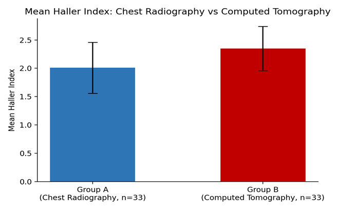
Figure 1. Compared Mean Haller Index: Chest Radiography (Group A) vs Computed Tomography (Group B).
Based on the CT derived Haller Index cut-off of ≥ 3.2 for severe pectus excavatum, 18.2% (6/33) of the patients in group B had severe disease and 81.8% (27/33) had non-severe disease as shown in Figure 2.
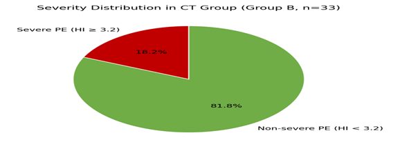
Figure 2. The figure illustrates the severity distribution in CT Group (Group B, 33 patients).
Table 3: The severity classification in computed tomography (Group B, n=33)
|
Severity (Haller Index) |
Frequency |
Percentage |
|
Severe (≥ 3.2) |
6 |
18.2% |
|
Non-severe (< 3.2) |
27 |
81.8% |
DISCUSSION :
This cross sectional comparison demonstrated that there is statistically significant difference between the mean Haller Index determined using chest radiography (2.01 ± 0.45) and using computed tomography (2.35 ± 0.39), (p<0.001). These results compare favorably with the findings of Kiani et al. whereby they found almost identical mean CT derived HL and chest radiography derived HL of 2.35 ± 0.39 vs 2.01 ± 0.45 respectively in a similar population in the city of Peshawar, KP, Pakistan with strong and statistical significant correlation between the two modalities (9).
Similarly, Khanna et al. evaluated patients for pectus excavatum and found correlation between the Haller Index values obtained from chest radiographs and those obtained from CT scans, although the radiographic values were always lower than those of the CT scans, and the two measures were satisfactory for use for screening purposes (8). Ewert et al. also showed that there was a meaningful correlation between external and radiographic chest wall measurements and CT-based measures in patients with chest wall deformities for those with less-radiation measurement methods, allowing for serial measurements (1).
Part of the explanation for the lessened Haller Index in chest radiography in this study may be the inherent differences between the methods of obtaining images: chest radiography is creating a 2D image of a 3D thoracic cavity, and cross sectional (true-axial) is obtained on CT. However, Messerli-Odermatt et al. investigated a chest X-ray dose-equivalent low-dose CT protocol with tin filtration as a potential compromise between radiation exposure and measurement reliability and concluded that ultra-low dose CT techniques may further reduce this difference and provide significant radiation reduction over current-dose CT (7).
In a comprehensive review of advances in the imaging of pectus excavatum for preoperative assessment, Coorens et al., noted that although the reference standard for data in definitive surgical assessment is CT, radiation-free options of chest radiography and surface scanning technologies were gradually introduced to screening and follow-up programs, especially in younger patients who would need multiple evaluations (6). This aligns with the result of the present study that even though the absolute Haller Index value was underestimated, it did identify the proportion of patients with clinically relevant deformity (as would be expected based on the CT based severity cut-offs).
In a population-based cohort study, Biavati et al reported prevalence for radiographic diagnosis of pectus excavatum that were largely consistent with the prevalence of severe cases observed in the current study, as they indicated that the radiographic indices could be useful for epidemiological and screening applications when CT cannot be applied to the entire population (5). Janssen et al also presented a narrative review of pectus excavatum and carinatum, pointing out significant heterogeneity in reported Haller Index values across different imaging modalities and patient populations, thus emphasizing the need for institution-specific comparative data like that of the present study (3).
This study has certain limitations. It was performed in one tertiary care center and the findings might not be applicable to other centers. Due to the different patients in both groups (A&B) being used, and the same patients not being used under both groups, it was not possible to directly compare them, nor use any diagnostic accuracy parameters like sensitivity or specificity. Further studies of patients with paired measurements and over a broader age range incorporating women may help facilitate a better understanding of the comparative diagnostic performance of chest radiography versus CT.
CONCLUSION :
In patients with pectus excavatum, computed tomography has a mean Haller Index which is higher than the mean index on chest radiography, but the difference is an overall systematic and significant one with a pattern of difference broadly consistent with earlier local and international data. Considering its significantly less radiation exposure and regular ease of availability, the use of chest radiography may be a useful screening tool for pectus excavatum severity with computed tomography reserved in patients who meet or are approaching surgical threshold.
ACKNOWLEDGMENT
The authors would like to acknowledge the department of Diagnostic radiology, PNS Shifa Hospital, Karachi for providing institutional support in conducting the study.
CONFLICT OF INTEREST
None.
FUNDING
None.
REFERENCES:
1. Ewert F, Syed J, Wagner S, Besendoerfer M, Carbon RT, Schulz-Drost S. In patient with chest wall deformity, is there a correlation between an external chest wall measurement and a CT measurement? J Pediatr Surg. 2017;52(10):1583-90.
2. Boettcher M, Klohs S, Kloth K, Bhullar J, Hempel M, Trah J, et al. Pectus excavatum – epidemiology based on clinical study and review of literature. Eur J Pediatr Surg. 2022;32(04):316-20.
3. Janssen N, Coorens NA, Franssen AJ, Daemen JH, Michels IL, Hulsewé KW and others. Pectus excavatum and carinatum: epidemiologic, etiopathogenic, clinical and classification aspects in a narrative review. J Thorac Dis. 2024;16(2):1687.
4. Notrica DM, McMahon LE, Jaroszewski, DE. Pectus disorders (excavatum, carinatum and arcuatum). Adv Pediatr. 2024;71(1):181-94.
5. Biavati M, Kozlitina J, Alder AC, Foglia R, McColl RW, Peshock RM, et al. Radiographic estimation of chest wall shape in an adult population-based cohort yields a prevalence for pectus excavatum. PLoS One. 2020;15(5):e0232575.
6. Coorens NA, Janssen N, Daemen JH, Franssen AJ, Hulsewé KW, Vissers YL, et al. Advancements in terms of preoperative imaging of pectus excavatum – a comprehensive review. J Thorac Dis. 2024;16(1):696.
7. Messerli-Odermatt O, Serrallach B, Gubser M, Leschka S, Bauer RW, Dubois J, et al. Chest X-ray dose equivalent low-dose CT with tin filtration: potential role for the assessment of pectus excavatum. Acad Radiol. 2020;27(5):644-50.
8. Khanna G.A, Jaju A., Don S, Keys T, Hildebolt CF. Comparison of Haller Index values calculated with chest radiographs versus CT for evaluation of pectus excavatum. Pediatr Radiol. 2010;40(11):1763-7.
9. Kiani AA, Haider S, Sharif S, tul Kubra K, Fatima K, Murtaza N. Comparison of Haller Index value calculated with Chest Radiograph and computed tomography in evaluation of pectus excavatum. Pak Armed Forces Med J. 2024;74(1):216-19.