Exploration of Endophytic Fungi Associated with the Medicinal Plant Centella asiatica
- Prathibha K Y , Professors, Department of Botany, Maharani Cluster University, Palace Road, Bengaluru, Karnataka, India, 560001
- Geethanjali R , Associate Professors, Department of Botany, Maharani Cluster University, Palace Road, Bengaluru, Karnataka, India, 560001
- Nalini T J , Associate Professors, Department of Botany, Maharani Cluster University, Palace Road, Bengaluru, Karnataka, India, 560001
- Suresh Kumar C4, , Professors, Department of Botany, Maharani Cluster University, Palace Road, Bengaluru, Karnataka, India, 560001
- Sannidhi M Bhat Alekh , B.Sc Students, Maharani Cluster University, Palace Road, Bengaluru, Karnataka, India, 560001.
- Shweta Shivanagouda Biradar , B.Sc Students, Maharani Cluster University, Palace Road, Bengaluru, Karnataka, India, 560001.
- Nagalli Umadevi , B.Sc Students, Maharani Cluster University, Palace Road, Bengaluru, Karnataka, India, 560001.
- Mythri T , B.Sc Students, Maharani Cluster University, Palace Road, Bengaluru, Karnataka, India, 560001.
Article Information:
Abstract:
Endophytic fungi are microorganisms that live internally within apparently healthy and asymptomatic plant hosts without causing visible disease symptoms. They are ubiquitous in nature and have been reported from nearly all plant species studied to date. The present study was undertaken to isolate and identify endophytic fungi associated with different tissues of Centella asiatica. Fresh and healthy leaves, stems, and roots of C. asiatica were collected from Byrasandra, Bangalore, India. Plant samples were surface sterilized and inoculated onto Potato Dextrose Agar (PDA) supplemented with ampicillin. Emerging fungal colonies were subcultured to obtain pure isolates and identified based on morphological characteristics using lactophenol cotton blue staining and microscopic observation. A total of sixteen endophytic fungal isolates were recovered from C. asiatica. Five isolates were obtained from leaves, namely Alternaria sp., Aspergillus nidulans, Aspergillus sp., Glomerella cingulata, and Penicillium sp. Eight isolates were recovered from stems, including Cladosporium sp., Cryptococci sp., Curvularia sp., Fusarium sp., two species of Penicillium, Talaromyces sp., and Trichoderma sp. Three isolates were obtained from roots, namely Aspergillus sp., Fusarium sp., and Talaromyces sp. The study revealed a diverse assemblage of endophytic fungi inhabiting different tissues of C. asiatica, suggesting that this medicinal plant serves as an important reservoir of fungal endophytes with potential biotechnological and pharmaceutical applications.
Keywords:
Article :
INTRODUCTION:
Endophytic fungi are microorganisms that live internally within apparently healthy and asymptomatic plant hosts. They inhabit plant tissues without causing visible disease symptoms or morphological damage to the host. These fungi are considered ubiquitous in nature, and studies have shown that almost every plant species examined so far hosts endophytes. In fact, no research has yet demonstrated the existence of a plant species completely devoid of endophytic microorganisms. One of the most remarkable characteristics of endophytic fungi is their high species diversity. Endophyte surveys frequently report complex assemblages consisting of more than 30 fungal species associated with a single host plant species.
This high diversity reflects the ecological significance of endophytic fungi in plant ecosystems. These fungi are recognized as important sources of cytotoxic compounds, including anticancer molecules, as well as antibacterial substances. In addition to these pharmacologically valuable compounds, endophytic fungi can also act as biostimulants that enhance essential oil biosynthesis in plants. Furthermore, they contribute to nutrient solubilization within the plant rhizosphere, thereby improving nutrient availability for plant uptake.
Endophytic fungi are also known to promote plant growth and can function as biological control agents that help protect plants from pathogens. In addition, they can activate systemic resistance mechanisms in plants, enabling them to tolerate both biotic stresses, such as pathogens and pests, and abiotic stresses, including environmental factors. Endophytic fungi reside within healthy plant tissues and form an integral part of the plant’s microecosystem. The population dynamics and diversity of these fungi are influenced by several factors, including the genetic composition of the host plant, the age of the plant, and environmental conditions. Through long-term associations with their hosts, endophytic fungi establish symbiotic relationships that can be beneficial to plant growth and survival. These fungi produce a variety of important bioactive compounds such as alkaloids, diterpenes, flavonoids, and isoflavonoids.
These compounds contribute to plant growth, enhance the plant’s resilience to environmental stresses, and strengthen defence mechanisms against pests and diseases. They also promote the accumulation of secondary metabolites within the host plant. An important feature of endophytic fungi is their ability to produce bioactive compounds that are chemically identical or very similar to those synthesized by their host plants. Notable examples include Taxol, capsaicin, and piperine. The discovery that fungal endophytes can produce these valuable compounds has opened new possibilities for sustainable production through in vitro cultivation of these microorganisms. Culturing endophytic fungi capable of producing such metabolites offers a practical alternative to harvesting large quantities of plants from natural populations. This approach reduces environmental pressure on medicinal plants, minimizes ecological damage, and ensures consistent production of bioactive compounds regardless of climatic conditions. As a result, endophytic fungi have gained considerable importance in biotechnology and industrial applications. They are currently being explored for their potential roles as biocontrol agents, antimicrobials, antitumor agents, antioxidants, antidiabetic agents, antibiotics, and insecticides.1,2,3.
Medicinal plants have long been used as important sources for the isolation and characterization of bioactive metabolites. Traditionally, many biologically active compounds used in medicine were obtained directly from plants. However, the discovery of fungal endophytes residing within these medicinal plants and their ability to produce the same or similar bioactive compounds has shifted scientific interest toward fungi as alternative sources of valuable metabolites. Consequently, bioactive natural products derived from endophytic fungi isolated from different plant species have attracted considerable attention from natural product chemists and biologists. This growing interest is clearly reflected in the steady increase in scientific publications devoted to endophytic fungi and their metabolites in recent years.
Plants, particularly perennial species, are colonized by a wide variety of endophytic microorganisms. These microorganisms live inside plant tissues either throughout their entire life cycle or during a specific phase of their life cycle without producing visible symptoms in the host plant. Endophytic microorganisms include both fungi and bacteria, and they often coexist with pathogenic microorganisms within the same host plant tissues. Despite the presence of pathogens, endophytes maintain a balanced interaction with their host plants that does not result in disease.1,2,3.
Centella asiatica (L.) Urban., also known as Gotu kola, Indian pennywort or Pegaga, is a clonal, perennial herbaceous creeper belonging to the family Apiaceae (Umbelliferae). The plant grows widely in tropical and subtropical regions of the world including India, Pakistan, Sri Lanka, Madagascar, equatorial Africa, Central America and tropical Oceania. It commonly occurs in moist and swampy habitats such as wetlands, rice paddies and other pantropical environments, and in India it can grow up to an altitude of about 1800 m. Around twenty related species occur in wet tropical regions, sometimes even in rocky areas at higher elevations.
The plant is characterized by small fan-shaped green leaves, white or light purple-to-pink flowers and small oval fruits. It is generally tasteless and odourless and thrives in and around water. The whole plant is used for medicinal purposes and has long been valued in many traditional medical systems. Centella asiatica has a long history of use in traditional medicine across Asia, including Ayurveda, traditional Chinese medicine and Southeast Asian ethnomedicine. Historical records indicate that it was used as a medicinal “panacea” in countries such as China, India, Africa, the Philippines, Sri Lanka and Madagascar for thousands of years. Due to its medicinal importance, the plant was included in the Indian Pharmacopoeia in the nineteenth century and later incorporated into several other pharmacopoeias such as the British Herbal Pharmacopoeia, Chinese Pharmacopoeia, German Pharmacopoeia and European Pharmacopoeia. Traditionally, the herb has been used for revitalizing nerves and brain cells, improving memory and promoting longevity. It has also been employed as a blood purifier and for managing high blood pressure.
In Eastern healing practices it was used to treat emotional disturbances such as depression, which were believed to be associated with physical conditions. In addition, the plant has been used to relieve anxiety, improve cognition and treat various ailments including fever, diarrhoea, amenorrhea and disorders of the female genitourinary system. In dermatological treatments, C. asiatica has been widely applied for wound healing and for managing several skin conditions such as eczema, psoriasis, lupus, varicose ulcers, burns and hypertrophic scars. It has also been used traditionally for minor itching, insect bites, rheumatic conditions and venous insufficiency. Earlier medicinal uses included treatment for epilepsy, hysteria and leprosy, and studies conducted in the twentieth century reported positive results when the plant and its alcohol extracts were used in leprosy treatment.
The therapeutic properties of Centella asiatica are mainly attributed to its bioactive compounds, particularly pentacyclic triterpenoids and saponins such as asiaticoside, madecassoside, asiatic acid and madecassic acid. The plant also contains flavonoids and essential oils that contribute to its pharmacological activity. These compounds promote fibroblast proliferation, stimulate collagen synthesis and increase intracellular fibronectin levels, thereby improving the tensile strength of newly formed skin and enhancing wound healing. They also help inhibit inflammatory processes involved in hypertrophic scars and keloids. Modern research has confirmed many of the traditional uses of C. asiatica, highlighting its anti-inflammatory, cognitive-enhancing and adaptogenic properties. The plant is also being investigated for applications in treating photoaging skin, cellulite and striae.4,5,6.
MATERIALS AND METHODS :
Samples of Centella asiatica, fresh and healthy stems, leaves and roots were collected from Byrasandra, Bangalore, India. The plant parts were thoroughly washed with running tap water, rinsing with sterile distilled water (SDW) and then treated with 20% commercial bleach with 0.1% Tween-20 for 5 min and later again rinsed in SDW. They were then soaked in 100 mL solution containing fungicide bavistin (30 mg), tetracycline (0.6 mg), rifampicin (0.6 mg) and 0.1% Tween-20 for 20 min and rinsed in SDW. All samples were finally surface sterilized with 0.1% mercuric chloride for 10 min, followed by five to six washings with SDW. The surface sterilized stem segments were cut in to small pieces (1–2 mm), leaves were cut into 1 cm2 pieces and placed on petri plates containing potato dextrose agar (PDA) supplemented with 100 mg/L ampicillin. The plates were labelled and incubated at 25 ± 2°C with 12 h photoperiod or in darkness. The fungal mycelia that emerged from the cut surface of the segments after several days were transferred on to fresh PDA plates.7 Fungi were mounted using lactophenol cotton blue method.8.
RESULTS:
A total of sixteen endophytic fungi were isolated from Centella asiatica: five from leaves, namely Alternaria sp. Aspergillus nidulans, Aspergillus sp. Glomerella cingulata, and Penicillium sp. eight from stems, namely Cladosporium sp. Cryptococci sp. Curvularia sp. Fusarium sp. two species of Penicillium, Talaromyces sp. and Trichoderma sp. and three from roots, namely Aspergillus sp. Fusarium sp. and Talaromyces sp. Alternaria sp. was isolated from the leaves of Centella asiatica in the present study.
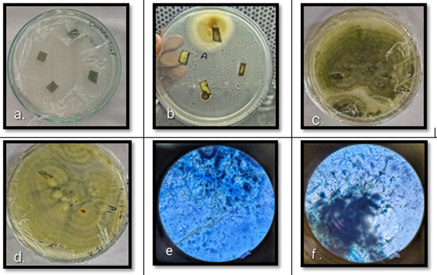
Fig 1; Cultured plates showing endophytic fungi isolated from Centella asiatica leaf: a: Inoculation of Centella asiatica leaf on culture media, b: Initial stage of fungal growth on the culture plate, c: Subcultured plates exhibiting active growth of Aspergillus nidulans, d: Backside view of the same culture plate, e: Microscopic observation of Aspergillus nidulans (10x), f: Microscopic observation of Aspergillus nidulans (40x).
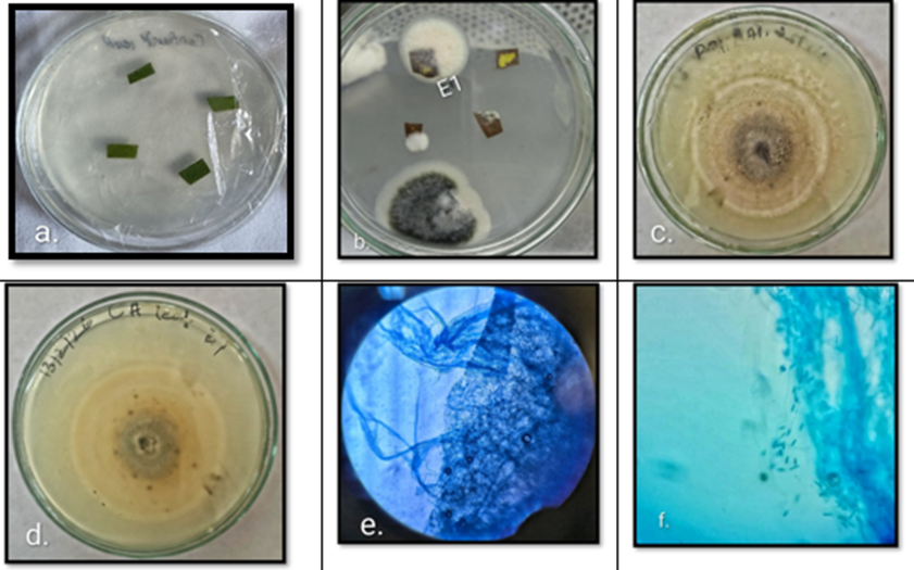
Fig 2: Cultured plates showing endophytic fungi isolated from Centella asiatica leaf: a: Inoculation of Centella asiatica leaf on culture media, b: Initial stage of fungal growth on the culture plate, c: Subcultured plates of E1 colony exhibiting active growth of Glomerella cingulata, d: Backside view of the same culture plate, e: Microscopic observation of Glomerella cingulata (10x), f: Microscopic observation of Glomerella cingulata (40x).
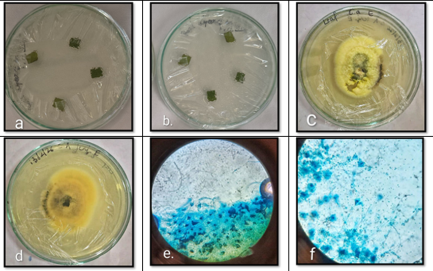
Fig 3: Cultured plates showing endophytic fungi isolated from Centella asiatica leaf: a: Inoculation of Centella asiatica leaf on culture media, b: Backside view of the inoculated culture plate, c: Subcultured plates exhibiting active growth of Penicillium sp. d: Backside view of the same culture plate, e: Microscopic observation of Penicillium sp. (10x), f: Microscopic observation of Penicillium sp. (40x).
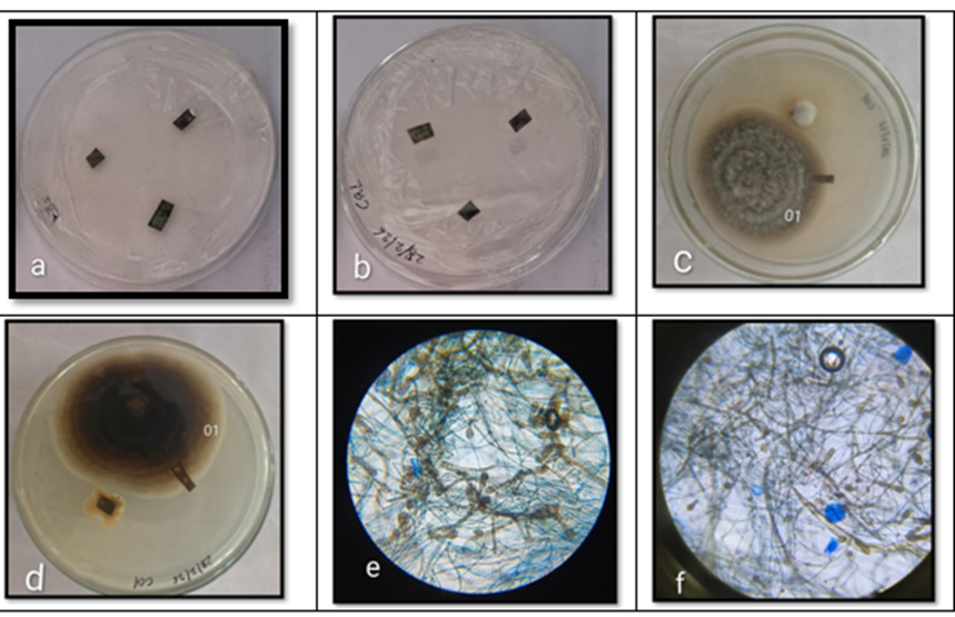
Fig 4: Cultured plates showing endophytic fungi isolated from Centella asiatica leaf: a: Inoculation of Centella asiatica leaf on culture media, b: Backside view of the inoculated culture plate, c: Fungal colony 01 showing growth characteristics of Alternaria sp. d: Backside view of the same culture plate, e: Microscopic observation of Alternaria sp. (10x), f: Microscopic observation of Alternaria sp. (40x)
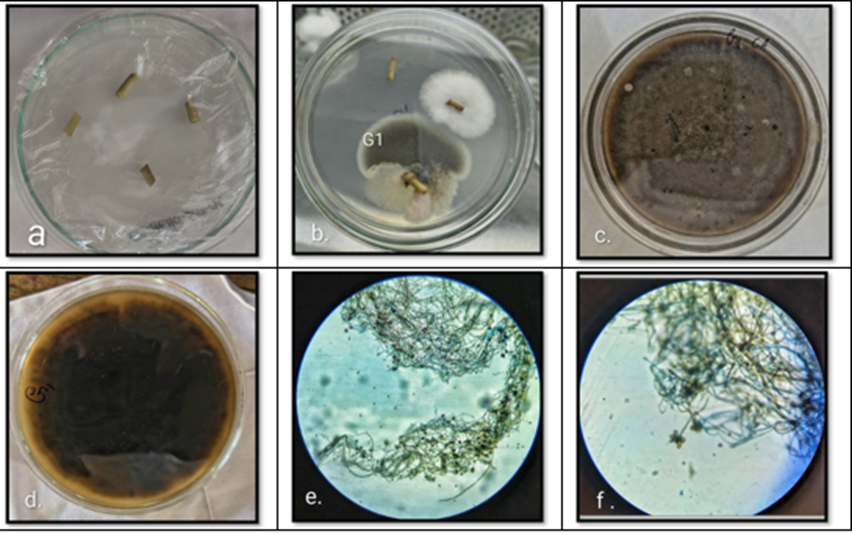
Fig 5: Cultured plates showing endophytic fungi isolated from Centella asiatica stem: a: Inoculation of Centella asiatica stem on culture media, b: Initial stage of fungal growth on the culture plate, c: Subcultured plates of G1 colony exhibiting active growth of Curvularia sp. d: Backside view of the same culture plate, e: Microscopic observation of Curvularia sp. (10x), f: Microscopic observation of Curvularia sp. (40x)
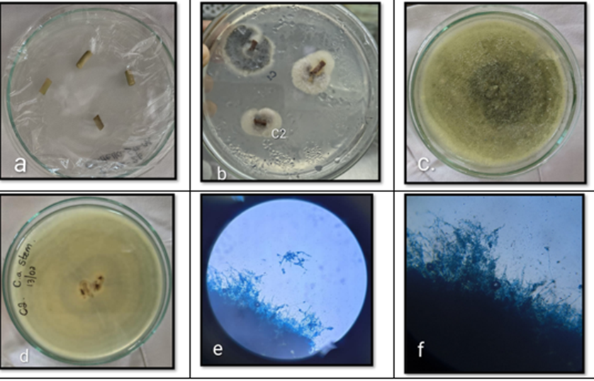
Fig 6: Cultured plates showing endophytic fungi isolated from Centella asiatica stem: a: Inoculation of Centella asiatica stem on culture media, b: Initial stage of fungal growth on the culture plate, c: Subcultured plates exhibiting active growth of Penicillium sp. d: Backside view of the same culture plate, e: Microscopic observation of Penicillium sp. (10x), f: Microscopic observation of Penicillium sp. (40x)
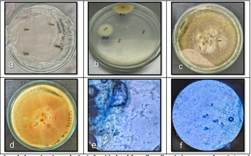
Fig 7: Cultured plates showing endophytic fungi isolated from Centella asiatica stem: a: Inoculation of Centella asiatica stem on culture media, b: Initial stage of fungal growth on the culture plate, c: Subcultured plates exhibiting active growth of Penicillium sp. d: Backside view of the same culture plate, e: Microscopic observation of Penicillium sp. (10x), f: Microscopic observation of Penicillium sp. (40x).
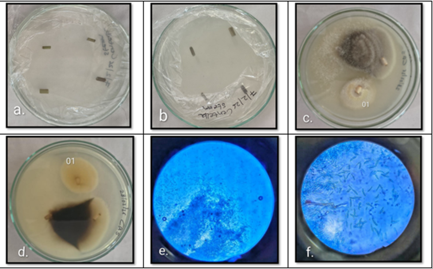
Fig 8: Cultured plates showing endophytic fungi isolated from Centella asiatica stem: a: Inoculation of Centella asiatica stem on culture media, b: Backside view of the inoculated culture plate, c: Fungal colony 01 showing growth characteristics of Fusarium sp. d: Backside view of the same culture plate, e: Microscopic observation of Fusarium sp. (10x), f: Microscopic observation of Fusarium sp. (40x).
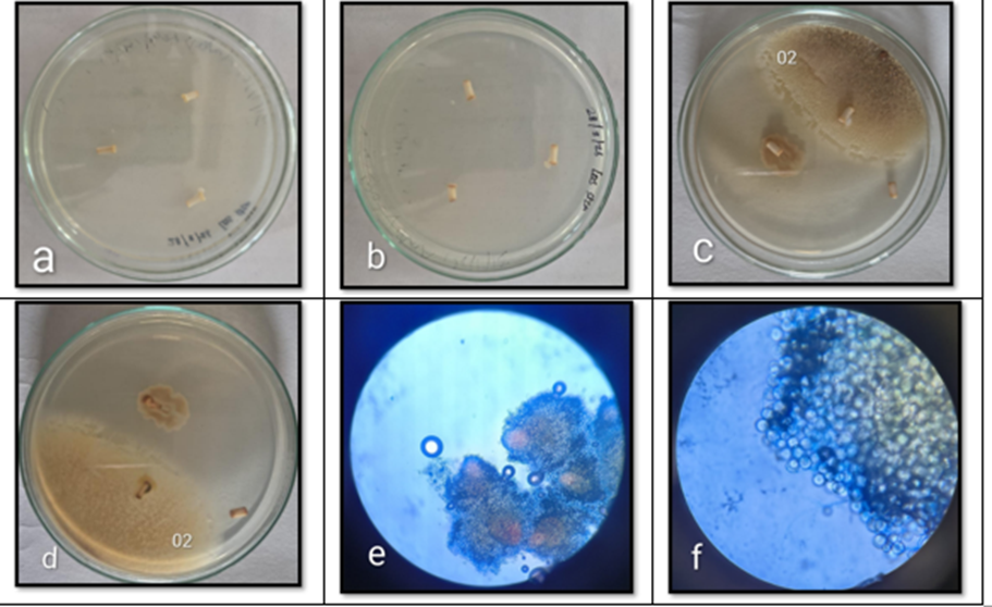
Fig 9: Cultured plates showing endophytic fungi isolated from Centella asiatica stem: a: Inoculation of Centella asiatica stem on culture media, b: Backside view of the inoculated culture plate, c: Fungal colony 02 showing growth characteristics of Cryptococci sp. d: Backside view of the same culture plate, e: Microscopic observation of Cryptococci sp. (10x), f: Microscopic observation of Cryptococci sp. (40x).
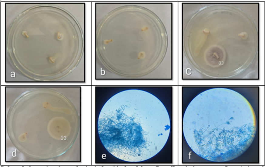
Fig 10: Cultured plates showing endophytic fungi isolated from Centella asiatica stem: a: Inoculation of Centella asiatica stem on culture media, b: Backside view of the inoculated culture plate, c: Fungal colony 03 showing growth characteristics of Talaromyces sp. d: Backside view of the same culture plate, e: Microscopic observation of Talaromyces sp. (10x), f: Microscopic observation of Talaromyces sp. (40x).
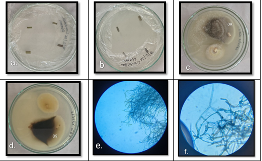
Fig 11: Cultured plates showing endophytic fungi isolated from Centella asiatica stem: a: Inoculation of Centella asiatica stem on culture media, b: Backside view of the inoculated culture plate, c: Fungal colony 09 showing growth characteristics of Trichoderma sp. d: Backside view of the same culture plate, e: Microscopic observation of Trichoderma sp. (10x), f: Microscopic observation of Trichoderma sp. (40x).
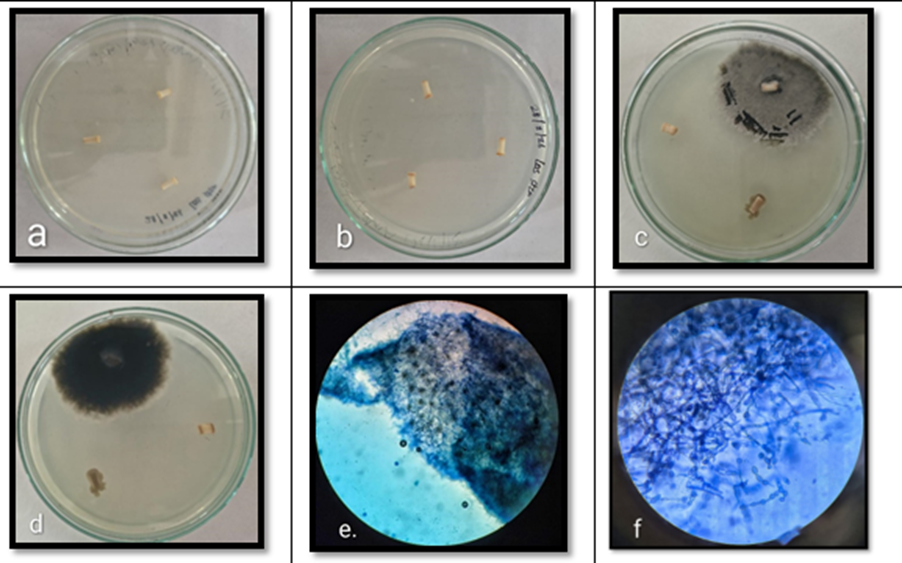
Fig 12: Cultured plates showing endophytic fungi isolated from Centella asiatica stem: a: Inoculation of Centella asiatica stem on culture media, b: Backside view of the inoculated culture plate, c: Fungal colony showing growth characteristics of Cladosporium sp. d: Backside view of the same culture plate, e: Microscopic observation of Cladosporium sp. (10x), f: Microscopic observation of Cladosporium sp. (40x).
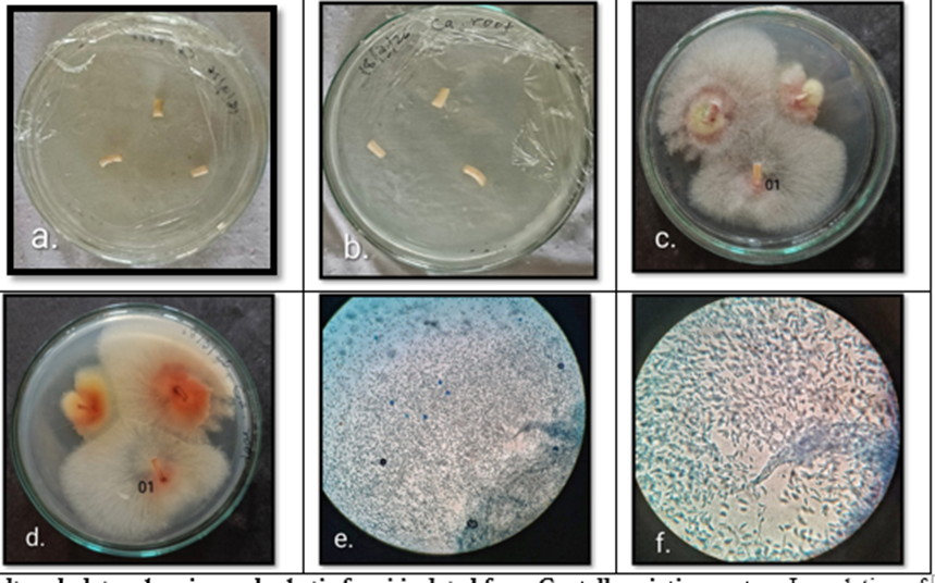
Fig 13:Cultured plates showing endophytic fungi isolated from Centella asiatica root: a: Inoculation of Centella asiatica root on culture media, b: Backside view of the inoculated culture plate, c: Fungal colony 01 showing growth characteristics of Fusarium sp. d: Backside view of the same culture plate, e: Microscopic observation of Fusarium sp. (10x), f: Microscopic observation of Fusarium sp. (40x).
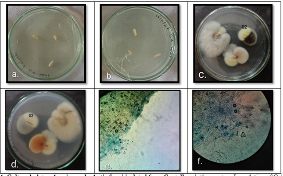
Fig 14: Cultured plates showing endophytic fungi isolated from Centella asiatica root: a: Inoculation of Centella asiatica root on culture media, b: Backside view of the inoculated culture plate, c: Fungal colony 03 showing growth characteristics of Talaromyces sp. d: Backside view of the same culture plate, e: Microscopic observation of Talaromyces sp. (10x), f: Microscopic observation of Talaromyces sp. (40x).
DISCUSSION :
A total of sixteen endophytic fungi were isolated from Centella asiatica: five from leaves, namely Alternaria sp. Aspergillus nidulans, Aspergillus sp. Glomerella cingulata, and Penicillium sp. eight from stems, namely Cladosporium sp. Cryptococci sp. Curvularia sp. Fusarium sp. two species of Penicillium, Talaromyces sp. and Trichoderma sp. and three from roots, namely Aspergillus sp. Fusarium sp. and Talaromyces sp. Alternaria sp. was isolated from the leaves of Centella asiatica in the present study; similarly, Kunasakdakul et al. (2015) isolated Alternaria brassicicola from Centella asiatica9, and Chaturvedi et al. (2017) reported Alternaria sp. among endophytic fungi from C. asiatica10. Aspergillus nidulans was isolated from leaves, and two other species of Aspergillus were isolated from leaves and roots of Centella asiatica in the present study; similarly, Nagda et al. (2017) reported Aspergillus sp. from Calotropis procera11, and Chaturvedi et al. (2017) also reported Aspergillus sp. from C. asiatica18. Cladosporium sp. was isolated from the stem of Centella asiatica in the present study; similarly, Nessa et al. (2023) isolated Cladosporium sp. from leaves, stems, and roots of Centella asiatica12. Cryptococci sp. was isolated from the stem of Centella asiatica in the present study; similarly, Isaeva et al. (2010) reported Cryptococci as endophytic yeast in plant tissues13. Curvularia sp. was isolated from the stem of Centella asiatica in the present study; similarly, Chaturvedi et al. (2017) reported Curvularia sp. from C. asiatica10. Fusarium sp. was isolated from the stem and root of Centella asiatica in the present study; similarly, Chaturvedi et al. (2017) reported Fusarium sp. from C. asiatica10. Glomerella cingulata was isolated from the leaves of Centella asiatica in the present study; similarly, Rakotoniriana et al. (2008) reported Glomerella sp. among endophytic fungi from leaves of Centella asiatica14. Penicillium sp. was isolated from the leaves and stems of Centella asiatica in the present study; similarly, Devi et al. (2012)15 and Nagda et al. (2017)11 reported Penicillium sp., and Holeyannavar et al. (2025) reported Penicillium chrysogenum from Centella asiatica16. Talaromyces sp. was isolated from the stems and roots of Centella asiatica in the present study; similarly, Holeyannavar et al. (2025) reported Talaromyces verruculosus16. Trichoderma sp. was isolated from the stem of Centella asiatica in the present study; similarly, Holeyannavar et al. (2024) reported Trichoderma harzianum17. Chaturvedi et al. (2017) also reported Colletotrichum gloeosporioides, Colletotrichum sp. Fusarium sp. Curvularia sp. Nigrospora sp. Alternaria sp. Aspergillus sp. and Fusarium equiseti from Centella asiatica10.
CONCLUSION :
The present study successfully isolated and identified endophytic fungi associated with different tissues of Centella asiatica. A total of sixteen fungal isolates were recovered from leaves, stems, and roots following surface sterilization and culture on Potato Dextrose Agar. The isolated fungi belonged to diverse genera, including Alternaria, Aspergillus, Cladosporium, Cryptococci, Curvularia, Fusarium, Glomerella, Penicillium, Talaromyces, and Trichoderma. The stem tissues harbored the highest number of endophytic fungi, followed by leaves and roots. Among the isolates, Alternaria sp. was obtained from leaf tissues of Centella asiatica. The results indicate that Centella asiatica serves as a rich reservoir of diverse endophytic fungal communities. The occurrence of different fungal genera in various plant tissues suggests tissue-specific colonization and adaptation. These endophytic fungi may represent a valuable source of bioactive metabolites with potential applications in agriculture, biotechnology, and pharmaceutical research. Further studies involving molecular identification and screening for bioactive compounds are recommended to explore their functional and therapeutic potential.
REFERENCES:
1. Nisa, H., Kamili, A. N., Nawchoo, I. A., Shafi, S., Shameem, N., & Bandh, S. A. (2015). Fungal endophytes as prolific source of phytochemicals and other bioactive natural products: A review. Microbial Pathogenesis, 82, 50–59. https://doi.org/10.1016/j.micpath.2015.04.001
2. Alam, B., Li, J., Ge, Q., Khan, M. A., Gong, J., Mehmood, S., Yuan, Y., & Gong, W. (2021). Endophytic fungi: From symbiosis to secondary metabolite communications or vice versa? Frontiers in Plant Science, 12, 791033. https://doi.org/10.3389/fpls.2021.791033
3. Mendieta-Brito, S., Sayed, M., Hamza, F. A., et al. (2025). Identification, characterization, antimicrobial activity and biocontrol potential of four endophytic fungi isolated from Amazonian plants. Scientific Reports, 15, 39361. https://doi.org/10.1038/s41598-025-26865-6
4. Gohil, K. J., Patel, J. A., & Gajjar, A. K. (2010). Pharmacological review on Centella asiatica: A potential herbal cure-all. Indian Journal of Pharmaceutical Sciences, 72(5), 546–556. https://doi.org/10.4103/0250-474X.78519
5. Bylka, W., Znajdek-Awiżeń, P., Studzińska-Sroka, E., & Brzezińska, M. (2013). Centella asiatica in cosmetology. Postępy Dermatologii I Alergologii, 30(1), 46–49. https://doi.org/10.5114/pdia.2013.33378
6. Hein, Z. M., Gopalakrishna, P. K., Kanuri, A. K., Thomas, W., Hussan, F., Naik, V. R., Shantakumari, N., Ramli, M. D. C., Moklas, M. A. M., Nassir, C. M. N. C. M., & Vishnumukkala, T. (2025). Centella asiatica: Advances in extraction technologies, phytochemistry, and therapeutic applications. Life, 15(7), 1081. https://doi.org/10.3390/life15071081
7. Naik, T., Chandrashekaraih Vanitha, S., Raykar, P. K., Chandrika, M., Kamalraj, S., & Jayabaskaran, C. (2017). Novel microbial sources of tropane alkaloids: First report of production by endophytic fungi isolated from Datura metel L. Current Microbiology. https://doi.org/10.1007/s00284-017-1367-y
8. Leck A. Preparation of lactophenol cotton blue slide mounts. Community Eye Health. 1999;12(30):24. PMID: 17491984; PMCID: PMC1706009.
9. Phuakjaiphaeo, C., & Kunasakdakul, K. (2015). Isolation and screening for inhibitory activity on Alternaria brassicicola of endophytic actinomycetes from Centella asiatica (L.) Urban. Journal of Agricultural Technology, 11(4), 903–912.
10. Gupta, S., & Chaturvedi, P. (2017). Foliar endophytic diversity of Centella asiatica (L.) Urban in relation to different seasons and leaf age. International Journal of Current Microbiology and Applied Sciences, 6(6), 468–477. https://doi.org/10.20546/ijcmas.2017.606.054
11. Nagda, V., Gahibye, A., & Kumar, D. (2017). Isolation and characterization of endophytic fungi from Calotropis procera for their antioxidant activity. Asian Journal of Pharmaceutical and Clinical Research, 10(3), 254–258. https://doi.org/10.22159/ajpcr.2017.v10i3.16125
12. Nessa, F., Hosen, S., & Shamsi, S. (2023). Seasonal variation and diversity of endophytic fungi from different parts of Centella asiatica (L.) Urban in Dhaka City, Bangladesh. Bioresearch Communications, 9(2), 1263–1269. https://doi.org/10.3329/brc.v9i2.67077
13. Isaeva, O. V., Glushakova, A. M., Garbuz, S. A., Kachalkin, A. V., & Chernov, I. Y. (2010). Endophytic yeast fungi in plant storage tissues. Biology Bulletin, 37, 26–34. https://doi.org/10.1134/S1062359010010048
14. Rakotoniriana EF, Munaut F, Decock C, Randriamampionona D, Andriambololoniaina M, Rakotomalala T, Rakotonirina EJ, Rabemanantsoa C, Cheuk K, Ratsimamanga SU, Mahillon J, El-Jaziri M, Quetin-Leclercq J, Corbisier AM. Endophytic fungi from leaves of Centella asiatica: occurrence and potential interactions within leaves. Antonie Van Leeuwenhoek. 2008 Jan-Feb;93(1-2):27-36. Doi: 10.1007/s10482-007-9176-0. Epub 2007 Jul 4. PMID: 17610142.
15. Devi, N. N., Prabakaran, J. J., & Wahab, F. (2012). Phytochemical analysis and enzyme analysis of endophytic fungi from Centella asiatica. Asian Pacific Journal of Tropical Biomedicine, 2(3, Suppl.), S1280–S1284. https://doi.org/10.1016/S2221-1691(12)60400-6
16. Holeyannavar, V., & Kumar, V. S. (2025). Biophysical characterization of exudates of endophytic fungi isolated from Centella asiatica as a source of bioactive compounds. International Journal of Pharmaceutical Sciences and Research, 16(7), 817–824. https://doi.org/10.13040/IJPSR.0975-8232.16(7).817-824
17. Holeyannavar, V., & Kumar, V. S. (2024). Effect of pH on extracellular enzyme activity of endophytic fungi isolated from Centella asiatica. Journal of Chemical Health Risks, 14(6), 1706–1718.