Diversity of Endophytic Fungi in Different Tissues of Datura stramonium and Datura sanguinea.
- Geethanjali R , Associate Professors, Department of Botany, Maharani Cluster University, Palace Road, Bengaluru, Karnataka, India, 560001
- Prathibha K Y , Professor, Department of Botany, Maharani Cluster University, Palace Road, Bengaluru, Karnataka, India, 560001
- Nalini T J , Associate Professors, Department of Botany, Maharani Cluster University, Palace Road, Bengaluru, Karnataka, India, 560001
- Sannidhi M Bhat Alekh , B.Sc Students, Maharani Cluster University, Palace Road, Bengaluru, Karnataka, India, 560001
- Shweta Shivanagouda Biradar , B.Sc Students, Maharani Cluster University, Palace Road, Bengaluru, Karnataka, India, 560001
- Nagalli Umadevi , B.Sc Students, Maharani Cluster University, Palace Road, Bengaluru, Karnataka, India, 560001
- Mythri T. , B.Sc Students, Maharani Cluster University, Palace Road, Bengaluru, Karnataka, India, 560001
- Kalpana J S. , B.Sc Students, Maharani Cluster University, Palace Road, Bengaluru, Karnataka, India, 560001
Article Information:
Abstract:
Endophytic fungi are microorganisms that inhabit internal plant tissues without causing apparent harm to their hosts. The present study aimed to isolate and identify endophytic fungi associated with Datura stramonium and Datura sanguinea collected from Bangalore, India. Surface-sterilized leaf, stem, and root segments were cultured on Potato Dextrose Agar supplemented with ampicillin and incubated under controlled conditions. Emerging fungal colonies were subcultured and identified based on morphological characteristics using lactophenol cotton blue staining and microscopic observation. A total of thirteen endophytic fungal taxa were isolated from D. stramonium, while four taxa were isolated from D. sanguinea. The isolated fungi belonged to genera including Aspergillus, Penicillium, Phoma, Talaromyces, Acremonium, Bipolaris, Glomerella, Fusarium, Alternaria, Ceratobasidium, Nigrospora, and Pythium. The results revealed greater endophytic diversity in D. stramonium and showed distinct tissue-specific fungal distribution patterns. These findings highlight the significance of Datura species as hosts of diverse endophytic fungi with potential biotechnological and pharmaceutical applications.
Keywords:
Article :
INTRODUCTION:
Endophytic fungi are microorganisms that live internally within apparently healthy and asymptomatic plant hosts. They inhabit plant tissues without causing visible disease symptoms or morphological damage to the host. These fungi are considered ubiquitous in nature, and studies have shown that almost every plant species examined so far hosts endophytes. In fact, no research has yet demonstrated the existence of a plant species completely devoid of endophytic microorganisms. One of the most remarkable characteristics of endophytic fungi is their high species diversity. Endophyte surveys frequently report complex assemblages consisting of more than 30 fungal species associated with a single host plant species. This high diversity reflects the ecological significance of endophytic fungi in plant ecosystems. These fungi are recognized as important sources of cytotoxic compounds, including anticancer molecules, as well as antibacterial substances. In addition to these pharmacologically valuable compounds, endophytic fungi can also act as biostimulants that enhance essential oil biosynthesis in plants. Furthermore, they contribute to nutrient solubilization within the plant rhizosphere, thereby improving nutrient availability for plant uptake. Endophytic fungi are also known to promote plant growth and can function as biological control agents that help protect plants from pathogens.
In addition, they can activate systemic resistance mechanisms in plants, enabling them to tolerate both biotic stresses, such as pathogens and pests, and abiotic stresses, including environmental factors. Endophytic fungi reside within healthy plant tissues and form an integral part of the plant’s microecosystem. The population dynamics and diversity of these fungi are influenced by several factors, including the genetic composition of the host plant, the age of the plant, and environmental conditions. Through long-term associations with their hosts, endophytic fungi establish symbiotic relationships that can be beneficial to plant growth and survival. These fungi produce a variety of important bioactive compounds such as alkaloids, diterpenes, flavonoids, and isoflavonoids. These compounds contribute to plant growth, enhance the plant’s resilience to environmental stresses, and strengthen defence mechanisms against pests and diseases. They also promote the accumulation of secondary metabolites within the host plant.
An important feature of endophytic fungi is their ability to produce bioactive compounds that are chemically identical or very similar to those synthesized by their host plants. Notable examples include Taxol, capsaicin, and piperine. The discovery that fungal endophytes can produce these valuable compounds has opened new possibilities for sustainable production through in vitro cultivation of these microorganisms. Culturing endophytic fungi capable of producing such metabolites offers a practical alternative to harvesting large quantities of plants from natural populations. This approach reduces environmental pressure on medicinal plants, minimizes ecological damage, and ensures consistent production of bioactive compounds regardless of climatic conditions. As a result, endophytic fungi have gained considerable importance in biotechnology and industrial applications. They are currently being explored for their potential roles as biocontrol agents, antimicrobials, antitumor agents, antioxidants, antidiabetic agents, antibiotics, and insecticides.1,2,3
Medicinal plants have long been used as important sources for the isolation and characterization of bioactive metabolites. Traditionally, many biologically active compounds used in medicine were obtained directly from plants. However, the discovery of fungal endophytes residing within these medicinal plants and their ability to produce the same or similar bioactive compounds has shifted scientific interest toward fungi as alternative sources of valuable metabolites. Consequently, bioactive natural products derived from endophytic fungi isolated from different plant species have attracted considerable attention from natural product chemists and biologists. This growing interest is clearly reflected in the steady increase in scientific publications devoted to endophytic fungi and their metabolites in recent years. Plants, particularly perennial species, are colonized by a wide variety of endophytic microorganisms. These microorganisms live inside plant tissues either throughout their entire life cycle or during a specific phase of their life cycle without producing visible symptoms in the host plant. Endophytic microorganisms include both fungi and bacteria, and they often coexist with pathogenic microorganisms within the same host plant tissues. Despite the presence of pathogens, endophytes maintain a balanced interaction with their host plants that does not result in disease.1,2,3
Datura is a genus of flowering medicinal herbs belonging to the Solanaceae family. It is widely distributed across Europe, Asia, America, South Africa, and many other tropical and subtropical regions. The plant can grow well in average soils but shows better growth in nutrient-rich, moist, or alkaline soils. Species of Datura are generally represented as annual or perennial herbs possessing glandular or sometimes simple hairs. Although the plant is well known for its intoxicant and hallucinogenic properties, it also possesses significant medicinal value and has long been used in traditional systems of medicine. Among the different species, Datura stramonium L., commonly known as Datura or Jimson weed, is one of the most widely recognized and studied species. The plant is an annual herb with an herbaceous base that is branched and glabrous, reaching a height of approximately one meter. The branches are firm, erect, leafy, and usually pale yellow or green in colour, often branching in a forked manner. The leaves are petiolate with simple leaf blades that are sinuate or completely dentate.
They measure about 10–20 cm in length and 5–18 cm in breadth and are covered with soft, short greyish hairs. The upper surface of the leaves is typically dark greyish-green and soft, while the lower surface appears paler and slightly wrinkled when dried. The flowers of Datura are solitary and occur in inflorescences located in the leaf axils or branch forks. These flowers are usually large and actinomorphic, supported by stout pedicels. The bracts, peduncles, and bracteoles are generally absent. The corolla is funnel-shaped and elongated with cuspidate lobes, giving the flower a distinctive appearance. In many cases, elongated anthers are present which dehisce longitudinally. The calyx is long, tubular, and swollen. The fruit is a dry capsule that is unevenly dehiscent and four-valved. It is typically unarmed and often delimited by the remnants of the persistent calyx. The fruits resemble walnuts and are covered with thorns, which is why the plant is often referred to as “thorn apple.” The seeds are numerous, dark, flat, and kidney-shaped, and they are laterally compressed with a curved embryo. The plant generally possesses 12 pairs of chromosomes.
Different parts of the Datura plant contain a wide range of bioactive constituents that contribute to its pharmacological activities. Phytochemical investigations of Datura extracts have revealed the presence of alkaloids, carbohydrates, cardiac glycosides, tannins, flavonoids, amino acids, and phenolic compounds. Among these constituents, toxic tropane alkaloids such as atropine, scopolamine, and hyoscyamine are particularly significant. These compounds are largely responsible for both the therapeutic effects and toxic properties of the plant. The various plant parts of Datura stramonium exhibit several pharmacological activities, including analgesic, antiviral, anti-diarrheal, and anti-inflammatory effects. Due to these biological properties, the plant has been traditionally used to treat numerous human ailments. It has been used in the treatment of ulcers, inflammation, wounds, rheumatism, gout, bruises, swellings, sciatica, fever, toothache, asthma, and bronchitis. In traditional medicinal systems such as Ayurveda, D. stramonium has been described as a valuable therapeutic plant used for treating wounds, ulcers, rheumatism, fever, inflammation, asthma, and toothache. Leaf extracts taken orally have been used to treat asthma and sinus infections, while stripped bark applied externally has been used to heal burns, swellings, and ulcers.
Apart from these medicinal uses, Datura species possess several other biological activities. Studies have shown that the plant exhibits antimicrobial, antidiabetic, anti-asthmatic, anti-inflammatory, antioxidant, analgesic, insecticidal, cytotoxic, wound healing, and neurological activities. The plant has also demonstrated larvicidal activity against insects such as the red flour beetle (Tribolium castaneum) and has mosquito repellent properties. In addition, Datura species have been used traditionally to relieve pain caused by animal bites such as snake bites. The species Datura stramonium is also known to be used for mystical and religious purposes in some cultures. Its seeds are sometimes smoked to produce hallucinogenic experiences.
Despite its wide range of therapeutic properties, Datura is also highly toxic. The consumption of any part of the plant may lead to severe anticholinergic effects that can result in toxicity. Although the entire plant is toxic to some degree, the seeds are considered the most toxic part. Importantly, drying or boiling the plant does not destroy its toxic properties. Because of these toxic effects, the plant is sometimes abused for recreational purposes, which has led to several cases of poisoning and toxic syndromes. The intake of large doses of Datura stramonium can severely affect the central nervous system. Symptoms associated with toxicity include confusion, hallucinations, amnesia, and unusual or bizarre behaviour. Additional symptoms of acute poisoning may include dryness of the lips and skin, dilation of the pupils, urinary retention, impaired vision, and rapid heartbeat. Numerous cases of accidental or intentional poisoning have been reported from different parts of the world, either through direct consumption of the plant or through decoctions prepared from herbal formulations due to its mind-altering properties.4.
MATERIALS AND METHOD :
Samples of Datura stramonium and Datura sanguinea, fresh and healthy stems, leaves and roots were collected from Byrasandra, Bangalore, India. The plant parts were thoroughly washed with running tap water, rinsing with sterile distilled water (SDW) and then treated with 20% commercial bleach with 0.1% Tween-20 for 5 min and later again rinsed in SDW. They were then soaked in 100 mL solution containing fungicide bavistin (30 mg), tetracycline (0.6 mg), rifampicin (0.6 mg) and 0.1% Tween-20 for 20 min and rinsed in SDW. All samples were finally surface sterilized with 0.1% mercuric chloride for 10 min, followed by five to six washings with SDW. The surface sterilized stem segments were cut in to small pieces (1–2 mm), leaves were cut into 1 cm2 pieces and placed on petri plates containing potato dextrose agar (PDA) supplemented with 100 mg/L ampicillin. The plates were labelled and incubated at 25 ± 2°C with 12 h photoperiod or in darkness. The fungal mycelia that emerged from the cut surface of the segments after several days were transferred on to fresh PDA plates.5 Fungi were mounted using lactophenol cotton blue method.6.
RESULTS:
A total of thirteen endophytic fungi were isolated from Datura stramonium from leaves, stems, and roots. From leaves of Datura stramonium, two species of Phoma and Talaromyces sp., Acremonium sp. and Aspergillus sp. were isolated; from stems, two species of Aspergillus, Bipolaris sp., two species of Penicillium, and Phoma sp., Glomerella sp., and Fusarium oxysporium were isolated; and from roots, three species of Aspergillus, Fusarium sp., and Talaromyces sp. were isolated.
A total of four endophytic fungi were isolated from Datura sanguinea. Alternaria sp. and Ceratobasidium sp. were isolated from the leaves of Datura sanguinea, and Nigrospora sp. and Pythium sp. were isolated from the stem of Datura sanguinea.
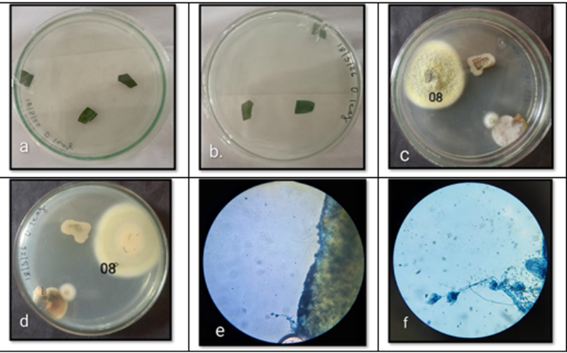
Fig 1; Cultured plates showing endophytic fungi isolated from Datura stramonium leaf: a: Inoculation of Datura stramonium leaf on culture media, b:Backside view of same plate, c: Fungal colony 08 showing growth characteristics of Talaromyces sp. d: Backside view of the same culture plate, e: Microscopic observation of Talaromyces sp. (10x), f: Microscopic observation of Talaromyces sp. (40x).
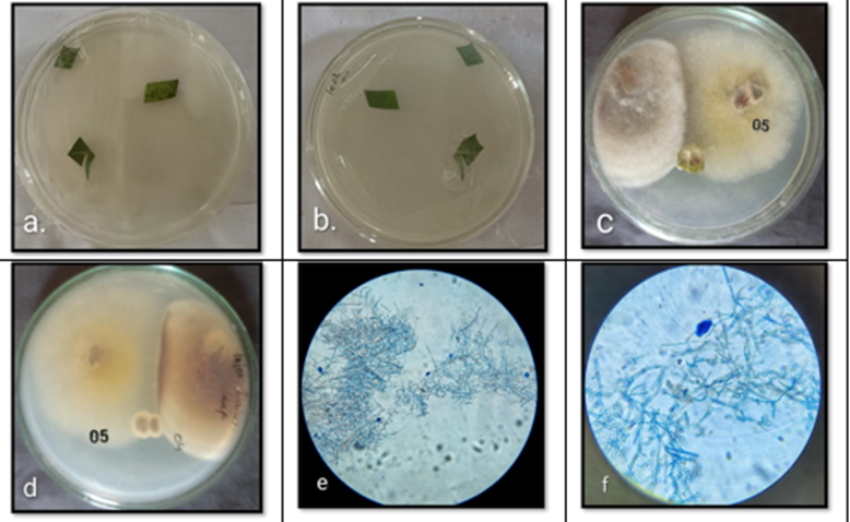
Fig 2; Cultured plates showing endophytic fungi isolated from Datura stramonium leaf: a: Inoculation of Datura stramonium leaf on culture media, b: Backside view of same plate, c: Fungal colony 05 showing growth characteristics of Acremonium sp. d: Backside view of the same culture plate, e: Microscopic observation of Acremonium sp. (10x), f: Microscopic observation of Acremonium sp. (40x).
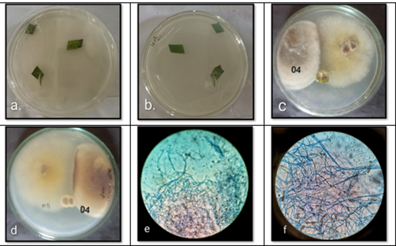
Fig 3; Cultured plates showing endophytic fungi isolated from Datura stramonium leaf: a: Inoculation of Datura stramonium leaf on culture media, b: Backside view of same plate, c: Fungal colony 04 showing growth characteristics of Phoma sp. d: Backside view of the same culture plate, e: Microscopic observation of Phoma sp. (10x), f: Microscopic observation of Phoma sp. (40x)
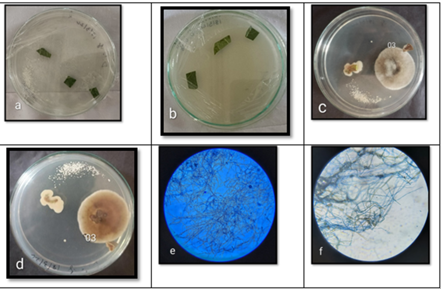
Fig 4; Cultured plates showing endophytic fungi isolated from Datura stramonium leaf: a: Inoculation of Datura stramonium leaf on culture media, b: Backside view of same plate, c: Fungal colony 03 showing growth characteristics of Phoma sp. d: Backside view of the same culture plate, e: Microscopic observation of Phoma sp. (10x), f: Microscopic observation of Phoma sp. (40x)
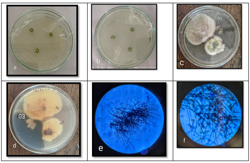
Fig 5; Cultured plates showing endophytic fungi isolated from Datura stramonium stem: a: Inoculation of Datura stramonium stem on culture media, b: Backside view of same plate, c: Fungal colony 03 showing growth characteristics of Bipolaris sp. d: Backside view of the same culture plate, e: Microscopic observation of Bipolaris sp. (10x), f: Microscopic observation of Bipolaris sp. (40x)
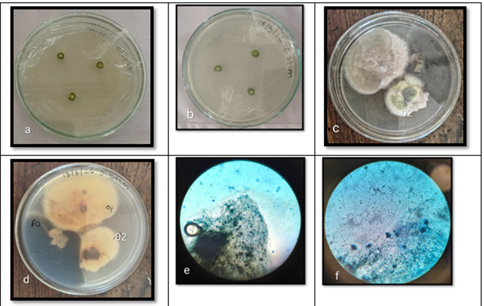
Fig 6; Cultured plates showing endophytic fungi isolated from Datura stramonium stem: a: Inoculation of Datura stramonium stem on culture media, b: Backside view of same plate, c: Fungal colony 02 showing growth characteristics of Penicillium sp. d: Backside view of the same culture plate, e and f: Microscopic observation of Penicillium sp. (10x) and (40x).
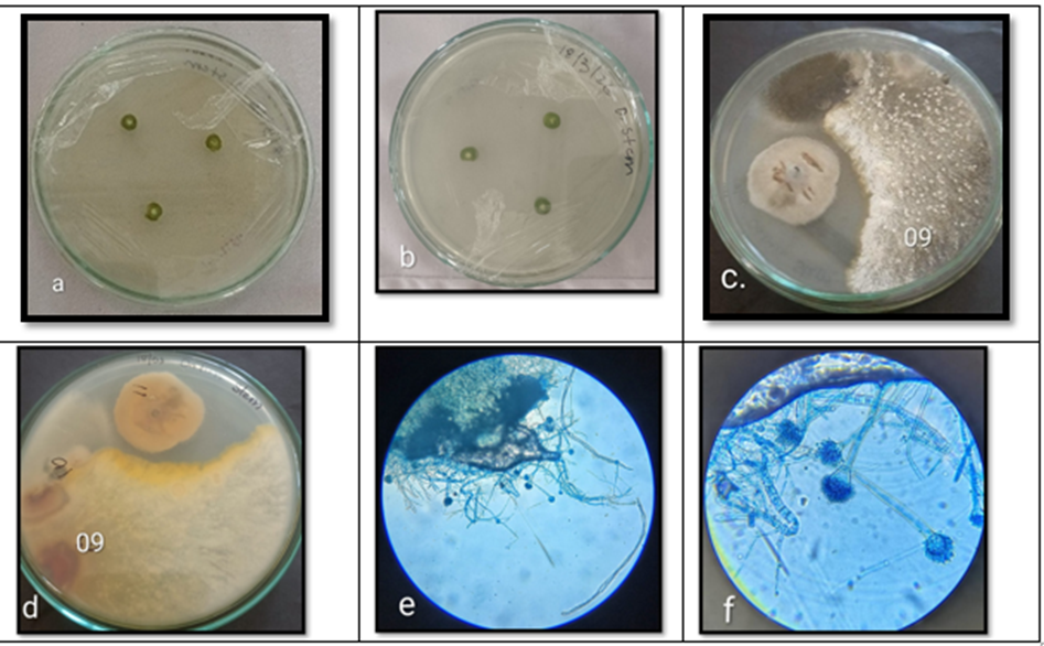
Fig 7; Cultured plates showing endophytic fungi isolated from Datura stramonium stem: a: Inoculation of Datura stramonium stem on culture media, b: Backside view of same plate, c: Fungal colony 09 showing growth characteristics of Aspergillus sp. d: Backside view of the same culture plate, e and f: Microscopic observation of Aspergillus sp. (10x) and (40x).
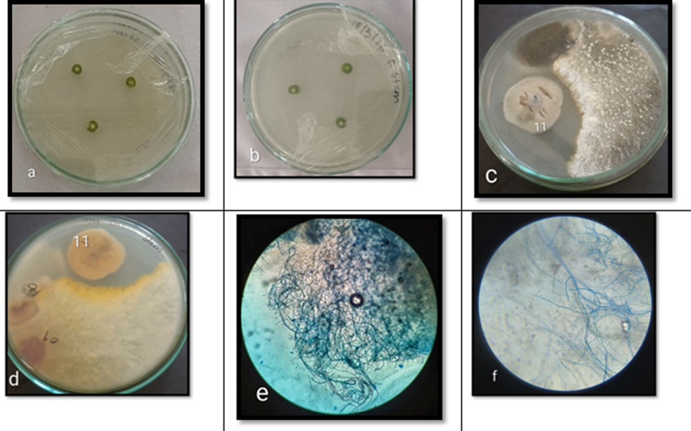
Fig 8; Cultured plates showing endophytic fungi isolated from Datura stramonium stem: a: Inoculation of Datura stramonium stem on culture media, b: Backside view of same plate, c: Fungal colony 11 showing growth characteristics of Glomerella sp. d: Backside view of the same culture plate, e and f: Microscopic observation of Glomerella sp. (10x) and (40x)
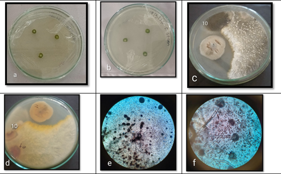
Fig 9; Cultured plates showing endophytic fungi isolated from Datura stramonium stem: a: Inoculation of Datura stramonium stem on culture media, b: Backside view of same plate, c: Fungal colony 10 showing growth characteristics of Aspergillus sp. d: Backside view of the same culture plate, e and f: Microscopic observation of Aspergillus sp. (10x) and (40x).
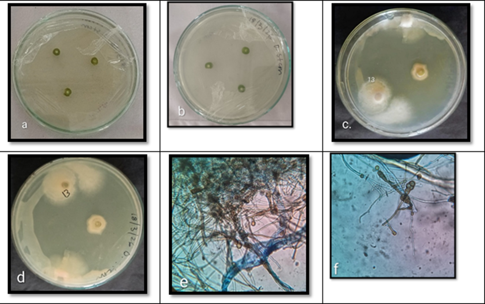
Fig 10; Cultured plates showing endophytic fungi isolated from Datura stramonium stem: a: Inoculation of Datura stramonium stem on culture media, b: Backside view of same plate, c: Fungal colony 13 showing growth characteristics of Phoma sp. d: Backside view of the same culture plate, e: Microscopic observation of Phoma sp. (10x), f: Microscopic observation of Phoma sp. (40x).
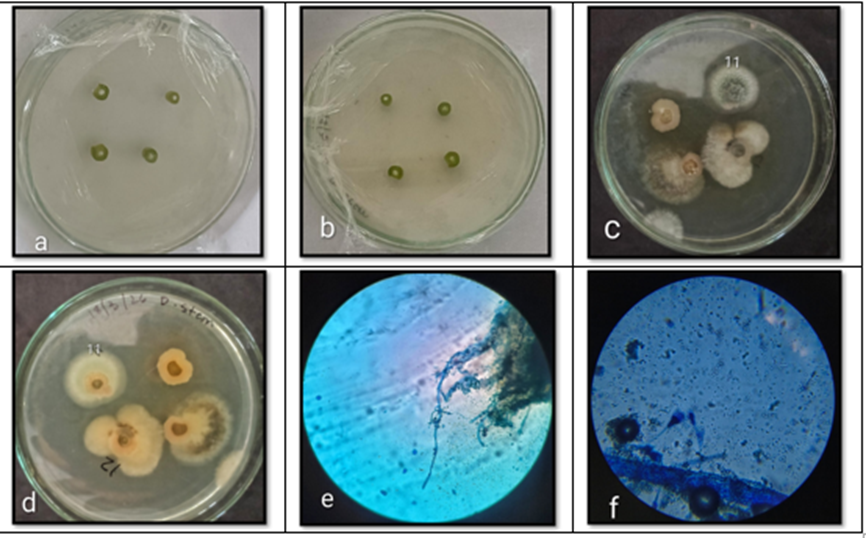
Fig 11; Cultured plates showing endophytic fungi isolated from Datura stramonium stem: a: Inoculation of Datura stramonium stem on culture media, b: Backside view of same plate, c: Fungal colony 11 showing growth characteristics of Penicillium sp. d: Backside view of the same culture plate, e: Microscopic observation of Penicillium sp. (10x), f: Microscopic observation of Penicillium sp. (40x).
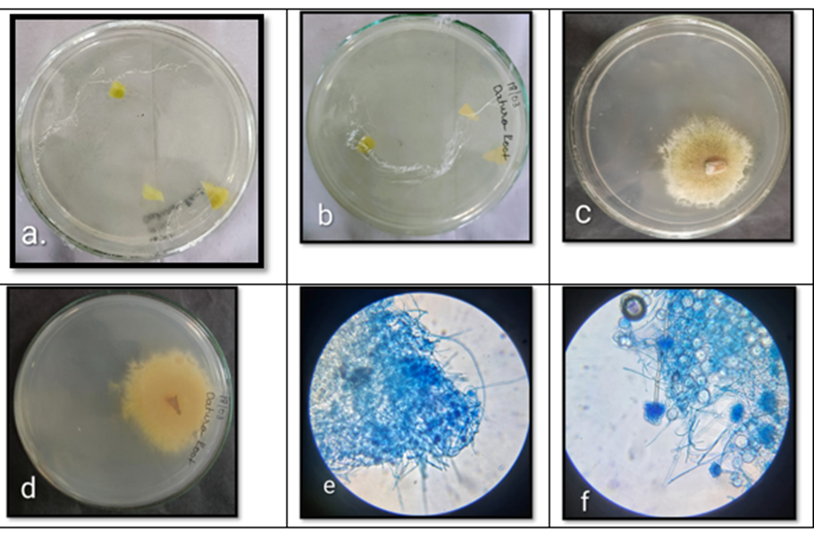
Fig 12; Cultured plates showing endophytic fungi isolated from Datura stramonium root: a: Inoculation of Datura stramonium root on culture media, b: Backside view of same plate, c: Fungal colony showing growth characteristics of Aspergillus sp. d: Backside view of the same culture plate, e and f: Microscopic observation of Aspergillus sp. (10x) and (40x)
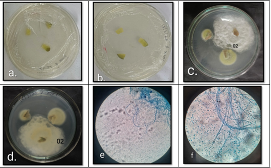
Fig 13; Cultured plates showing endophytic fungi isolated from Datura stramonium root: a: Inoculation of Datura stramonium root on culture media, b: Backside view of same plate, c: Fungal colony showing growth characteristics of Fusarium sp. d: Backside view of the same culture plate, e and f: Microscopic observation of Fusarium sp. (10x) and (40x).
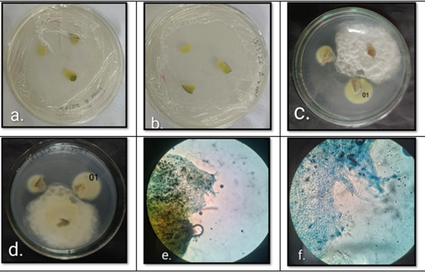
Fig 14; Cultured plates showing endophytic fungi isolated from Datura stramonium root: a: Inoculation of Datura stramonium root on culture media, b: Backside view of same plate, c: Fungal colony showing growth characteristics of Talaromyces sp. d: Backside view of the same culture plate, e and f: Microscopic observation of Talaromyces sp. (10x) and (40x)
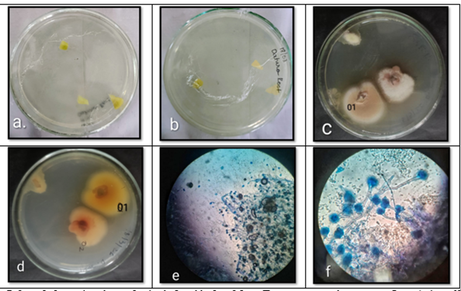
Fig 15; Cultured plates showing endophytic fungi isolated from Datura stramonium root: a: Inoculation of Datura stramonium root on culture media, b: Backside view of same plate, c: Fungal colony showing growth characteristics of Aspergillus sp. d: Backside view of the same culture plate, e and f: Microscopic observation of Aspergillus sp. (10x) and (40x)
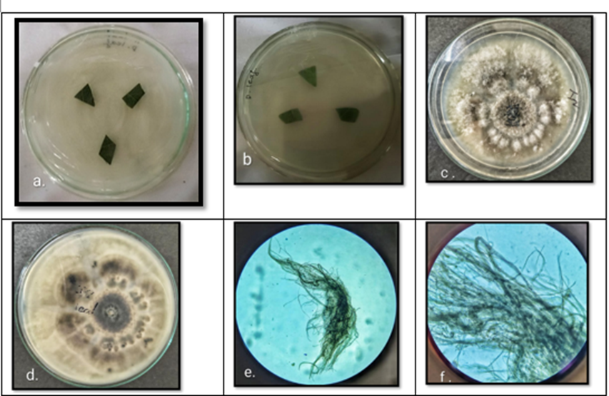
Fig 16; Cultured plates showing endophytic fungi isolated from Datura sanguinea leaf: a: Inoculation of Datura sanguinea leaf on culture media, b: backside view of same plate, c: Fungal colony showing growth characteristics of Alternaria sp. d: Backside view of the same culture plate, e: Microscopic observation of Alternaria sp. (10x), f: Microscopic observation of Alternaria sp. (40x).
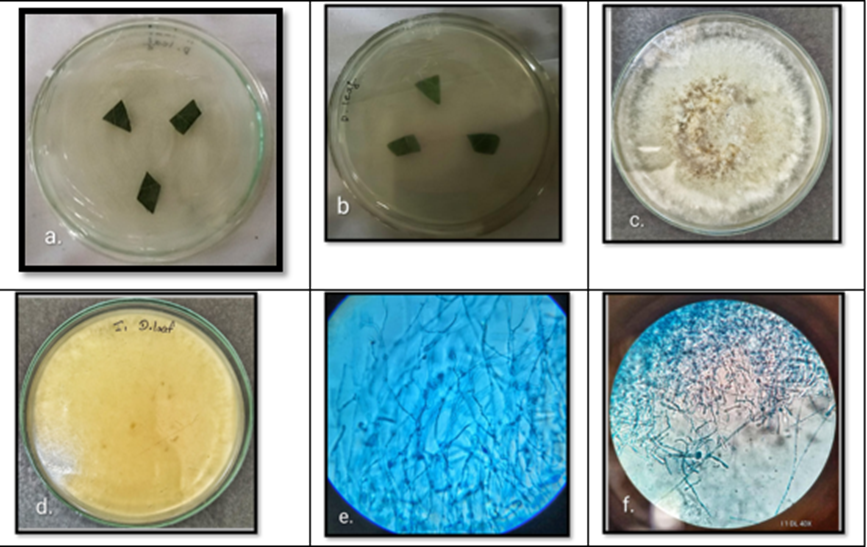
Fig 17; Cultured plates showing endophytic fungi isolated from Datura sanguinea leaf: a: Inoculation of Datura sanguinea leaf on culture media, b: backside view of same plate, c: Fungal colony showing growth characteristics of Ceratobasidium sp. d: Backside view of the same culture plate, e: Microscopic observation of Ceratobasidium sp. (10x), f: Microscopic observation of Ceratobasidium sp. (40x).
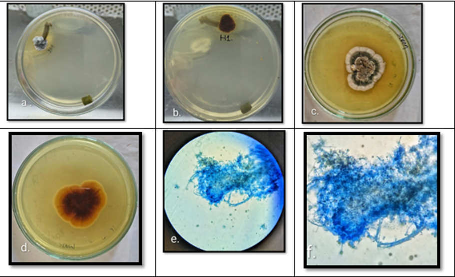
Fig 18; Cultured plates showing endophytic fungi isolated from Datura sanguinea stem: a: Inoculation of Datura sanguinea stem on culture media, b: backside view of same plate, c: Fungal colony showing growth characteristics of Pythium sp. d: Backside view of the same culture plate, e: Microscopic observation of Pythium sp. (10x), f: Microscopic observation of Pythium sp. (40x)
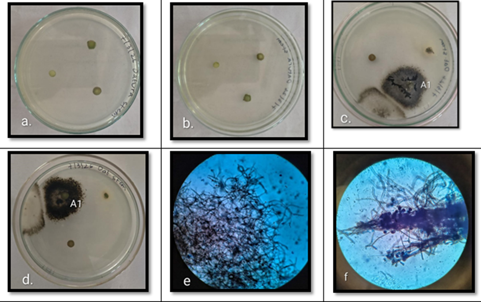
Fig 19; Cultured plates showing endophytic fungi isolated from Datura sanguinea stem: a: Inoculation of Datura sanguinea stem on culture media, b: backside view of same plate, c: Fungal colony A1 showing growth characteristics of Nigrospora sp. d: Backside view of the same culture plate, e: Microscopic observation of Nigrospora sp. (10x), f: Microscopic observation of Nigrospora sp. (40x)
DISCUSSION :
A total of thirteen endophytic fungi were isolated from Datura stramonium from leaves, stems, and roots. From leaves of Datura stramonium, two species of Phoma and Talaromyces sp., Acremonium sp. and Aspergillus sp. were isolated; from stems, two species of Aspergillus, Bipolaris sp., two species of Penicillium, and Phoma sp., Glomerella sp., and Fusarium oxysporium were isolated; and from roots, three species of Aspergillus, Fusarium sp., and Talaromyces sp. were isolated. Aspergillus sp. (six species) was isolated from the stem, leaf and root of Datura stramonium in the present study; similarly, Nagda et al. (2017) reported Aspergillus sp. from Calotropis procera7.
Bipolaris sp. was isolated from the stem of Datura stramonium in the present study; similarly, Tapfuma et al. (2019) reported Bipolaris setariae from Datura stramonium8. Fusarium sp. was isolated from the root and Fusarium oxysporium was isolated from the stem of Datura stramonium in the present study; similarly, Jayabhaskaran et al. (2018) reported Fusarium solani from Datura metel5. Penicillium sp. (two species) were isolated from the stem of Datura stramonium in the present study; similarly, Nagda et al. (2017) reported Penicillium sp. from Calotropis procera7. Phoma sp. (three species) was isolated from the leaves and stem of Datura stramonium in the present study; similarly, Yang et al. (2024) reported Phoma sp. from Gastrodia elata9. Talaromyces sp. was isolated from the leaves and root of Datura stramonium in the present study. Tapfuma et al. (2019) also reported Gyroporus sp., Alternaria tenuissima, Alternaria alternata, Colletotrichum sp., Talaromyces sp., Alternaria sp., Sporothrix schenckii, Trichoderma sp. Pilobolus crystallinus, Rhodotorula mucilaginosa, and Bipolaris setariae from Datura stramonium8.
A total of four endophytic fungi were isolated from Datura sanguinea. Alternaria sp. and Ceratobasidium sp. were isolated from the leaves of Datura sanguinea, and Nigrospora sp. and Pythium sp. were isolated from the stem of Datura sanguinea. Alternaria sp. was isolated from the leaves of Datura sanguinea in the present study; similarly, Spurr et al. (1975) reported Alternaria sp. from Nicotiana sp.10, and Tapfuma et al. (2019) reported Alternaria tenuissima from Datura stramonium8. Ceratobasidium sp. was isolated from the leaves of Datura sanguinea in the present study; similarly, Abdul Rahman et al. (2019) reported Ceratobasidium ramicola from Curcuma mangga11, and Verma et al. (2024) reported Ceratobasidium sp. from Bergenia ciliata12. Nigrospora sp. was isolated from the stem of Datura sanguinea in the present study; similarly, Chaturvedi et al. (2017) reported Nigrospora sp. from Centella asiatica13. Pythium sp. was isolated from the stem of Datura sanguinea in the present study.
CONCLUSION :
The present study successfully isolated and identified endophytic fungi from different tissues of Datura stramonium and Datura sanguinea collected from Bangalore, India. A greater diversity of endophytic fungi was observed in Datura stramonium compared to Datura sanguinea. Thirteen fungal taxa belonging to genera such as Aspergillus, Penicillium, Phoma, Talaromyces, Acremonium, Bipolaris, Glomerella, and Fusarium were isolated from D. stramonium, whereas only four fungal taxa, namely Alternaria, Ceratobasidium, Nigrospora, and Pythium, were isolated from D. sanguinea. The distribution of endophytes varied among leaves, stems, and roots, indicating tissue-specific colonization patterns. The findings demonstrate that both Datura species serve as important reservoirs of diverse endophytic fungi, with D. stramonium exhibiting higher endophytic richness. These fungal endophytes may represent potential sources of biologically active metabolites and warrant further molecular characterization and screening for pharmaceutical and agricultural applications.
REFERENCES:
1. Nisa, H., Kamili, A. N., Nawchoo, I. A., Shafi, S., Shameem, N., & Bandh, S. A. (2015). Fungal endophytes as prolific source of phytochemicals and other bioactive natural products: A review. Microbial Pathogenesis, 82, 50–59. https://doi.org/10.1016/j.micpath.2015.04.001
2. Alam, B., Li, J., Ge, Q., Khan, M. A., Gong, J., Mehmood, S., Yuan, Y., & Gong, W. (2021). Endophytic fungi: From symbiosis to secondary metabolite communications or vice versa? Frontiers in Plant Science, 12, 791033. https://doi.org/10.3389/fpls.2021.791033
3. Mendieta-Brito, S., Sayed, M., Hamza, F. A., et al. (2025). Identification, characterization, antimicrobial activity and biocontrol potential of four endophytic fungi isolated from Amazonian plants. Scientific Reports, 15, 39361. https://doi.org/10.1038/s41598-025-26865-6
4. Sharma, M., Dhaliwal, I., Rana, K., Delta, A. K., & Kaushik, P. (2021). Phytochemistry, pharmacology, and toxicology of Datura species—A review. Antioxidants, 10(8), 1291. https://doi.org/10.3390/antiox10081291
5. Naik, T., Chandrashekaraih Vanitha, S., Raykar, P. K., Chandrika, M., Kamalraj, S., & Jayabaskaran, C. (2017). Novel microbial sources of tropane alkaloids: First report of production by endophytic fungi isolated from Datura metel L. Current Microbiology. https://doi.org/10.1007/s00284-017-1367-y
6. Leck A. Preparation of lactophenol cotton blue slide mounts. Community Eye Health. 1999;12(30):24. PMID: 17491984; PMCID: PMC1706009.
7. Nagda, V., Gahibye, A., & Kumar, D. (2017). Isolation and characterization of endophytic fungi from Calotropis procera for their antioxidant activity. Asian Journal of Pharmaceutical and Clinical Research, 10(3), 254–258. https://doi.org/10.22159/ajpcr.2017.v10i3.16125
8. Tapfuma, K. I., Uche-Okereafor, N., Sebola, T. E., Hussan, R., Mekuto, L., Makatini, M. M., Green, E., & Mavumengwana, V. (2019). Cytotoxic activity of crude extracts from Datura stramonium fungal endophytes against A549 lung carcinoma and UMG87 glioblastoma cell lines and LC-QTOF-MS/MS-based metabolite profiling. BMC Complementary and Alternative Medicine, 19(1), 330. https://doi.org/10.1186/s12906-019-2752-9
9. Yang, Y., Li, G.-D., Shao, Y.-T., Sun, Z.-W., Li, L.-W., Li, W., & Li, H.-T. (2024). Fungal polyketides produced by an endophytic fungus Phoma sp. Associated with Gastrodia elata. Fitoterapia, 173, 105793. https://doi.org/10.1016/j.fitote.2023.105793
10. Spurr, H. W. (1975). Characterization of endophytic fungi in healthy leaves of Nicotiana spp. Phytopathology, 65, 417. https://doi.org/10.1094/Phyto-65-417
11. Abdul Rahman, K. A. M., & Ibrahim, D. (2019). Ceratobasidium ramicola IBRLCM127, an endophytic fungus isolated from Curcuma mangga Valeton & Zijp with strong anti-candidal activity. Journal of Applied Pharmaceutical Science, 9(11), 086–092. https://doi.org/10.7324/JAPS.2019.91111
12. Verma, H., Parki, P., Paliwal, P. C., Chandola, R., & Khulbe, K. (2024). Ceratobasidium as a new endophyte from Bergenia ciliata: Its chemical composition, enzymatic activity, and antimicrobial potential. Vegetos. https://doi.org/10.1007/s42535-024-01140-7
13. Gupta, S., & Chaturvedi, P. (2017). Foliar endophytic diversity of Centella asiatica (L.) Urban in relation to different seasons and leaf age. International Journal of Current Microbiology and Applied Sciences, 6(6), 468–477. https://doi.org/10.20546/ijcmas.2017.606.054