B-Cell Activating Factor (BAFF): Implications for Immune Regulation and Novel Therapeutic Strategies in Autoimmune Diseases.
- Nikolay Shakhpazyan , Avtsyn Research Institute of Human Morphology, Petrovsky National Research Center of Surgery, 119435, Moscow, Russia;
- Nikolay Sadykhov , Avtsyn Research Institute of Human Morphology, Petrovsky National Research Center of Surgery, 119435, Moscow, Russia;
- Liudmila Mikhaleva , Avtsyn Research Institute of Human Morphology, Petrovsky National Research Center of Surgery, 119435, Moscow, Russia;
- Arkady Bedzhanyan , Department of Abdominal Surgery and Oncology II (Coloproctology and Uro-Gynecology), Petrovsky National Research Center of Surgery, 119435, Moscow, Russia;
- Alexandra Konyukova , Avtsyn Research Institute of Human Morphology, Petrovsky National Research Center of Surgery, 119435, Moscow, Russia;
- Ksenia Maslyonkina , Avtsyn Research Institute of Human Morphology, Petrovsky National Research Center of Surgery, 119435, Moscow, Russia;
- Andrey Kontorschikov , Avtsyn Research Institute of Human Morphology, Petrovsky National Research Center of Surgery, 119435, Moscow, Russia;
- Alexander L. Golovyuk , VInstitute of General Pathology and Pathophysiology, 8, Baltiiskaya Street, 125315 Moscow, Russia;
- Natalia Vladimirovna Elizova , Institute of General Pathology and Pathophysiology, 8, Baltiiskaya Street, 125315 Moscow, Russia;
- Anastasia Poznyak , Institute for Atherosclerosis Research, Moscow 121096, Russia;
- Alexander Orekhov , Institute for Atherosclerosis Research, Moscow 121096, Russia;
Article Information:
Abstract:
This review article examines the role of B-lymphocyte activating factor (BAFF) in immune regulation and its implications for autoimmune diseases. BAFF, a member of the TNF superfamily, is crucial for B-cell survival, maturation, and differentiation. We detail the mechanisms by which BAFF interacts with its receptors—BAFF receptor, TNFRSF13B, and BCMA—impacting various signaling pathways essential for humoral immunity. Elevated BAFF levels are associated with autoimmune conditions such as systemic lupus erythematosus, rheumatoid arthritis, and primary Sjögren’s syndrome, where it contributes to B-cell dysregulation and autoinflammatory responses. We discuss how therapies targeting BAFF and associated signaling pathways, such as belimumab and atacicept, show promise in modulating immune responses and offering new treatment avenues. This review synthesizes current findings to highlight the potential of BAFF as a therapeutic target and emphasizes the need for additional studies to clarify its role in the pathogenesis and treatment of autoimmune disorders.
Keywords:
Article :
INTRODUCTION:
Cytokines are low molecular weight proteins, typically smaller than 30 kDa. Cytokines are generated and released during immune responses, whether innate or adaptive, serving as signals that facilitate complex interactions among lymphoid cells, inflammatory cells, and hematopoietic stem cells [1,2]. Cytokines bind to particular receptors on cellular membranes and trigger a series of cell signaling, changing the expression of genes and modulating critical biophysiological mechanisms, e.g., cell activation, cell differentiation or cellular death [3,4].
Recent studies have shown that B-lymphocyte activating factor (BAFF) plays a crucial role in maintaining homeostasis, supporting the maturation and survival of peripheral B cells, and facilitating the development of the lymphatic system. BAFF induces pleiotropic responses by interacting with receptors such as TNF, TNFRSF13B, BCMA, and the BAFF receptor, which are expressed primarily on B cells and T cells [5-8].
The crucial role of BAFF has been demonstrated in mouse models, where its deficiency leads to a reduction in the number of peripheral B lymphocytes and a decrease in humoral immunity. Excessive expression of B-lymphocytes activating factor was related to autoimmune diseases hematological neoplasms progression. Elevated concentrations of B-lymphocytes activating factor was related to presence of AAbs, e.g., anti-DS-DNA Abs in SLE, anti-Ro Abs in SjS and RF in RA [9,10].
In this review, we focus on the available data regarding the activity of BAFF, the processes associated with its expression, its receptors, and its potential applications in the treatment of autoimmune disorders, including its use in targeted therapies.
Contribution to the Field: Our findings underscore BAFF's multifaceted role in autoimmune pathogenesis, highlighting its importance as a therapeutic target. We propose that future research should investigate the underlying mechanisms of BAFF dysregulation in various autoimmune diseases, explore the impact of genetic factors on BAFF signaling, and assess the long-term effects of BAFF-targeting therapies. Furthermore, comparative studies examining BAFF's role in different autoimmune conditions may reveal novel insights into its function and lead to tailored therapeutic strategies.
B-lymphocytes activating factor and its receptors
B-lymphocytes activating factor is a member of TNF superfamily. Its expression takes place on surface of DCs, monocytes, neutrophils, MSCs, activated T-lymphocytes, cancer B cells and ECs. B-lymphocytes activating factor is binding to various receptors, such as B-lymphocytes activating factor receptor, TNFRSF13B, and BCMA [11,12]. Expression of these receptors is spread in time in the process of B-lymphocyte development. BAFF is removed from the cell surface and enters circulation as a soluble, active homotrimer. It is then binding to B-lymphocytes activating factor receptor. A part of B-lymphocytes activating factor assembles into 20-trimer multimers which then are binding to TNFRSF13B triggering its activation [13,14]. A proliferation-inducing ligand molecule is binding to TNFRSF13B and BCMA, however it is not binding to B-lymphocytes activating factor receptor. Activation of TNFRSF13B is triggered by B-lymphocytes activating factor multimerization or binding to membrane and a proliferation-inducing ligand multimerization. Homotrimers, on the contrary, do not stimulate its activation. The B-cell maturation antigen (BCMA) is a high-affinity receptor for APRIL [15,16]. B-cell maturation antigen is binding low-affinity B-lymphocytes activating factor in humans. Although, in murine model, B-cell maturation antigen is specific for a proliferation-inducing ligand (APRIL), which interplays with sulfated proteoglycans accumulating APRIL on cellular membrane. TNFRSF13B is binding to Sdcs via a region separated from B-lymphocytes activating factor binding region. The importance of these processes is still not fully studied [17,18].
Expression of B-lymphocytes activating factor, TNFRSF13B and B-cell maturation antigen was not detected until transition stage of B lymphocytes, where B-lymphocytes activating factor receptor is prevalent on naive B lymphocytes and memory B cells (MBCs), TNFRSF13B is prevalent on short-lived effector B cells, and B-cell maturation antigen is prevalent on long-lived effector B cells. Every receptor triggers activation of a particular series of signaling pathways [19]. The only receptor for BAFF that activates the NF-kappa B pathway is the BAFF receptor. BAFF supports the survival of long-lived B cells primarily through this pathway, increasing the levels of anti-apoptotic proteins. Ligation of B-lymphocytes activating factor receptor leads to signaling via Pim2 which improves metabolic performance by causing a metabolic shift to glycolysis. Hereby, B-lymphocytes response to B-cell receptor activation is stimulated. TNFRSF13B and B-cell maturation antigen signaling is carried out via traditional nuclear factor-kappa beta pathway and via MEK pathway to enhance anti-apoptosis pathways and suppress pro-apoptosis pathways, as well as to promote class switching via JNK/p-38 [20,21].
In summary, BAFF interacts with its receptors in a complex manner that regulates B-lymphocyte survival, proliferation, and differentiation, with distinct pathways activated depending on the receptor engaged
BAFF in the Innate Immune System
Role in Innate Immunity
BAFF affects the innate immune response by stimulating conventional dendritic cells via type 1 interferons and enhances the expression of Toll-like receptors (TLR). BAFF enhances B-cell survival and differentiation alongside interleukin 6 [36-38]. It plays a pivotal role in mediating B-cells' interaction with other immune cells, promoting recruitment to inflammatory regions and modulating cellular responses.
Signaling Pathways and Mechanisms
Cellular toll-like receptor activation in B-cells enhances BAFF receptor expression, amplifying both B-cell receptor mediated signaling and cellular sensitivity to apoptosis. This creates a positive feedback loop facilitating IgG antibody synthesis independently of T-lymphocytes [39,40]. The role of BAFF on non-B-cell lineages, especially T-lymphocytes, is still under investigation.
To summarize, BAFF plays a critical role in modulating the innate immune response by enhancing B-cell function and supporting interactions with other immune cells, thereby contributing to the overall immune activation
BAFF in the Adaptive Immune System
Role in Adaptive Immunity
In the adaptive immune system, BAFF is essential for the development, survival, and functionality of B cells. It affects B-cell maturation at various stages, with its receptors being differentially expressed throughout [19]. Enhanced competition for BAFF promotes the elimination of autoreactive B-cells, while excess BAFF can lead to the survival of such cells, contributing to autoimmunity.
Signaling Mechanisms
BAFF and APRIL signaling pathways influence B-cell receptor survival signals and class switching. For instance, in mouse models, excessive levels of BAFF lead to the emergence of lupus-like phenotypes, highlighting its critical role in the adaptive immune response [22-33]. By facilitating the differentiation into effector cells, BAFF holds a substantial impact on antibody production and overall immune regulation.
In conclusion, BAFF is integral to the adaptive immune response, promoting B-cell maturation and influencing the survival of autoreactive B cells, which is crucial for the development of autoimmunity.
Functions of B-lymphocytes activating factor and APRIL
A number of critical B-lymphocytes functions are mediated by B-lymphocytes activating factor and a proliferation-inducing ligand. Interplay between B-lymphocytes activating factor with B-lymphocytes activating factor receptor is crucial for B2 cells surviving T1 stage, considering a small influence of TNFRSF13B and no influence of a proliferation-inducing ligand or B-cell maturation antigen [22,23]. BAFF acts as a rheostat to select B lymphocytes; increased competition for BAFF enhances the deletion of autoreactive B lymphocytes. Conversely, in the context of B lymphocyte lymphopenia or elevated BAFF levels in the bloodstream, decreased competition for BAFF weakens the selection process for B lymphocytes and promotes the expression of autoreactive naive B lymphocytes [24,25]. B-cell receptor signal level regulates the B-lymphocytes activating factor signaling since it produces p-100, which is the substrate for non-traditional nuclear factor-kappa beta pathway utilized by B-lymphocytes activating factor receptor. P-100 enables B-lymphocytes activating factor receptor signaling causing B-lymphocyte surviving. P-100 deficiency leads to B-lymphocyte death due to not sufficient B-lymphocytes activating factor receptor signal [26]. B-cell receptor signaling produces small amount of p-100 in T 1 cells since their rafts are not mature and do not comprise enough cholesterol. Therefore, T1 cells are subject to negative selection. Conversely, p-100 produced by B-cell receptor signal is enough for B-lymphocytes activating factor instructed surviving of B-lymphocytes, both follicular and transitional, and without B-lymphocytes activating factor present, surviving of these cells is significantly reduced [27,28]. The expression of BAFF receptors is increased by B cell receptors (BCR) ligation in mature B lymphocytes. That is the prevalent receptor which is released on classical MBCs. Although, it is evident that MBCs surviving and re-activating does not depend on B-lymphocytes activating factor. Effector B cells release TNFRSF13B and B-cell maturation antigen. Their surviving might be facilitated by APRIL or B-lymphocytes activating factor produced by various cells inside lymph glands or bone marrow. Conversely, B 1 cells surviving does not depend on APRIL or B-lymphocytes activating factor [29,30].
B-lymphocytes activating factor is crucial for humoral immune response. Type 2 responses independent of T-lymphocyte need the interplay of B-lymphocytes activating factor sixtymer or membrane B-lymphocytes activating factor with TNFRSF13B, which is also necessary for IgM responses that depend on T-lymphocytes [31]. Immunoglobulin G response depends on B-lymphocytes activating factor to a lesser extent. In murine model of B-lymphocytes activating factor deficit, FDCs network was found to be undeveloped which reduced and destabilized germinal centers (GCs) where SHM and class-switching take place, although with decreased immunoglobulin G and secondary responses. Class-switching to immunoglobulin A was reported to depend on multimerized APRIL interplay with TNFRSF13B [32,33]. Immunoglobulin A deficit is an important aberrance in context of APRIL deficit in murine model. This implies that it might play a role in mucosal immune system regulation. TNFRSF13B regulation function requires further study, since its deficiency in murine model leads to development of autoimmune diseases and lymphomas. A number of other explanations were given for this phenomenon [34,35].
B-lymphocytes activating factor is of great importance for innate immunity. It is stimulated in conventional dendritic cells by type 1 interferons. B-lymphocytes activating factor contributes to the expression of TLR, stimulates surviving of B-lymphocytes and stimulates immunoglobulin class switching and effector B cell differentiation together with interleukin 6 [36-38]. Cellular toll-like receptor activation in B-lymphocytes by immunity complexes comprising nucleic acids (NAs) enhances expression of B-lymphocytes activating factor receptor, specifically TNFRSF13B, as well as enhances B-cell receptor mediated signal. Conversely, B-lymphocytes activation through toll-like receptor 4 enhances B-lymphocytes activating factor receptor and elevates cellular sensitivity to apoptotic processes mediated by Fas receptor. Together, toll-like receptors, type 1 interferons and B-lymphocytes activating factor make an enhancement loop which promotes synthesis of immunoglobulin G AAbs to NAs without influence of T-lymphocytes [39,40].
B-lymphocytes activating factor role on cells apart from B-lymphocytes is yet to investigate. Whereas the expression of B-lymphocytes activating factor receptor takes place on T- lymphocytes, its impact on T-lymphocyte co-stimulation is not fully understood. Levels of T-lymphocytes are normal in murine model of B-lymphocytes activating factor deficit. Dendritic cells can be expressing B-lymphocytes activating factor receptors as well (mostly TNFRSF13B) [41,42]. When dendritic cells are transduced with small interfering RNA which is silencing B-lymphocytes activating factor, they stay immature and cannot generate interleukin 6 needed for Th17 differentiation. In humans, conventional dendritic cells (cDCs) promoted by B-lymphocytes activating factor upregulate co-stimulation molecules, stop their phagocytic functioning and synthesize proinflammatory cytokines and chemotactic cytokines such as interleukin 1, interleukin 6, CCL-2 and CCL-5, in vitro facilitating type 1 helper T cell responses [43,44]. These findings imply that B-lymphocytes activating factor activity on dendritic cells stimulates recruitment of immunity cells to inflammation regions and promotes pro-inflammatory function of T-lymphocytes. It is yet to investigate if this is an autocrine activity of dendritic cells in inflammation [45,46].
B-lymphocyte activating factor (BAFF) and a proliferation-inducing ligand (APRIL) play vital roles in regulating B-cell functions and maintaining immune homeostasis. BAFF acts as a critical survival factor for B cells, particularly influencing the selection and differentiation of B2 cells. Elevated BAFF levels can lead to the survival of autoreactive B lymphocytes, contributing to autoimmune conditions, while reduced competition for BAFF in lymphopenic states can weaken B-cell selection. Additionally, BAFF stimulates immunoglobulin production, with implications for both T-cell dependent and independent responses. APRIL also supports B-cell survival and facilitates immunoglobulin class switching, particularly to IgA. Beyond B cells, BAFF influences dendritic cell activity, enhancing pro-inflammatory responses and their recruitment to sites of inflammation. Collectively, these insights underscore the complexity of BAFF and APRIL signaling in both the adaptive and innate immune systems, warranting further exploration of their roles in health and disease.
Human BAFF receptor deficit
To date, there were reported only 2 cases of BAFF receptor deficit in humans caused by total inactivation of BAFF receptor protein encoding gene TNF receptor superfamily member 13C. In these cases, autosomal recessive homozygous 24 base pair deletion was removing sequences of highly conserved 8 amino acids (L V L A L V L V) from membrane site of BAFF receptor, that expands from remnants [47]. After truncation, the BAFF receptor protein becomes unstable; however, computer modeling suggests it may form a new membrane site between remnants that partially overlaps with the wild-type protein's membrane site. When BAFF receptor is not sufficiently expressed, it can inhibit differentiation of B-lymphocytes at transition from CD-10+ Tr1 B or immature B lymphocytes to naive or Tr2 B and MZ B lymphocytes [48,49]. While heterozygous mutation is not distinguishable from control group, homozygous one has total penetrance. In BAFF receptor-/- murine model, a number of immune features of human BAFF receptor deficit were found [50].
These characteristics mostly involve reduced levels of B-lymphocytes in bloodstream, lowered immunoglobulin M and immunoglobulin G AB titre, but elevated immunoglobulin A in serum. Nonetheless, human and mouse BAFF receptor deficiency are in many ways different. Firstly, TNF receptor superfamily member 13C inactivation in mouse model does not suppress B1 B lymphocytes development, while in humans nothing like B1 B lymphocytes were found [51,52]. Although, these cells could disappear in subjects with BAFF receptor deficit the same way they do in ageing mice. At the same time, suppressed development of MZ and follicular B lymphocytes could have facilitated the extension of B1 B lymphocytes. These cells could be able then to compensate for the deficiency of B2 B cells and to grow into immunoglobulin A releasing effector B cells in the intestine, as it was reported for murine model [53].
It is noteworthy that in subjects with BAFF receptor deficiency there was detected an efficient release of immature bone marrow B lymphocytes with high amount of TrB cells. Unlike BAFF receptor -/- mouse model, humans with that deficit displayed serious lymphocytopenia. This could also be explained by ageing or pool size difference [54-56]. Whereas immunoglobulin M+ CD-27+ MZ B lymphocytes regulate encapsulated bacterial infections, without MZ B lymphocytes present, T-independent responses to vaccine Pneumovax were inadequate. Interestingly, weak immunoglobulin G response to tetanus toxoid, elevated serum immunoglobulin A level and immunoglobulin A+ effector B cells in the gut demonstrated that BAFF receptor deficit can still allow B lymphocytes to transform into plasma cells. The same was reported for BAFF receptor-/- mouse model where BAFF receptor deficient B lymphocytes finished GC reaction and progressed into switched MBCs and effector B cells that are capable of survival in BAFF receptor deficit [57,58]. With B-lymphocytes activating factor drained by therapy with Belimumab and Benlysta or Atacicept, long-lived MBCs not depending on BAFF receptor were found in subjects with lupus. Those trials demonstrated a more than 75 percent decline in the amount of naive B lymphocytes and an elevation of switched MBCs. Immunoglobulin G AB levels, accumulated before the B-lymphocytes activating factor-neutralizing treatment, did not change, while the elevation of AB titre against influenza was considerably poorer that in healthy subjects. Another trial also did not show changes in switched MBCs during 6 months therapy of RA subjects with atacicept [59-61].
Apart from BAFF receptor deletion, various MMs (missense mutations) were detected for BAFF receptor. Such mutations were detected in subjects with CVID, a disorder featured by reduced serum immunoglobulin M, immunoglobulin G, and immunoglobulin A, reduced switched MBCs in blood and absent effector B cells in blood. The BAFF receptor MMs alter amino acid remnants in BAFF receptor, and they have an impact on progression and surviving of B lymphocytes in a manner comparable to the BAFF receptor deletion mutation [62,63]. Hereby, their role in common variable immunodeficiency progression is yet to be elucidated. One exception is represented by P21-R BAFF receptor variant, that is encoded by a SNP. Proline 21 is found in a little loop upstream of B-lymphocytes activating factor binding site. Various trials demonstrated that this little loop is vital for BAFF receptor protein chains assembly into multimers in a ligand independent manner. Hereby, it mirrors pre-ligand assembling site of BAFF receptor [64]. Whereas BAFF receptor complex defect associated with P21-R decreases by half the number of B-lymphocytes activating factor molecules that are capable of binding to BAFF receptor on B lymphocyte surface, it does not have an impact on TrB cells transformation into naïve mature b lymphocytes. As BAFF receptor multimerization induces B-lymphocytes activating factor binding, B-lymphocytes with P21-R progress into immunoglobulin M releasing plasmablasts with lower efficiency [47,65]. Furthermore, homozygous P21-R shows total resistance to B-lymphocytes activating factor-induced BAFF receptor processing by CDw156. This mutation appears to have compensatory effect for the decreased capacity of binding B-lymphocytes activating factor. This characteristic could partly disguise the disrupted differentiation of P21-R+ B lymphocytes into immunoglobulin M releasing plasmablasts as well as help avoid progression of immunocompromisation [66].
Human BAFF receptor deficiency, caused by total inactivation of the TNF receptor superfamily member 13C gene, has been reported in only two cases. These cases involve a significant genetic alteration leading to the instability of the BAFF receptor protein, impacting B-lymphocyte differentiation and resulting in immune dysregulation. The deficiency is characterized by markedly reduced levels of circulating B-lymphocytes and altered immunoglobulin profiles. While the murine BAFF receptor knockout model exhibits variations in B-cell populations compared to humans, both models show impaired development of certain B-cell subsets. Notably, BAFF receptor deficiency in humans leads to severe lymphocytopenia and an inefficient immune response to specific vaccines.
Additionally, missense mutations (MMs) in the BAFF receptor have been associated with Common Variable Immunodeficiency (CVID), further complicating B-cell survival and progression. Emerging research on these mutations, including the P21-R variant, highlights their impact on BAFF receptor function and B-cell maturation, underscoring the complexity of immune responses in BAFF receptor-deficient individuals. Understanding these mechanisms may provide insights for targeted therapies in autoimmune disorders linked to BAFF receptor dysregulation.
Primary Sjögren's syndrome (SjS)
SjS is a chronic auto-immune condition which mostly involves exocrine glands, causes their damage and changes their functioning. It is featured by mononuclear infiltration located around ducts [67].
SjS underlying processes involve overexpression of B-lymphocytes activating factor. This alteration is primarily attributed to the overproduction of interferon alpha in infiltrating cells, which is driven by apoptotic processes in endothelial cells that induce cytokine expression—such as BAFF—and promote autoreactive B cells [68,69]. These discoveries imply that ECs might be important for development of auto-immune lesions in SjS and they possibly could autocrinely synthesize B-lymphocytes activating factor that makes environment favorable for interplay of cytokines, ECs, DCs, hyperactive T cells and B cells, and that together leads to aberrances of immune regulation in organs [70,71].
Abnormal GC-like lymphocyte deposition happens in seventeen percent of SjS subjects. It is an intricate mechanism influenced by interplay of various factors, such as B-lymphocytes activating factor. B-lymphocytes activating factor is a major mediator of GC genesis in SjS. Hereby, there was detected an enhancement of B cell signaling which stimulates their differentiation into AB-generating effector B cells and proliferation. B-lymphocytes activating factor concentrations are in correlation with anti-Ro and rheumatoid factor titre [72,73]. That implies that B-lymphocytes activating factor is an important player in regulation of AAbs synthesis.
B-lymphocytes activating factor transgenic mice develop lupus-like and consequently demonstrate gland infiltrations and symptoms similar to SjS. With ageing, these mice demonstrate how B-lymphocytes activating factor causes upregulated signaling of surviving of auto-reactive B cells, missing the spleen checkpoint [74,75].
In a physiological SjS model, B-lymphocytes activating factor is released by ECs after being promoted by type 1 interferon, that is affected by viruses. That implies that infections could be the agents stimulating the disorder. Hereby, ECs take part in expression of auto-antigens and also are able to simultaneously activate B cells by local secretion of B-lymphocytes activating factor. These discoveries are crucial for SjS treatment development, since B cells are one of the major targets in SjS. Neutralization of B-lymphocytes activating factor could become a new promising therapeutical approach [76].
Primary Sjögren's syndrome (SjS) is characterized by autoimmune damage predominantly affecting the exocrine glands, leading to functional impairment. Central to its pathophysiology is the overexpression of B-lymphocyte activating factor (BAFF), driven by interferon alpha production from infiltrating cells, which facilitates the promotion of autoreactive B cells. This dysregulation supports the formation of abnormal germinal center-like structures and enhances B cell activity, contributing to the synthesis of autoantibodies, such as anti-Ro and rheumatoid factors. Moreover, BAFF’s role is underscored by findings in transgenic mouse models, which exhibit lupus-like symptoms and gland infiltration. The potential autocrine production of BAFF by endothelial cells further supports a microenvironment conducive to autoimmune responses. Consequently, targeting BAFF presents a promising therapeutic strategy for managing SjS and its associated symptoms.
Rheumatoid arthritis (RA)
B lymphocytes contribute to the pathogenesis of rheumatoid arthritis by regulating the synthesis of autoantibodies, such as rheumatoid factor, antibodies against type 3 collagen, and ACPA. B lymphocytes are also crucial for cytokine genesis and T cells promotion. In a BL-deficient murine model, collagen-induced rheumatoid arthritis was not observed, and treatment with anti-CD20 antibodies proved to be effective. These findings imply that B lymphocytes are particularly important in RA pathogenesis [77,78].
Numerous trials have demonstrated that elevated B-lymphocytes activating factor are in correlation with RA progression. B-lymphocytes activating factor in serum was higher in nineteen to forty percent of subjects. Immunosuppressive drugs and different dosages might account for it. Both B-lymphocyte activating factor and APRIL play roles in both upregulating and downregulating processes involved in rheumatoid arthritis [79,80]. In rheumatoid arthritis, B-lymphocytes facilitate the formation of lymphoid tissue microarchitecture within the synovium during inflammation, characterized by ectopic germinal centers, accumulation of T-B lymphocytes without germinal center reactions, and disorganized diffuse infiltrations.
By phenotype, synovitis related to ectopic GC formation is featured by elevated APRIL release by CD-83+ DCs, whereas B-lymphocytes activating factor has the same concentrations in different tissues and is obtained only from CD-68+ macrophages. This suggests that APRIL, rather than B-lymphocyte activating factor, correlates with differences in the functional state of B lymphocytes within the tissue [81].
Considering receptors expression, it has been reported that they do not depend on synovitis type. B-lymphocytes activating factor expression is higher than TNFRSF13B expression. Although, its correlation with lymphoid structure was not detected. At the same time, TNFRSF13B is detected in T lymphocytes in lymphoid accumulations, but not in GC synovitis. B-cell maturation antigen expression takes place in B lymphocyte and T lymphocyte infiltrations [82,83].
In the context of synovitis treatment, atacicept reduced germinal center formation and suppressed interferon production, emphasizing the roles of B-lymphocyte activating factor and APRIL in lymphoid organogenesis. However, this effect was not observed in synovitis without germinal center formations, where T cells expressing TNFRSF13B were present, alongside increased production of inflammatory cytokines and interferon-gamma [84]. Thus, B-lymphocyte activating factor and APRIL can regulate inflammatory processes in the synovium in rheumatoid arthritis by modulating the functions of both B lymphocytes and T lymphocytes, exhibiting pro-inflammatory and anti-inflammatory activities. The anti-inflammatory function is modulated by T lymphocytes with TNFRSF13B receptors [85].
In the early stages of rheumatoid arthritis, TNFRSF13B expression, B-lymphocyte activating factor concentrations, and the expression of the B-lymphocyte activating factor gene are observed in the synovium within the first weeks after disease onset. Then along with the disorder progression they are gradually reduced. In contrast, the expression of the B-lymphocyte activating factor receptor is significantly elevated in advanced rheumatoid arthritis [86].
B lymphocytes are integral to the pathogenesis of rheumatoid arthritis (RA), contributing to autoantibody synthesis and T cell activation. Elevated levels of B-lymphocyte activating factor (BAFF) are associated with disease progression, highlighting its significant role alongside APRIL in modulating inflammatory processes within the synovium. Inflammatory synovitis is characterized by the formation of ectopic germinal centers and the accumulation of B and T lymphocytes, with BAFF and APRIL influencing both pro-inflammatory and anti-inflammatory pathways. Treatment strategies targeting these factors, such as atacicept, demonstrate potential in attenuating RA-related synovitis by reducing germinal center formation and cytokine production. Understanding the dynamic roles of BAFF and APRIL may offer new therapeutic avenues for managing RA and improving clinical outcomes.
Scleroderma
Constantly activated B lymphocytes contribute to AAbs stimulation and to progression of skin sclerosis in scleroderma. Whereas its pathophysiological processes are yet to be fully studied, it had been reported that polyclonal B lymphocyte activation and B lymphocyte aberrances featured by synthesis of AAbs, that are in correlation with some symptoms of the disorder, are major players [87].
High B-lymphocytes activating factor concentration was detected in scleroderma. This increase is in correlation with expansion of skin fibrosis assessed by modified Rodnan criteria, manifestation or progression of the organic damage and with diffuse diversity, in comparison to restricted, and is subsequently reduced with corticosteroid therapy [88]. These findings demonstrate that B-lymphocytes activating factor is related to the disorder development.
APRIL is increased as well in comparison to the healthy donors. It was found to be in correlation with more frequent occurrence of lung fibrosis, although not with concentrations of B-lymphocytes activating factor [89].
These discoveries indicate that it is possible to assess the gravity and subjects’ profiles using B-lymphocytes activating factor and APRIL concentration in serum, where the former is a biomarker of serious skin sclerosis and the latter is a biomarker of lung fibrosis.
In summary, the activation of B lymphocytes plays a critical role in the pathogenesis of scleroderma, contributing to the production of autoantibodies and disease progression, particularly skin sclerosis. Elevated levels of B-lymphocyte activating factor (BAFF) and APRIL have been associated with the severity of skin fibrosis and lung complications, respectively. Monitoring serum concentrations of these factors may provide valuable biomarkers for assessing disease severity and guiding therapeutic approaches in scleroderma management. Further research is needed to fully elucidate the underlying mechanisms by which B lymphocytes and their regulatory factors influence scleroderma pathology.
Systemic Lupus Erythematosus (SLE)
Systemic lupus erythematosus (SLE) is a complex autoimmune disease characterized by the production of a variety of autoantibodies, resulting in systemic inflammation and potential damage to multiple organ systems, including the kidneys, skin, joints, and central nervous system. One of the key features of SLE pathogenesis is the dysregulation of B-cell activity, with a notable role played by B-lymphocyte activating factor (BAFF).
Elevated levels of BAFF have been consistently observed in patients with SLE, correlating with disease activity and the presence of specific autoantibodies, particularly anti-dsDNA antibodies, which are strongly associated with renal involvement. BAFF enhances the survival and proliferation of B cells, including autoreactive B cells, thereby contributing to the pathogenesis of SLE. This overabundance of BAFF provides a survival signal to these autoreactive B cells, enabling their persistence even in the presence of self-reactivity [9,10].
BAFF signals through its receptors on B cells, primarily the BAFF receptor (BR3), TNFRSF13B, and BCM. The engagement of these receptors leads to the activation of several downstream signaling pathways, including NF-κB and MAPK pathways, which promote B-cell survival and differentiation. In SLE, the aberrant signaling resulting from elevated BAFF levels leads to enhanced differentiation of B cells into antibody-producing plasma cells, further driving the autoantibody response [20,21].
The critical role of BAFF in SLE has prompted investigations into therapies that target this factor. Belimumab, a monoclonal antibody that neutralizes soluble BAFF, has shown effectiveness in reducing disease activity and the production of pathogenic autoantibodies. Clinical trials have demonstrated that belimumab treatment results in decreased flares and improved patient outcomes, highlighting BAFF's potential as a therapeutic target in the management of SLE [95,96].
In summary, BAFF plays a pivotal role in the pathogenesis of systemic lupus erythematosus by supporting the survival and activity of autoreactive B cells. Understanding the mechanisms surrounding BAFF dysregulation in SLE is essential for developing targeted therapies aimed at improving disease management and patient outcomes.
Table 1: Role of BAFF in Autoimmune Diseases
|
Disease |
Impact of Elevated BAFF |
Therapeutic Approaches |
References |
|
Systemic Lupus Erythematosus (SLE) |
Associated with anti-dsDNA and other autoantibodies; promotes B-cell dysregulation. |
Belimumab (BAFF-neutralizing Ab) effective in reducing symptoms. |
95,96 |
|
Rheumatoid Arthritis (RA) |
Elevated BAFF correlates with disease activity; influences B-cell and T-cell interactions. |
Atacicept (dual BAFF/APRIL inhibitor) shows promise. |
113-116 |
|
Primary Sjögren’s Syndrome (SjS) |
Increases B-cell activation and autoantibody production; impacts glandular function. |
BAFF-targeting therapies may improve outcomes. |
67-76 |
|
Scleroderma |
Correlation with severity of skin fibrosis; BAFF levels reflect disease activity. |
Ongoing research on BAFF-targeting treatments. |
87-89 |
Therapies targeted against B-lymphocytes activating factor in autoimmune disorders
Above mentioned discoveries demonstrate the important role of B-lymphocytes activating factor/APRIL system as well as of its receptors. Hereby, anti-B-lymphocytes activating factor/APRIL treatment could be a promising therapeutic approach in such disorders [90].
Belimumab
Human immunoglobulin G1 lambda is targeting dissoluble B-lymphocytes activating factor, suppressing its function and decreasing its levels. This triggers apoptotic processes of auto-reactive B lymphocytes, averting cellular self-reproduction and proliferation.
Belimumab binds to soluble B-lymphocyte activating factor (BAFF), preventing its association with TNFRSF13B, B-cell maturation antigen, and the BAFF receptor. This medicine does not affect membrane-bounded B-lymphocytes activating factor or other tumor necrosis factor superfamily members [91,92].
This selectivity leads to a reduction in the number of CD-20+ B cells, naïve B cells, activated B lymphocytes, and plasmablasts, while the number of effector B cells remains unchanged. Conversely, it elevated number of MBCs by 28th day of therapy, but by 52nd week of therapy they go back to baseline. This discovery is linked to elevated expression of TNFRSF13B by these cells [93]. Belimumab therapy also considerably reduces concentrations of various immunoglobulins and elevates complement, which implies that this is a bioactive product that has a response profile. Belimumab half-life is thirteen to seventeen days, clearance 4±1,56 ml/day/kg, totally eliminated after eight months, and a baseline of 68±20,8 ml/kg. Its pharmacokinetics depend on dosage [94].
Belimumab efficiency in systemic lupus erythematosus treatment was observed in two phase 3 double blind RCTs in subjects with lupus (SLEDAI>6), after which it was approved by the FDA as an agent for lupus treatment [95,96].
Data on its safety and effectiveness over seven years were collected from a cohort of 1,745 subjects with annual follow-ups. A decrease in ABs and steroid-sparing effect have been detected in these subjects [97].
In rheumatoid arthritis, results from a phase 2 multicenter double-blind randomized controlled trial (RCT) are now available, focusing on dosing in subjects with severe rheumatoid arthritis who had failed other 24-week therapies. Its results showed an acceptable tolerability with amelioration in ACR20 at twenty-four weeks, particularly in subjects with positive rheumatoid factor, elevated disorder activity, with no previous anti-tumor necrosis factor therapy and in which methotrexate already failed [98,99]. Although, it did not show a considerable amelioration in ACR-50 and ACR-70 responses with no dosage/response interconnection.
In primary Sjögren's syndrome (SjS), the efficacy and safety of belimumab at a dosage of 10 mg/kg administered at weeks 0, 2, and 4, followed by monthly doses for a total of 24 weeks, were evaluated in a one-year prospective trial aimed at improving sicca symptoms, systemic functions, and B-lymphocyte markers. In 60% of subjects, the trial met its goals, demonstrating a significant reduction in ESSDAI scores from 8.8 to 6.3 (p=0.0015) and improvement in gland function and parotitis. However, there was no significant improvement in salivary gland function or Schirmer's test results, warranting further randomized controlled trials (RCTs) [100-102].
In a cohort study in ten subjects of ten to fifty-two weeks therapy, types of B lymphocytes and B-lymphocytes activating factor and its receptor expression were evaluated. The results indicated an increase in B cells in the bloodstream, along with elevated levels of naïve B cells and transitional B cells (TrB), increased BAFF levels, and a reduction in the expression of the BAFF receptor. Additionally, there was a decrease in immunoglobulin concentrations, rheumatoid factors, and antinuclear antibodies (ANAs), along with an elevation of complement C4, suggesting a normalization of B-lymphocyte occurrence, phenotype, and function [103].
Overall, belimumab demonstrates significant clinical efficacy in modulating the immune response across various autoimmune disorders, particularly systemic lupus erythematosus, rheumatoid arthritis, and primary Sjögren's syndrome. Its mechanism of selectively targeting soluble BAFF effectively reduces auto-reactive B-cell populations while preserving effector B cells, highlighting its potential to alter disease activity and improve patient outcomes. Clinical trials have reported favorable safety profiles, improvements in disease markers, and specific symptom alleviation, underscoring the need for continued exploration of belimumab's applications in autoimmune conditions. Future studies should focus on optimizing treatment protocols and further elucidating its long-term effects and benefits in diverse patient populations.
Atacicept
Atacicept is a fusion protein with conformation by extra-cellular TNFRSF13B receptor associated with Fc site of human immunoglobulin G1, with affinity for B-lymphocytes activating factor and APRIL. Upon its binding to hetero-trimers and homo-trimers of B-lymphocytes activating factor and APRIL, it drains mature B-lymphocytes, effector B cells and certain ABs in serum [104,105]. After 16 hours of administration, it reaches its maximum serum concentration. The concentration over time increases proportionally to the dosage; however, the clinical effect is relatively modest, while the safety profile is robust, with only a 1.6% increase in infection rates reported. In autoimmune disorders, this drug is linked to fast decrease in immunoglobulin concentrations in bloodstream, particularly immunoglobulin M after the first administration [106,107]. In contrast to belimumab, the suppression of APRIL leads to a greater reduction in immunoglobulin concentrations.
Atacicept safety in systemic lupus erythematosus was assessed in phase 1b trials [108,109]. Immunoglobulin A, M and G concentrations and amount of B lymphocytes were reduced depending on dosage. Although, it was not confirmed in phase 2/3 trials with administration of corticosteroids and mycophenolate mofetil in subjects with SLE nephritis. Those trials were terminated because of an elevation in infections frequency and a major reduction in immunoglobulin concentrations [110-112].
In rheumatoid arthritis AUGUST trial assessed the efficacy of this drug in subjects in which anti-tumor necrosis factor alpha or methotrexate has failed. The results demonstrated decreased concentrations of immunoglobulin M, A, and G in serum, as well as reduced levels of three types of rheumatoid factor, erythrocyte sedimentation rate (ESR), C-reactive protein (CRP), mature B cells, and effector B cells, with no changes observed in anti-cyclic citrullinated peptide concentrations [113,114]. Levels of T lymphocytes or NK cells were not changed as well. After the sixteenth week of therapy, levels of effector B cells increased, while there were no changes observed in mature B lymphocyte levels. This may be due to the selective mobilization of effector B cells or the rapid transformation of mature B cells into effector B cells independently of BAFF and APRIL signaling [115,116]. Immunoglobulin and rheumatoid factor titre were reduced at first, although they returned to baseline after thirteen weeks of therapy. There was detected a higher decrease in immunoglobulin G and A of the rheumatoid factor in comparison to the concentrations of the same class of serum immunoglobulin. The increased susceptibility of antibodies to suppression by BAFF/APRIL may explain this phenomenon [117,118].
Despite these findings, a more favorable response in ACR20 and CRP levels was not observed during the first twenty-six weeks when compared to the placebo group, indicating that atacicept does not induce a sufficient humoral immune response to achieve clinical significance.
In rheumatoid arthritis, this drug has not reached it outcomes, it has however showed good safety profile and a considerable bioactivity. This may be attributed to the inability of the drug to achieve sufficient concentrations in joint fluid compared to serum, indicating a need for additional local inhibition strategies. Currently there are several trials being conducted on this drug in various disorders [119,120].
In summary, Atacicept demonstrates potent activity as a fusion protein targeting BAFF and APRIL, with a robust safety profile but modest clinical efficacy in treating autoimmune disorders such as systemic lupus erythematosus and rheumatoid arthritis. While it effectively reduces immunoglobulin concentrations and B-cell populations, its impact on clinical outcomes has been limited, necessitating consideration of additional strategies to enhance its therapeutic effectiveness. Ongoing trials will further elucidate its potential across various autoimmune conditions.
Tabalumab
Tabalumab is an immunoglobulin G4 monoclonal antibody that neutralizes both soluble and membrane-bound forms of B-lymphocyte activating factor. Its half-life is approximately twenty-five days. Its effects include an increase in both mature and total B-lymphocytes, followed by a gradual reduction starting from the sixteenth week of therapy as serum drug levels decrease and remain stable until approximately the twenty-fourth week [121,122]. It shows a higher selectivity for naïve B cells in about sixty percent of cases, while preserving mature B cells. Immunoglobulin M levels are considerably reduced compared to immunoglobulin G and A. No considerable alterations in CRP or ESR were detected. Currently, trials are underway for rheumatoid arthritis, systemic lupus erythematosus, end-stage kidney disease (ESKD), and multiple myeloma [123,124].
In rheumatoid arthritis, this drug has not achieved its intended outcomes by the sixteenth week of therapy. A clinical response has been detected at the ninth week, related to the highest concentration of tabalumab. DAS28-CRP scores were reduced, alongside a decrease in its efficacy and serum levels, in a dose-independent manner. Its efficiency compared to placebo group ranges from the same to mildly higher after sixteenth week of therapy in subjects in which disease-modifying antirheumatic drugs and anti-tumor necrosis factor drugs have failed [125,126].
Tabalumab is a monoclonal antibody targeting both soluble and membrane-bound forms of BAFF, demonstrating a favorable half-life and selectivity for naïve B cells. While it increases overall B-lymphocyte counts initially, these levels decline after several weeks of therapy. Current investigations are exploring its efficacy in various autoimmune conditions, including rheumatoid arthritis and systemic lupus erythematosus. Despite some initial clinical responses observed in rheumatoid arthritis, its overall effectiveness in achieving primary therapeutic goals remains limited, warranting further research into its long-term impact and applications.
Blisibimod
Blisibimod is a fusion protein produced using Escherichia coli that targets both soluble and membrane-bound B-lymphocyte activating factor. It consists of a new binding region connected to N-terminal end of immunoglobulin constant domain. Blisibimod effectiveness has been assessed in subjects with active systemic lupus erythematosus (SLEDAI>6) in double-blind phase 2b RCT named PEARL-SC (n=547) [127-129]. The primary outcomes were defined at twenty-four weeks of therapy, aiming for an improvement in the SLE response index greater than 5. This outcome was not fully achieved compared to the placebo group; however, subjects receiving a higher dosage displayed a greater response than those in the placebo group (p=0.02). A significant reduction of protein in urine (p<0.01) and anti-DNA levels was also observed, along with an improvement in complement levels. Phase 3 trials are yet to be conducted [130,131].
Blisibimod is an innovative therapeutic agent designed to inhibit both soluble and membrane-bound B-lymphocyte activating factor (BAFF), showing promise in the treatment of systemic lupus erythematosus (SLE). In the phase 2b PEARL-SC clinical trial, while the primary outcome of improved SLE response was not fully met, higher dosages of Blisibimod resulted in a statistically significant response compared to placebo. Notable improvements included reduced protein levels in urine, decreased anti-DNA antibody levels, and enhanced complement levels. These findings suggest the potential for Blisibimod as a valuable treatment option, warranting further investigation in upcoming phase 3 trials to establish its efficacy and safety profile in SLE management.
Effects of the anti-CD-20 depletion on B-lymphocytes activating factor concentrations
Rituximab
Rituximab is a chimeric monoclonal antibody that consists of a human immunoglobulin G1κ heavy chain paired with regions derived from mouse immunoglobulin, specifically targeting human CD-20. CD-20 is expressed from the stage of pre-B lymphocytes through to memory B cells, but it is not found on mature effector B cells. Rituximab has been approved for B-lymphocyte lymphoma therapy. In rheumatoid arthritis it was approved for treatment of subjects in which anti-tumor necrosis factor drugs have failed [132,133]. In systemic lupus erythematosus, its effectiveness varies and is particularly inconsistent in individuals who do not respond to other therapies. This drug was assessed in phase 2 studies (EXPLORER) and phase 3 studies (LUNAR), although the results were not favorable. The differences in assessment methods compared to those used in belimumab trials may explain these results, along with the heterogeneity among subjects and variations in concomitant treatments that could obscure the therapeutic effects [134,135].
Most subjects who received rituximab therapy exhibit the following phases of B-lymphocyte kinetics:
In the first phase, B lymphocytes are completely depleted, which correlates with a reduction in memory B cells and a gradual increase in naïve B cells and transitional B cells (trB) due to elevated concentrations of B-lymphocyte activating factor. This elevation occurs as a response to decreased expression of BAFF receptors on B lymphocytes and slower modulation of BAFF messenger RNA. The clinical effectiveness of this drug results from the reduction in the populations of transitional B cells, naïve B cells, activated B cells, and short-lived effector B cells. The duration of this first phase varies based on the concentrations of B-lymphocyte activating factor and the restoration of B-lymphocyte counts, typically lasting between six to twelve months [136].
The second phase, known as the reconstitution phase, is characterized by the re-emergence of memory B cells, which may indicate a recurrence of disease and the need for retreatment in lupus patients [137,138].
It has been hypothesized that by depleting long-lived effector B cells with rituximab and reducing autoimmune B lymphocytes with belimumab, the repopulation of the B lymphocyte compartment with autoimmune effector B cells may be prevented. This leads to a conclusion that such combined treatment can have complementary activity. However, these treatments may also prove to be incompatible, as the risk of infection is increased in individuals with depleted B lymphocytes. This theory is yet to be assessed in clinical trials [139,140].
In summary, rituximab is a chimeric monoclonal antibody targeting CD-20, approved for treating B-lymphocyte lymphoma and rheumatoid arthritis in cases unresponsive to anti-tumor necrosis factor therapies. Its efficacy in systemic lupus erythematosus is inconsistent and varies among patients. The drug induces a two-phase response in B-lymphocyte populations: an initial depletion phase followed by a reconstitution phase marked by the resurgence of memory B cells, potentially leading to disease recurrence. The interplay between rituximab and other treatments, such as belimumab, suggests a need for further investigation to optimize therapeutic strategies while considering the heightened risk of infections in patients with reduced B-cell numbers.
Table 2: Therapies Targeting B-lymphocyte Activating Factor in Autoimmune Disorders
|
Therapy |
Mechanism of Action |
Clinical Findings |
Indications |
References |
|
Belimumab |
Monoclonal antibody targeting soluble BAFF, reducing auto-reactive B cell survival |
- Reduces CD20+ B cells, naive B cells, and activates B cells. - Elevates MBCs initially, but levels normalize by 52 weeks. - Reduces immunoglobulin levels and increases complement |
Approved for SLE; phase 2 trials in RA and SjS |
91-103 |
|
Atacicept |
Fusion protein targeting BAFF and APRIL, reducing mature B cells and certain antibodies in serum |
- Quick decrease in immunoglobulin levels, particularly IgM. - Solid safety profile with minimal increase in infection risk. - Poor clinical efficacy seen in trials |
Assessed in SLE and RA; safety issues in SLE nephritis trials |
104-120 |
|
Tabalumab |
Monoclonal antibody neutralizing BAFF, affecting both soluble and membrane-bound forms |
- Elevates total B cells and naive B cells but reduces IgM. - No significant alterations in CRP or ESR; variable efficacy in RA and SLE trials |
Under investigation for multiple autoimmune diseases |
121-126 |
|
Blisibimod |
Fusion protein targeting both forms of BAFF; not fully effective in reducing autoimmune symptoms |
- Promising results in high-dose subjects, with improved anti-DNA responses; however, primary outcomes not fully achieved. - Reduces protein in urine significantly |
Phase 2b trial for SLE; further studies needed |
127-131 |
|
Rituximab |
Chimeric monoclonal antibody targeting CD-20, depleting B cells |
- Two phases of B cell response observed: initial depletion followed by gradual return of memory B cells. - Inconsistent effectiveness in SLE and RA |
Approved for B-cell lymphoma; used in refractory RA |
132-140 |
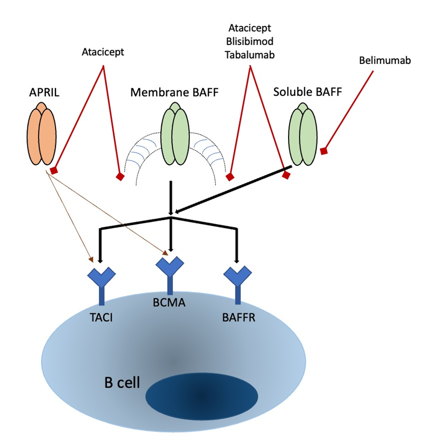
Figure 1. Targets of BAFF and APRIL. Belimumab specifically inhibits soluble BAFF, which is capable of interacting with all three receptors. Blisibimod addresses both membrane-bound and soluble forms of BAFF, while Atacicept targets both BAFF and APRIL, leading to the downregulation of signaling through TACI, BCMA, and BAFF-R.
CONCLUSION :
In conclusion, this review highlights the significant impact of B-lymphocyte activating factor (BAFF) on the development and progression of autoimmune diseases. By facilitating B-cell survival and activity, BAFF plays a central role in orchestrating the immune response; however, its dysregulation can lead to pathological conditions. Targeting BAFF presents a promising therapeutic strategy, as evidenced by emerging treatments that effectively modulate B-cell function in diseases like systemic lupus erythematosus and rheumatoid arthritis.
Looking ahead, additional research is crucial to fully understand the complex interactions between BAFF, APRIL, and their respective receptors, as well as how genetic and environmental factors influence BAFF expression and function. Understanding the long-term effects of BAFF modulation through current and future therapies will be vital for optimizing treatment regimens and minimizing potential side effects. Additionally, exploring the role of BAFF in other autoimmune disorders and its contribution to comorbid conditions could provide valuable insights into broader therapeutic applications.
Ultimately, a deeper insight into BAFF’s role in immune regulation could pave the way for innovative therapies, potentially leading to more targeted and effective interventions that enhance patient outcomes and improve the quality of life for individuals affected by autoimmune disorders. As our understanding of BAFF continues to evolve, it may well provide the foundation for a new era in the management of autoimmune diseases.
List of Abbreviations
BAFF – B-lymphocyte activating factor
TNF – tumor necrosis factor
TNFRSF13B – tumor necrosis factor receptor superfamily, member 13B
BCMA – B-cell maturation antigen
AAbs – Autoantibodies
anti-DS-DNA antibodies – Anti-double stranded DNA antibodies
Abs – antibodies
SLE – systemic lupus erythematosus
anti-Ro Abs - Anti-Ro antibodies
SjS - Sjögren syndrome
RF – rheumatoid factor
RA – rheumatoid arthritis
DCs – dendritic cells
MSCs – mesenchymal stem cells
ECs – endothelial cells
APRIL – A proliferation-inducing ligand
MBCs – memory B cells
NF-kappa – Nuclear factor kappa-light-chain-enhancer of activated B cells
JNK Jun – N-terminal kinase
FDCs – Follicular dendritic cells
GCs – granulosa cells
IgM - Immunoglobulin M
TLR – Toll-like Receptor
NAs – natural antibodies
cDCs – Classical dendritic cells
CCL-2 – chemokine ligand 2
MZ B lymphocytes – Marginal-zone B cells
Tr2 B lymphocytes – Transitional B cells type 2
Tr1 B lymphocytes – Transitional B cells type 1
MMs – missense mutations
FDA – “Food and Drug Administration”
RCT – randomized controlled trial
ACR – American College of Rheumatology
SDAI – Simplified Disease Activity Index
ANAs – Anti-nuclear antibodies
ESR – erythrocyte sedimentation rate
NK cells - natural killer cells
CRP – C-reactive protein
ESKD – End Stage Kidney Disease
DAS28 – Disease Activity Score 28.
REFERENCES:
1. Mantovani, A., Dinarello, C. A., Molgora, M., & Garlanda, C. (2019). Interleukin-1 and Related Cytokines in the Regulation of Inflammation and Immunity. Immunity, 50(4), 778–795. https://doi.org/10.1016/j.immuni.2019.03.012
2. Arango Duque, G., & Descoteaux, A. (2014). Macrophage cytokines: involvement in immunity and infectious diseases. Frontiers in immunology, 5, 491. https://doi.org/10.3389/fimmu.2014.00491
3. Mousa, A., & Bakhiet, M. (2013). Role of cytokine signaling during nervous system development. International journal of molecular sciences, 14(7), 13931–13957. https://doi.org/10.3390/ijms140713931
4. Delgoffe, G. M., Murray, P. J., & Vignali, D. A. (2011). Interpreting mixed signals: the cell's cytokine conundrum. Current opinion in immunology, 23(5), 632–638. https://doi.org/10.1016/j.coi.2011.07.013
5. Saito, Y., Miyagawa, Y., Onda, K., Nakajima, H., Sato, B., Horiuchi, Y., Okita, H., Katagiri, Y. U., Saito, M., Shimizu, T., Fujimoto, J., & Kiyokawa, N. (2008). B-cell-activating factor inhibits CD20-mediated and B-cell receptor-mediated apoptosis in human B cells. Immunology, 125(4), 570–590. https://doi.org/10.1111/j.1365-2567.2008.02872.x
6. Min, Y. N., Wang, C. Y., Li, X. X., Hou, Y., Qiu, J. H., Ma, J., Shao, L. L., Zhang, X., Wang, Y. W., Peng, J., Hou, M., & Shi, Y. (2016). Participation of B-cell-activating factor receptors in the pathogenesis of immune thrombocytopenia. Journal of thrombosis and haemostasis : JTH, 14(3), 559–571. https://doi.org/10.1111/jth.13246
7. Mackay, F., & Tangye, S. G. (2004). The role of the BAFF/APRIL system in B cell homeostasis and lymphoid cancers. Current opinion in pharmacology, 4(4), 347–354. https://doi.org/10.1016/j.coph.2004.02.009
8. Dubey, L., Karempudi, P., Luther, S. A., et al. (2017). Interactions between fibroblastic reticular cells and B cells promote mesenteric lymph node lymphangiogenesis. Nature Communications, 8, 367. https://doi.org/10.1038/s41467-017-00504-9
9. Tobón, G. J., Izquierdo, J. H., & Cañas, C. A. (2013). B lymphocytes: development, tolerance, and their role in autoimmunity-focus on systemic lupus erythematosus. Autoimmune diseases, 2013, 827254. https://doi.org/10.1155/2013/827254
10. Yu, Y., Lu, C., Yu, W., Lei, Y., Sun, S., Liu, P., Bai, F., Chen, Y., & Chen, J. (2024). B Cells Dynamic in Aging and the Implications of Nutritional Regulation. Nutrients, 16(4), 487. https://doi.org/10.3390/nu16040487
11. Giordano, D., Kuley, R., Draves, K. E., Elkon, K. B., Giltiay, N. V., & Clark, E. A. (2023). B cell-activating factor (BAFF) from dendritic cells, monocytes and neutrophils is required for B cell maturation and autoantibody production in SLE-like autoimmune disease. Frontiers in immunology, 14, 1050528. https://doi.org/10.3389/fimmu.2023.1050528
12. Rihacek, M., Bienertova-Vasku, J., Valik, D., Sterba, J., Pilatova, K., & Zdrazilova-Dubska, L. (2015). B-Cell Activating Factor as a Cancer Biomarker and Its Implications in Cancer-Related Cachexia. BioMed research international, 2015, 792187. https://doi.org/10.1155/2015/792187
13. Carrillo-Ballesteros, F. J., Oregon-Romero, E., Franco-Topete, R. A., Govea-Camacho, L. H., Cruz, A., Muñoz-Valle, J. F., Bustos-Rodríguez, F. J., Pereira-Suárez, A. L., & Palafox-Sánchez, C. A. (2019). B-cell activating factor receptor expression is associated with germinal center B-cell maintenance. Experimental and therapeutic medicine, 17(3), 2053–2060. https://doi.org/10.3892/etm.2019.7172
14. Cyster, J. G., & Allen, C. D. C. (2019). B Cell Responses: Cell Interaction Dynamics and Decisions. Cell, 177(3), 524–540. https://doi.org/10.1016/j.cell.2019.03.016
15. Cascalho, M., & Platt, J. L. (2023). TNFRSF13B in B cell responses to organ transplantation. Human immunology, 84(1), 27–33. https://doi.org/10.1016/j.humimm.2022.09.006
16. Cascalho, M., & Platt, J. L. (2021). TNFRSF13BDiversification Fueled by B Cell Responses to Environmental Challenges-A Hypothesis. Frontiers in immunology, 12, 634544. https://doi.org/10.3389/fimmu.2021.634544
17. Rickert, R. C., Jellusova, J., & Miletic, A. V. (2011). Signaling by the tumor necrosis factor receptor superfamily in B-cell biology and disease. Immunological reviews, 244(1), 115–133. https://doi.org/10.1111/j.1600-065X.2011.01067.x
18. Xu, S., & Lam, K. P. (2020). Transmembrane Activator and CAML Interactor (TACI): Another Potential Target for Immunotherapy of Multiple Myeloma?. Cancers, 12(4), 1045. https://doi.org/10.3390/cancers12041045
19. Guldenpfennig, C., Teixeiro, E., & Daniels, M. (2023). NF-kB's contribution to B cell fate decisions. Frontiers in immunology, 14, 1214095. https://doi.org/10.3389/fimmu.2023.1214095
20. Gerondakis, S., & Siebenlist, U. (2010). Roles of the NF-kappaB pathway in lymphocyte development and function. Cold Spring Harbor perspectives in biology, 2(5), a000182. https://doi.org/10.1101/cshperspect.a000182
21. Guo, Q., Jin, Y., Chen, X., et al. (2024). NF-ќЇB in biology and targeted therapy: New insights and translational implications. Signal Transduction and Targeted Therapy, 9, 53. https://doi.org/10.1038/s41392-024-01757-9
22. Cano, R. L. E., & Lopera, H. D. E. (2013). Introduction to T and B lymphocytes. In J. M. Anaya, Y. Shoenfeld, A. Rojas-Villarraga, et al. (Eds.), Autoimmunity: From Bench to Bedside (Chapter 5). Bogota (Colombia): El Rosario University Press. Available from: https://www.ncbi.nlm.nih.gov/books/NBK459471/
23. Gözmen, S., Karapnar, T. H., Tüfekçi, Ö., Vergin, C., Yüksel, F., İrken, G., & Ören, H. (2016). B-cell-activating factor, a proliferation inducing ligand and co-stimulatory molecules in the pathogenesis of immune thrombocytopenia in childhood. Blood coagulation & fibrinolysis : an international journal in haemostasis and thrombosis, 27(5), 494–499. https://doi.org/10.1097/MBC.0000000000000144
24. Zhang, Y., Tian, J., Xiao, F., Zheng, L., Zhu, X., Wu, L., Zhao, C., Wang, S., Rui, K., Zou, H., & Lu, L. (2022). B cell-activating factor and its targeted therapy in autoimmune diseases. Cytokine & growth factor reviews, 64, 57–70. https://doi.org/10.1016/j.cytogfr.2021.11.004
25. Ibrahim, H.Y., Sulaiman, G.M., Ad’hiah, A.H. et al. B-lymphocyte-activating factor is a potential biomarker associated with susceptibility to Graves’ disease in Iraqi women.Egypt J Med Hum Genet 24, 76 (2023). https://doi.org/10.1186/s43042-023-00455-5
26. Zinatizadeh, M. R., Schock, B., Chalbatani, G. M., Zarandi, P. K., Jalali, S. A., & Miri, S. R. (2020). The Nuclear Factor Kappa B (NF-kB) signaling in cancer development and immune diseases. Genes & diseases, 8(3), 287–297. https://doi.org/10.1016/j.gendis.2020.06.005
27. Gupta, N., & DeFranco, A. L. (2007). Lipid rafts and B cell signaling. Seminars in cell & developmental biology, 18(5), 616–626. https://doi.org/10.1016/j.semcdb.2007.07.009
28. Erazo-Oliveras, A., Muñoz-Vega, M., Mlih, M. et al. Mutant APC reshapes Wnt signaling plasma membrane nanodomains by altering cholesterol levels via oncogenic β-catenin.Nat Commun 14, 4342 (2023). https://doi.org/10.1038/s41467-023-39640-w
29. Carrillo-Ballesteros, F. J., Oregon-Romero, E., Franco-Topete, R. A., Govea-Camacho, L. H., Cruz, A., Muñoz-Valle, J. F., Bustos-Rodríguez, F. J., Pereira-Suárez, A. L., & Palafox-Sánchez, C. A. (2019). B-cell activating factor receptor expression is associated with germinal center B-cell maintenance. Experimental and therapeutic medicine, 17(3), 2053–2060. https://doi.org/10.3892/etm.2019.7172
30. Yam-Puc, J. C., Zhang, L., Zhang, Y., & Toellner, K. M. (2018). Role of B-cell receptors for B-cell development and antigen-induced differentiation. F1000Research, 7, 429. https://doi.org/10.12688/f1000research.13567.1
31. Tafalla, C., González, L., Castro, R., & Granja, A. G. (2017). B Cell-Activating Factor Regulates Different Aspects of B Cell Functionality and Is Produced by a Subset of Splenic B Cells in Teleost Fish. Frontiers in immunology, 8, 295. https://doi.org/10.3389/fimmu.2017.00295
32. Roco, J. A., Mesin, L., Binder, S. C., Nefzger, C., Gonzalez-Figueroa, P., Canete, P. F., Ellyard, J., Shen, Q., Robert, P. A., Cappello, J., Vohra, H., Zhang, Y., Nowosad, C. R., Schiepers, A., Corcoran, L. M., Toellner, K. M., Polo, J. M., Meyer-Hermann, M., Victora, G. D., & Vinuesa, C. G. (2019). Class-Switch Recombination Occurs Infrequently in Germinal Centers. Immunity, 51(2), 337–350.e7. https://doi.org/10.1016/j.immuni.2019.07.001
33. Stavnezer, J., & Schrader, C. E. (2014). IgH chain class switch recombination: mechanism and regulation. Journal of immunology (Baltimore, Md. : 1950), 193(11), 5370–5378. https://doi.org/10.4049/jimmunol.1401849
34. Nagaishi, T., Watabe, T., Kotake, K., Kumazawa, T., Aida, T., Tanaka, K., Ono, R., Ishino, F., Usami, T., Miura, T., Hirakata, S., Kawasaki, H., Tsugawa, N., Yamada, D., Hirayama, K., Yoshikawa, S., Karasuyama, H., Okamoto, R., Watanabe, M., Blumberg, R. S., … Adachi, T. (2022). Immunoglobulin A-specific deficiency induces spontaneous inflammation specifically in the ileum. Gut, 71(3), 487–496. https://doi.org/10.1136/gutjnl-2020-322873
35. Breedveld, A., & van Egmond, M. (2019). IgA and FcαRI: Pathological Roles and Therapeutic Opportunities. Frontiers in immunology, 10, 553. https://doi.org/10.3389/fimmu.2019.00553
36. Kiefer, K., Oropallo, M. A., Cancro, M. P., & Marshak-Rothstein, A. (2012). Role of type I interferons in the activation of autoreactive B cells. Immunology and cell biology, 90(5), 498–504. https://doi.org/10.1038/icb.2012.10
37. McNab, F., Mayer-Barber, K., Sher, A. et al. Type I interferons in infectious disease. Nat Rev Immunol 15, 87–103 (2015). https://doi.org/10.1038/nri3787
38. Duan, T., Du, Y., Xing, C., Wang, H. Y., & Wang, R. F. (2022). Toll-Like Receptor Signaling and Its Role in Cell-Mediated Immunity. Frontiers in immunology, 13, 812774. https://doi.org/10.3389/fimmu.2022.812774
39. Cascalho, M., & Platt, J. L. (2023). TNFRSF13B in B cell responses to organ transplantation. Human immunology, 84(1), 27–33. https://doi.org/10.1016/j.humimm.2022.09.006
40. Hua, Z., & Hou, B. (2013). TLR signaling in B-cell development and activation. Cellular & molecular immunology, 10(2), 103–106. https://doi.org/10.1038/cmi.2012.61
41. Rastogi, I., Jeon, D., Moseman, J. E., Muralidhar, A., Potluri, H. K., & McNeel, D. G. (2022). Role of B cells as antigen presenting cells. Frontiers in immunology, 13, 954936. https://doi.org/10.3389/fimmu.2022.954936
42. Syeda, M.Z., Hong, T., Huang, C. et al. B cell memory: from generation to reactivation: a multipronged defense wall against pathogens. Cell Death Discov. 10, 117 (2024). https://doi.org/10.1038/s41420-024-01889-5
43. Heink, S., Yogev, N., Garbers, C., Herwerth, M., Aly, L., Gasperi, C., Husterer, V., Croxford, A. L., Möller-Hackbarth, K., Bartsch, H. S., Sotlar, K., Krebs, S., Regen, T., Blum, H., Hemmer, B., Misgeld, T., Wunderlich, T. F., Hidalgo, J., Oukka, M., Rose-John, S., … Korn, T. (2017). Trans-presentation of IL-6 by dendritic cells is required for the priming of pathogenic TH17 cells. Nature immunology, 18(1), 74–85. https://doi.org/10.1038/ni.3632
44. Salah, A., Wang, H., Li, Y., Ji, M., Ou, W. B., Qi, N., & Wu, Y. (2021). Insights Into Dendritic Cells in Cancer Immunotherapy: From Bench to Clinical Applications. Frontiers in cell and developmental biology, 9, 686544. https://doi.org/10.3389/fcell.2021.686544
45. Vazquez, M. I., Catalan-Dibene, J., & Zlotnik, A. (2015). B cells responses and cytokine production are regulated by their immune microenvironment. Cytokine, 74(2), 318–326. https://doi.org/10.1016/j.cyto.2015.02.007
46. Liu, J., Zhang, X., Cheng, Y. et al. Dendritic cell migration in inflammation and immunity.Cell Mol Immunol 18, 2461–2471 (2021). https://doi.org/10.1038/s41423-021-00726-4
47. Warnatz, K., Salzer, U., Rizzi, M., Fischer, B., Gutenberger, S., Böhm, J., Kienzler, A. K., Pan-Hammarström, Q., Hammarström, L., Rakhmanov, M., Schlesier, M., Grimbacher, B., Peter, H. H., & Eibel, H. (2009). B-cell activating factor receptor deficiency is associated with an adult-onset antibody deficiency syndrome in humans. Proceedings of the National Academy of Sciences of the United States of America, 106(33), 13945–13950. https://doi.org/10.1073/pnas.0903543106
48. Smulski, C. R., & Eibel, H. (2018). BAFF and BAFF-Receptor in B Cell Selection and Survival. Frontiers in immunology, 9, 2285. https://doi.org/10.3389/fimmu.2018.02285
49. D Lempicki, M., Paul, S., Serbulea, V., Upchurch, C. M., Sahu, S., Gray, J. A., Ailawadi, G., Garcia, B. L., McNamara, C. A., Leitinger, N., & Meher, A. K. (2022). BAFF antagonism via the BAFF receptor 3 binding site attenuates BAFF 60-mer-induced classical NF-κB signaling and metabolic reprogramming of B cells. Cellular immunology, 381, 104603. Advance online publication. https://doi.org/10.1016/j.cellimm.2022.104603
50. Consonni, F., Gambineri, E., & Favre, C. (2022). ALPS, FAS, and beyond: From inborn errors of immunity to acquired immunodeficiencies. Annals of Hematology, 101, 469-484. https://doi.org/10.1007/s00277-022-04761-7
51. Hoffman, W., Lakkis, F. G., & Chalasani, G. (2016). B Cells, Antibodies, and More. Clinical journal of the American Society of Nephrology : CJASN, 11(1), 137–154. https://doi.org/10.2215/CJN.09430915
52. Smulski, C. R., & Eibel, H. (2018). BAFF and BAFF-Receptor in B Cell Selection and Survival. Frontiers in immunology, 9, 2285. https://doi.org/10.3389/fimmu.2018.02285
53. Cox, E. M., El-Behi, M., Ries, S., Vogt, J. F., Kohlhaas, V., Michna, T., Manfroi, B., Al-Maarri, M., Wanke, F., Tirosh, B., Pondarre, C., Lezeau, H., Yogev, N., Mittenzwei, R., Descatoire, M., Weller, S., Weill, J. C., Reynaud, C. A., Boudinot, P., Jouneau, L., … Fillatreau, S. (2023). AKT activity orchestrates marginal zone B cell development in mice and humans. Cell reports, 42(4), 112378. https://doi.org/10.1016/j.celrep.2023.112378
54. Rowland, S. L., Leahy, K. F., Halverson, R., Torres, R. M., & Pelanda, R. (2010). BAFF receptor signaling aids the differentiation of immature B cells into transitional B cells following tonic BCR signaling. Journal of immunology (Baltimore, Md. : 1950), 185(8), 4570–4581. https://doi.org/10.4049/jimmunol.1001708
55. Sevdali, E., Block, V., Lataretu, M., Li, H., Smulski, C. R., Briem, J. S., Heitz, Y., Fischer, B., Ramirez, N. J., Grimbacher, B., Jäck, H. M., Voll, R. E., Hölzer, M., Schneider, P., & Eibel, H. (2022). BAFFR activates PI3K/AKT signaling in human naive but not in switched memory B cells through direct interactions with B cell antigen receptors. Cell reports, 39(13), 111019. https://doi.org/10.1016/j.celrep.2022.111019
56. Müller-Winkler, J., Mitter, R., Rappe, J. C. F., Vanes, L., Schweighoffer, E., Mohammadi, H., Wack, A., & Tybulewicz, V. L. J. (2021). Critical requirement for BCR, BAFF, and BAFFR in memory B cell survival. The Journal of experimental medicine, 218(2), e20191393. https://doi.org/10.1084/jem.20191393
57. Weller, S., Sterlin, D., Fadeev, T., Coignard, E., Verge de Los Aires, A., Goetz, C., Fritzen, R., Bahuaud, M., Batteux, F., Gorochov, G., Weill, J. C., & Reynaud, C. A. (2023). T-independent responses to polysaccharides in humans mobilize marginal zone B cells prediversified against gut bacterial antigens. Science immunology, 8(79), eade1413. https://doi.org/10.1126/sciimmunol.ade1413
58. Hafraoui, K., Moutschen, M., Smet, J., Mascart, F., Schaaf-Lafontaine, N., & Fillet, G. (2009). Selective defect of anti-pneumococcal IgG in a patient with persistent polyclonal B cell lymphocytosis. European journal of internal medicine, 20(3), e62–e65. https://doi.org/10.1016/j.ejim.2008.08.005
59. Ramsköld, D., Parodis, I., Lakshmikanth, T., Sippl, N., Khademi, M., Chen, Y., Zickert, A., Mikeš, J., Achour, A., Amara, K., Piehl, F., Brodin, P., Gunnarsson, I., & Malmström, V. (2019). B cell alterations during BAFF inhibition with belimumab in SLE. EBioMedicine, 40, 517–527. https://doi.org/10.1016/j.ebiom.2018.12.035
60. Furie, R., Stohl, W., Ginzler, E. M., Becker, M., Mishra, N., Chatham, W., Merrill, J. T., Weinstein, A., McCune, W. J., Zhong, J., Cai, W., Freimuth, W., & Belimumab Study Group. (2008). Biologic activity and safety of belimumab, a neutralizing anti-B-lymphocyte stimulator (BLyS) monoclonal antibody: A phase I trial in patients with systemic lupus erythematosus. Arthritis Research & Therapy, 10(5), R109. https://doi.org/10.1186/ar2506
61. Cheekati, M., & Murakhovskaya, I. (2024). Anti-B-Cell-Activating Factor (BAFF) Therapy: A Novel Addition to Autoimmune Disease Management and Potential for Immunomodulatory Therapy in Warm Autoimmune Hemolytic Anemia. Biomedicines, 12(7), 1597. https://doi.org/10.3390/biomedicines12071597
62. Block, V., Sevdali, E., Recher, M., Abolhassani, H., Hammarstrom, L., Smulski, C. R., Baronio, M., Plebani, A., Proietti, M., Speletas, M., Warnatz, K., Voll, R. E., Lougaris, V., Schneider, P., & Eibel, H. (2023). CVID-Associated B Cell Activating Factor Receptor Variants Change Receptor Oligomerization, Ligand Binding, and Signaling Responses. Journal of clinical immunology, 43(2), 391–405. https://doi.org/10.1007/s10875-022-01378-3
63. Sircana, M. C., Vidili, G., Gidaro, A., Delitala, A. P., Filigheddu, F., Castelli, R., & Manetti, R. (2023). Common Variable Immunodeficiency and Selective IgA Deficiency: Focus on Autoimmune Manifestations and Their Pathogenesis. International Journal of Translational Medicine, 3, 432-460. https://doi.org/10.3390/ijtm3040031
64. Sevdali, E., Block Saldana, V., Speletas, M., & Eibel, H. (2021). BAFF receptor polymorphisms and deficiency in humans. Current opinion in immunology, 71, 103–110. https://doi.org/10.1016/j.coi.2021.06.008
65. Germinaro, M., Reynolds, P., Knight, V., & Alam, R. (2015). Association of B-cell activating factor receptor deficiency with the P21R polymorphism and common variable immunodeficiency. Annals of allergy, asthma & immunology : official publication of the American College of Allergy, Asthma, & Immunology, 115(1), 82–83. https://doi.org/10.1016/j.anai.2015.04.020
66. Ntellas, P., Dardiotis, E., Sevdali, E., Siokas, V., Aloizou, A. M., Tsinti, G., Germenis, A. E., Hadjigeorgiou, G. M., Eibel, H., & Speletas, M. (2020). TNFRSF13C/BAFFR P21R and H159Y polymorphisms in multiple sclerosis. Multiple sclerosis and related disorders, 37, 101422. https://doi.org/10.1016/j.msard.2019.101422
67. Negrini, S., Emmi, G., Greco, M., Borro, M., Sardanelli, F., Murdaca, G., Indiveri, F., & Puppo, F. (2022). Sjögren's syndrome: a systemic autoimmune disease. Clinical and experimental medicine, 22(1), 9–25. https://doi.org/10.1007/s10238-021-00728-6
68. Domeier, P. P., Chodisetti, S. B., Schell, S. L., Kawasawa, Y. I., Fasnacht, M. J., Soni, C., & Rahman, Z. S. M. (2018). B-Cell-Intrinsic Type 1 Interferon Signaling Is Crucial for Loss of Tolerance and the Development of Autoreactive B Cells. Cell reports, 24(2), 406–418. https://doi.org/10.1016/j.celrep.2018.06.046
69. Thoreau, B., Chaigne, B., & Mouthon, L. (2022). Role of B-Cell in the Pathogenesis of Systemic Sclerosis. Frontiers in immunology, 13, 933468. https://doi.org/10.3389/fimmu.2022.933468
70. Ganguly, D., Haak, S., Sisirak, V., & Reizis, B. (2013). The role of dendritic cells in autoimmunity. Nature reviews. Immunology, 13(8), 566–577. https://doi.org/10.1038/nri3477
71. Senécal, J. L., Hoa, S., Yang, R., & Koenig, M. (2020). Pathogenic roles of autoantibodies in systemic sclerosis: Current understandings in pathogenesis. Journal of scleroderma and related disorders, 5(2), 103–129. https://doi.org/10.1177/2397198319870667
72. Chan, V. S., Tsang, H. H., Tam, R. C., Lu, L., & Lau, C. S. (2013). B-cell-targeted therapies in systemic lupus erythematosus. Cellular & molecular immunology, 10(2), 133–142. https://doi.org/10.1038/cmi.2012.64
73. Dema, B., & Charles, N. (2016). Autoantibodies in SLE: Specificities, Isotypes and Receptors. Antibodies (Basel, Switzerland), 5(1), 2. https://doi.org/10.3390/antib5010002
74. Gao, Y., Chen, Y., Zhang, Z., Yu, X., & Zheng, J. (2020). Recent Advances in Mouse Models of Sjögren's Syndrome. Frontiers in immunology, 11, 1158. https://doi.org/10.3389/fimmu.2020.01158
75. Park, Y. S., Gauna, A. E., & Cha, S. (2015). Mouse Models of Primary Sjogren's Syndrome. Current pharmaceutical design, 21(18), 2350–2364. https://doi.org/10.2174/1381612821666150316120024
76. Tolomeo, M., Cavalli, A., & Cascio, A. (2022). STAT1 and Its Crucial Role in the Control of Viral Infections. International journal of molecular sciences, 23(8), 4095. https://doi.org/10.3390/ijms23084095
77. Fillatreau S. (2018). B cells and their cytokine activities implications in human diseases. Clinical immunology (Orlando, Fla.), 186, 26–31. https://doi.org/10.1016/j.clim.2017.07.020
78. Orange, D. E., & Donlin, L. T. (2020). Advancing Rheumatoid Arthritis Synovial Biopsy Analysis: One B Cell at a Time. Arthritis & rheumatology (Hoboken, N.J.), 72(5), 702–703. https://doi.org/10.1002/art.41185
79. Volkov, M., van Schie, K. A., & van der Woude, D. (2020). Autoantibodies and B Cells: The ABC of rheumatoid arthritis pathophysiology. Immunological reviews, 294(1), 148–163. https://doi.org/10.1111/imr.12829
80. Chekol Abebe, E., Asmamaw Dejenie, T., Mengie Ayele, T., Dagnew Baye, N., Agegnehu Teshome, A., & Tilahun Muche, Z. (2021). The Role of Regulatory B Cells in Health and Diseases: A Systemic Review. Journal of inflammation research, 14, 75–84. https://doi.org/10.2147/JIR.S286426
81. Wu, F., Gao, J., Kang, J., Wang, X., Niu, Q., Liu, J., & Zhang, L. (2021). B Cells in Rheumatoid Arthritis:Pathogenic Mechanisms and Treatment Prospects. Frontiers in immunology, 12, 750753. https://doi.org/10.3389/fimmu.2021.750753
82. de Mattos Barbosa, M. G., Lefferts, A. R., Huynh, D., Liu, H., Zhang, Y., Fu, B., Barnes, J., Samaniego, M., Bram, R. J., Geha, R. S., Shikanov, A., Prak, E. T. L., Farkash, E. A., Platt, J. L., & Cascalho, M. (2021). TNFRSF13B genotypes control immune-mediated pathology by regulating the functions of innate B cells. JCI insight, 6(17), e150483. https://doi.org/10.1172/jci.insight.150483
83. Platt, J. L., de Mattos Barbosa, M. G., Huynh, D., Lefferts, A. R., Katta, J., Kharas, C., Freddolino, P., Bassis, C. M., Wobus, C., Geha, R., Bram, R., Nunez, G., Kamada, N., & Cascalho, M. (2021). TNFRSF13B polymorphisms counter microbial adaptation to enteric IgA. JCI insight, 6(14), e148208. https://doi.org/10.1172/jci.insight.148208
84. Kaegi, C., Steiner, U. C., Wuest, B., Crowley, C., & Boyman, O. (2020). Systematic Review of Safety and Efficacy of Atacicept in Treating Immune-Mediated Disorders. Frontiers in immunology, 11, 433. https://doi.org/10.3389/fimmu.2020.00433
85. Carvalho-Santos, A., Ballard Kuhnert, L. R., Hahne, M., Vasconcellos, R., Carvalho-Pinto, C. E., & Villa-Verde, D. M. S. (2024). Anti-inflammatory role of APRIL by modulating regulatory B cells in antigen-induced arthritis. PloS one, 19(5), e0292028. https://doi.org/10.1371/journal.pone.0292028
86. Jang, S., Kwon, E. J., & Lee, J. J. (2022). Rheumatoid Arthritis: Pathogenic Roles of Diverse Immune Cells. International journal of molecular sciences, 23(2), 905. https://doi.org/10.3390/ijms23020905
87. Thoreau, B., Chaigne, B., & Mouthon, L. (2022). Role of B-Cell in the Pathogenesis of Systemic Sclerosis. Frontiers in immunology, 13, 933468. https://doi.org/10.3389/fimmu.2022.933468
88. Melissaropoulos, K., Iliopoulos, G., Sakkas, L. I., & Daoussis, D. (2022). Pathogenetic Aspects of Systemic Sclerosis: A View Through the Prism of B Cells. Frontiers in immunology, 13, 925741. https://doi.org/10.3389/fimmu.2022.925741
89. Beesley, C. F., Goldman, N. R., Taher, T. E., Denton, C. P., Abraham, D. J., Mageed, R. A., & Ong, V. H. (2023). Dysregulated B cell function and disease pathogenesis in systemic sclerosis. Frontiers in immunology, 13, 999008. https://doi.org/10.3389/fimmu.2022.999008
90. Vincent, F. B., Saulep-Easton, D., Figgett, W. A., Fairfax, K. A., & Mackay, F. (2013). The BAFF/APRIL system: emerging functions beyond B cell biology and autoimmunity. Cytokine & growth factor reviews, 24(3), 203–215. https://doi.org/10.1016/j.cytogfr.2013.04.003
91. Struemper, H., Kurtinecz, M., Edwards, L., Freimuth, W. W., Roth, D. A., & Stohl, W. (2022). Reductions in circulating B cell subsets and immunoglobulin G levels with long-term belimumab treatment in patients with SLE. Lupus science & medicine, 9(1), e000499. https://doi.org/10.1136/lupus-2021-000499
92. Hofmann, K., Clauder, A. K., & Manz, R. A. (2018). Targeting B Cells and Plasma Cells in Autoimmune Diseases. Frontiers in immunology, 9, 835. https://doi.org/10.3389/fimmu.2018.00835
93. Pavlasova, G., & Mraz, M. (2020). The regulation and function of CD20: an "enigma" of B-cell biology and targeted therapy. Haematologica, 105(6), 1494–1506. https://doi.org/10.3324/haematol.2019.243543
94. Stohl, W., Hiepe, F., Latinis, K. M., Thomas, M., Scheinberg, M. A., Clarke, A., Aranow, C., Wellborne, F. R., Abud-Mendoza, C., Hough, D. R., Pineda, L., Migone, T. S., Zhong, Z. J., Freimuth, W. W., Chatham, W. W., BLISS-52 Study Group, & BLISS-76 Study Group (2012). Belimumab reduces autoantibodies, normalizes low complement levels, and reduces select B cell populations in patients with systemic lupus erythematosus. Arthritis and rheumatism, 64(7), 2328–2337. https://doi.org/10.1002/art.34400
95. Singh, J. A., Shah, N. P., & Mudano, A. S. (2021). Belimumab for systemic lupus erythematosus. The Cochrane database of systematic reviews, 2(2), CD010668. https://doi.org/10.1002/14651858.CD010668.pub2
96. Guerreiro Castro, S., & Isenberg, D. A. (2017). Belimumab in systemic lupus erythematosus (SLE): evidence-to-date and clinical usefulness. Therapeutic advances in musculoskeletal disease, 9(3), 75–85. https://doi.org/10.1177/1759720X17690474
97. Meier, J., & Sturm, A. (2011). Current treatment of ulcerative colitis. World journal of gastroenterology, 17(27), 3204–3212. https://doi.org/10.3748/wjg.v17.i27.3204
98. Wang, Z., Huang, J., Xie, D., He, D., Lu, A., & Liang, C. (2021). Toward Overcoming Treatment Failure in Rheumatoid Arthritis. Frontiers in immunology, 12, 755844. https://doi.org/10.3389/fimmu.2021.755844
99. Rubbert-Roth, A., Furst, D. E., Nebesky, J. M., Jin, A., & Berber, E. (2018). A Review of Recent Advances Using Tocilizumab in the Treatment of Rheumatic Diseases. Rheumatology and therapy, 5(1), 21–42. https://doi.org/10.1007/s40744-018-0102-x
100. Wallace, D. J., Ginzler, E. M., Merrill, J. T., Furie, R. A., Stohl, W., Chatham, W. W., Weinstein, A., McKay, J. D., McCune, W. J., Petri, M., Fettiplace, J., Roth, D. A., Ji, B., & Heath, A. (2019). Safety and Efficacy of Belimumab Plus Standard Therapy for Up to Thirteen Years in Patients With Systemic Lupus Erythematosus. Arthritis & rheumatology (Hoboken, N.J.), 71(7), 1125–1134. https://doi.org/10.1002/art.40861
101. Mariette, X., Seror, R., Quartuccio, L., Baron, G., Salvin, S., Fabris, M., Desmoulins, F., Nocturne, G., Ravaud, P., & De Vita, S. (2015). Efficacy and safety of belimumab in primary Sj√ґgren's syndrome: Results of the BELISS open-label phase II study. Annals of the Rheumatic Diseases, 74(3), 526-531. https://doi.org/10.1136/annrheumdis-2013-203991
102. Wang, D., Shan, C., Liu, J., Zhang, R., Zhu, G., Gao, T., Chang, H., Gao, S., Bai, C., Nie, N., Zhang, Q., & Lin, Y. (2022). Efficacy and safety of belimumab for the treatment of refractory childhood-onset systemic lupus erythematosus: A single-center, real-world, retrospective study. Frontiers in immunology, 13, 1067721. https://doi.org/10.3389/fimmu.2022.1067721
103. LeBien, T. W., & Tedder, T. F. (2008). B lymphocytes: how they develop and function. Blood, 112(5), 1570–1580. https://doi.org/10.1182/blood-2008-02-078071
104. Hartung, H. P., & Kieseier, B. C. (2010). Atacicept: targeting B cells in multiple sclerosis. Therapeutic advances in neurological disorders, 3(4), 205–216. https://doi.org/10.1177/1756285610371146
105. Kaegi, C., Steiner, U. C., Wuest, B., Crowley, C., & Boyman, O. (2020). Systematic Review of Safety and Efficacy of Atacicept in Treating Immune-Mediated Disorders. Frontiers in immunology, 11, 433. https://doi.org/10.3389/fimmu.2020.00433
106. Jonklaas, J., Bianco, A. C., Bauer, A. J., Burman, K. D., Cappola, A. R., Celi, F. S., Cooper, D. S., Kim, B. W., Peeters, R. P., Rosenthal, M. S., Sawka, A. M., & American Thyroid Association Task Force on Thyroid Hormone Replacement (2014). Guidelines for the treatment of hypothyroidism: prepared by the american thyroid association task force on thyroid hormone replacement. Thyroid : official journal of the American Thyroid Association, 24(12), 1670–1751. https://doi.org/10.1089/thy.2014.0028
107. Jonklaas, J., Bianco, A. C., Cappola, A. R., Celi, F. S., Fliers, E., Heuer, H., McAninch, E. A., Moeller, L. C., Nygaard, B., Sawka, A. M., Watt, T., & Dayan, C. M. (2021). Evidence-Based Use of Levothyroxine/Liothyronine Combinations in Treating Hypothyroidism: A Consensus Document. Thyroid : official journal of the American Thyroid Association, 31(2), 156–182. https://doi.org/10.1089/thy.2020.0720
108. Cogollo, E., Silva, M. A., & Isenberg, D. (2015). Profile of atacicept and its potential in the treatment of systemic lupus erythematosus. Drug design, development and therapy, 9, 1331–1339. https://doi.org/10.2147/DDDT.S71276
109. Pitsiu, M., Yalkinoglu, Ö., Farrell, C., Girard, P., Vazquez-Mateo, C., & Papasouliotis, O. (2023). Population pharmacokinetics of atacicept in systemic lupus erythematosus: An analysis of three clinical trials. CPT: pharmacometrics & systems pharmacology, 12(8), 1157–1169. https://doi.org/10.1002/psp4.12982
110. Fassbinder, T., Saunders, U., Mickholz, E., Jung, E., Becker, H., Schlüter, B., & Jacobi, A. M. (2015). Differential effects of cyclophosphamide and mycophenolate mofetil on cellular and serological parameters in patients with systemic lupus erythematosus. Arthritis research & therapy, 17(1), 92. https://doi.org/10.1186/s13075-015-0603-8
111. Lee, W. S., & Amengual, O. (2020). B cells targeting therapy in the management of systemic lupus erythematosus. Immunological medicine, 43(1), 16–35. https://doi.org/10.1080/25785826.2019.1698929
112. Lee, D.S.W., Rojas, O.L. & Gommerman, J.L. B cell depletion therapies in autoimmune disease: advances and mechanistic insights. Nat Rev Drug Discov 20, 179–199 (2021). https://doi.org/10.1038/s41573-020-00092-2
113. Horton, S., Buch, M. H., & Emery, P. (2010). Efficacy, tolerability and safety of biologic therapy in rheumatoid disease: patient considerations. Drug, healthcare and patient safety, 2, 101–119. https://doi.org/10.2147/DHPS.S6317
114. Kim, M. J., Lee, S. K., Oh, S., Kim, H. A., Park, Y. B., Lee, S. S., & Shin, K. (2022). Efficacy of Abatacept Versus Tumor Necrosis Factor Inhibitors in Anti-citrullinated Protein Antibody-Positive Patients with Rheumatoid Arthritis: Results from a Korean Nationwide Biologics Registry. Rheumatology and therapy, 9(4), 1143–1155. https://doi.org/10.1007/s40744-022-00467-4
115. Cook, K. D., Waggoner, S. N., & Whitmire, J. K. (2014). NK cells and their ability to modulate T cells during virus infections. Critical reviews in immunology, 34(5), 359–388. https://doi.org/10.1615/critrevimmunol.2014010604
116. Abel, A. M., Yang, C., Thakar, M. S., & Malarkannan, S. (2018). Natural Killer Cells: Development, Maturation, and Clinical Utilization. Frontiers in immunology, 9, 1869. https://doi.org/10.3389/fimmu.2018.01869
117. Ingegnoli, F., Castelli, R., & Gualtierotti, R. (2013). Rheumatoid factors: clinical applications. Disease markers, 35(6), 727–734. https://doi.org/10.1155/2013/726598
118. Zhu, L. N., Hou, H. M., Wang, S., Zhang, S., Wang, G. G., Guo, Z. Y., & Wu, J. (2023). FcRn inhibitors: a novel option for the treatment of myasthenia gravis. Neural regeneration research, 18(8), 1637–1644. https://doi.org/10.4103/1673-5374.363824
119. van Vollenhoven, R. F., Kinnman, N., Vincent, E., Wax, S., & Bathon, J. (2011). Atacicept in patients with rheumatoid arthritis and an inadequate response to methotrexate: results of a phase II, randomized, placebo-controlled trial. Arthritis and rheumatism, 63(7), 1782–1792. https://doi.org/10.1002/art.30372
120. Lin, Y. J., Anzaghe, M., & Schülke, S. (2020). Update on the Pathomechanism, Diagnosis, and Treatment Options for Rheumatoid Arthritis. Cells, 9(4), 880. https://doi.org/10.3390/cells9040880
121. Witcher, J., Fleischmann, R., Chindalore, V. L., Hansen, R. J., Hu, L., Radtke, D., Voelker, J., Gomez, E., & McColm, J. (2016). Pharmacokinetics and safety of single doses of tabalumab in subjects with rheumatoid arthritis or systemic lupus erythematosus. British journal of clinical pharmacology, 81(5), 908–917. https://doi.org/10.1111/bcp.12860
122. Manetta, J., Bina, H., Ryan, P., Fox, N., Witcher, D. R., & Kikly, K. (2014). Generation and characterization of tabalumab, a human monoclonal antibody that neutralizes both soluble and membrane-bound B-cell activating factor. Journal of inflammation research, 7, 121–131. https://doi.org/10.2147/JIR.S67751
123. Zhang, Z., Jara, C. J., Singh, M., Xu, H., Goodnow, C. C., Jackson, K. J., & Reed, J. H. (2021). Human transitional and IgMlow mature naïve B cells preserve permissive B-cell receptors. Immunology and cell biology, 99(8), 865–878. https://doi.org/10.1111/imcb.12478
124. Struemper, H., Kurtinecz, M., Edwards, L., Freimuth, W. W., Roth, D. A., & Stohl, W. (2022). Reductions in circulating B cell subsets and immunoglobulin G levels with long-term belimumab treatment in patients with SLE. Lupus science & medicine, 9(1), e000499. https://doi.org/10.1136/lupus-2021-000499
125. Ward, M. M., Guthrie, L. C., & Alba, M. I. (2015). Clinically important changes in individual and composite measures of rheumatoid arthritis activity: thresholds applicable in clinical trials. Annals of the rheumatic diseases, 74(9), 1691–1696. https://doi.org/10.1136/annrheumdis-2013-205079
126. Nasir, N., Majid, H., Khan, A. H., Awan, S., & Mehmood Riaz, M. (2022). Disease activity correlates and functionality in patients with rheumatoid arthritis - real-world experience from a South Asian country. Reumatologia, 60(3), 183–191. https://doi.org/10.5114/reum.2022.117838
127. Lenert, A., Niewold, T. B., & Lenert, P. (2017). Spotlight on blisibimod and its potential in the treatment of systemic lupus erythematosus: evidence to date. Drug design, development and therapy, 11, 747–757. https://doi.org/10.2147/DDDT.S114552
128. Merrill, J. T., Shanahan, W. R., Scheinberg, M., Kalunian, K. C., Wofsy, D., & Martin, R. S. (2018). Phase III trial results with blisibimod, a selective inhibitor of B-cell activating factor, in subjects with systemic lupus erythematosus (SLE): results from a randomised, double-blind, placebo-controlled trial. Annals of the rheumatic diseases, 77(6), 883–889. https://doi.org/10.1136/annrheumdis-2018-213032
129. Khambhati, K., Bhattacharjee, G., Gohil, N., Braddick, D., Kulkarni, V., & Singh, V. (2019). Exploring the Potential of Cell-Free Protein Synthesis for Extending the Abilities of Biological Systems. Frontiers in bioengineering and biotechnology, 7, 248. https://doi.org/10.3389/fbioe.2019.00248
130. Weimer, K., Colloca, L., & Enck, P. (2015). Placebo eff ects in psychiatry: mediators and moderators. The lancet. Psychiatry, 2(3), 246–257. https://doi.org/10.1016/S2215-0366(14)00092-3
131. Kato, T., Ishigooka, J., Miyajima, M., Watabe, K., Fujimori, T., Masuda, T., Higuchi, T., & Vieta, E. (2020). Double-blind, placebo-controlled study of lurasidone monotherapy for the treatment of bipolar I depression. Psychiatry and clinical neurosciences, 74(12), 635–644. https://doi.org/10.1111/pcn.13137
132. Selewski, D. T., Shah, G. V., Mody, R. J., Rajdev, P. A., & Mukherji, S. K. (2010). Rituximab (Rituxan). AJNR. American journal of neuroradiology, 31(7), 1178–1180. https://doi.org/10.3174/ajnr.A2142
133. Igarashi, T., Ogura, M., Itoh, K., Taniwaki, M., Ando, K., Kuroda, Y., Yamamoto, K., Uike, N., Tomita, A., Nagai, H., Kurosawa, M., Mori, S., Nawano, S., Terauchi, T., Ohashi, Y., & Tobinai, K. (2016). Japanese phase II study of rituximab maintenance for untreated indolent B-cell non-Hodgkin lymphoma with high tumor burden. International journal of hematology, 104(6), 700–708. https://doi.org/10.1007/s12185-016-2097-9
134. van Schaik, M., Arends, E. J., Soonawala, D., van Ommen, E., de Leeuw, K., Limper, M., van Paassen, P., Huizinga, T. W. J., Toes, R. E. M., van Kooten, C., Rotmans, J. I., Rabelink, T. J., & Teng, Y. K. O. (2022). Efficacy of belimumab combined with rituximab in severe systemic lupus erythematosus: study protocol for the phase 3, multicenter, randomized, open-label Synbiose 2 trial. Trials, 23(1), 939. https://doi.org/10.1186/s13063-022-06874-w
135. Mahieu, M. A., Strand, V., Simon, L. S., Lipsky, P. E., & Ramsey-Goldman, R. (2016). A critical review of clinical trials in systemic lupus erythematosus. Lupus, 25(10), 1122–1140. https://doi.org/10.1177/0961203316652492
136. Claverie, R., Perriguey, M., Rico, A., Boutiere, C., Demortiere, S., Durozard, P., Hilezian, F., Dubrou, C., Vely, F., Pelletier, J., Audoin, B., & Maarouf, A. (2023). Efficacy of Rituximab Outlasts B-Cell Repopulation in Multiple Sclerosis: Time to Rethink Dosing?. Neurology(R) neuroimmunology & neuroinflammation, 10(5), e200152. https://doi.org/10.1212/NXI.0000000000200152
137. Kraaij, T., Arends, E. J., van Dam, L. S., Kamerling, S. W. A., van Daele, P. L. A., Bredewold, O. W., Ray, A., Bakker, J. A., Scherer, H. U., Huizinga, T. J. W., Rabelink, T. J., van Kooten, C., & Teng, Y. K. O. (2021). Long-term effects of combined B-cell immunomodulation with rituximab and belimumab in severe, refractory systemic lupus erythematosus: 2-year results. Nephrology, dialysis, transplantation : official publication of the European Dialysis and Transplant Association - European Renal Association, 36(8), 1474–1483. https://doi.org/10.1093/ndt/gfaa117
138. Yuan, X., & Sun, L. (2022). Stem Cell Therapy in Lupus. Rheumatology and immunology research, 3(2), 61–68. https://doi.org/10.2478/rir-2022-0011
139. Pillai, S., Mattoo, H., & Cariappa, A. (2011). B cells and autoimmunity. Current opinion in immunology, 23(6), 721–731. https://doi.org/10.1016/j.coi.2011.10.007
140. Kraaij, T., Arends, E. J., van Dam, L. S., Kamerling, S. W. A., van Daele, P. L. A., Bredewold, O. W., Ray, A., Bakker, J. A., Scherer, H. U., Huizinga, T. J. W., Rabelink, T. J., van Kooten, C., & Teng, Y. K. O. (2021). Long-term effects of combined B-cell immunomodulation with rituximab and belimumab in severe, refractory systemic lupus erythematosus: 2-year results. Nephrology, dialysis, transplantation : official publication of the European Dialysis and Transplant Association - European Renal Association, 36(8), 1474–1483. https://doi.org/10.1093/ndt/gfaa117.