Histomorphometric Alterations of the Umbilical Cord and Vessels in Intrauterine Growth Restriction and Their Association with Perinatal Outcomes: A North Indian Case-Control Study
- S.M. Badar Hayat , Reasearch Scholar, Department of Anatomy, NIMS University, Jaipur, India
- Kumar Satish Ravi , Professor, Department of Anatomy, NIMS University, Jaipur, India
- Upendra Kumar Gupta , Assistant Professor, Department of Anatomy, Shree Narayan Medical Institute and Hospital, Saharsa, India
- Manoj Kumar , Assistant Professor, Department of Obstetrics & Gynecology, Shree Narayan Medical Institute and Hospital, Saharsa, India
- Kumari Anamika , Assistant Professor, Department of Obstetrics & Gynecology, Shree Narayan Medical Institute and Hospital, Saharsa, India
Article Information:
Abstract:
Subsequently, Bruch et al. demonstrated significant reductions in umbilical vessel dimensions and cord area in pregnancies complicated by FGR (12). More recently, Peyter et al. reported structural alterations of umbilical vessels and impaired nitric oxide-mediated vasodilation in growth-restricted fetuses, highlighting the contribution of vascular dysfunction to abnormal fetoplacental circulation (13).Material And Methods: Histological Processing were done following fixation, tissue specimens were processed by routine paraffin embedding. Transverse sections of 3–5 μm thickness were prepared using a rotary microtome.Results: Two umbilical arteries and one umbilical vein typically make up UC. In this investigation, we found a single HUA in one UC of the AGA group and two UC of the IUGR group. In both instances, no true UC knots were seen.Conclusion: These structural abnormalities are accompanied by adverse perinatal outcomes, including lower birth weight, increased NICU admissions, and fetal distress. Umbilical cord morphometry may therefore serve as a useful morphological marker of placental insufficiency and fetal compromise in growth-restricted pregnancies.
Keywords:
Article :
INTRODUCTION:
Fetal growth restriction (FGR), previously referred to as intrauterine growth restriction (IUGR), is a major obstetric complication characterized by the inability of the fetus to achieve its genetically determined growth potential due to pathological disturbances within the maternal–placental–fetal unit. FGR affects approximately 5–10% of pregnancies worldwide and remains a leading cause of perinatal morbidity, mortality, preterm birth, neonatal intensive care unit (NICU) admission, and long-term neurodevelopmental impairment (1–3). In addition, evidence from the developmental origins of health and disease (DOHaD) paradigm suggests that impaired fetal growth is associated with an increased risk of cardiovascular disease, hypertension, type 2 diabetes mellitus, and metabolic syndrome in adulthood (4,5).
Placental insufficiency is recognized as the principal pathological mechanism underlying most cases of FGR. Impaired placental perfusion results in chronic fetal hypoxia, reduced nutrient delivery, and adaptive fetal circulatory redistribution, ultimately leading to restricted fetal growth and adverse perinatal outcomes (1,6,7). Recent international guidelines from the International Society of Ultrasound in Obstetrics and Gynecology (ISUOG) and the American College of Obstetricians and Gynecologists (ACOG) emphasize the importance of placental dysfunction and fetoplacental hemodynamic alterations in the pathogenesis and management of FGR (1,8).
The umbilical cord serves as the lifeline between the fetus and placenta and is essential for maintaining uninterrupted fetoplacental circulation. Structurally, the normal umbilical cord consists of two arteries and one vein embedded within a specialized mucoid connective tissue known as Wharton's jelly. This extracellular matrix-rich tissue protects the vessels from compression, torsion, and kinking, thereby ensuring adequate blood flow between the fetus and placenta (9). Histologically, Wharton's jelly contains mesenchymal stromal cells, collagen fibers, proteoglycans, and hyaluronic acid, all of which contribute to the biomechanical properties of the cord (10).
Accumulating evidence suggests that structural alterations of the umbilical cord reflect abnormalities in placental function and fetal growth. Sonographic and histomorphometric studies have demonstrated that growth-restricted fetuses frequently exhibit a lean umbilical cord characterized by reduced cord diameter, decreased cross-sectional area, diminished Wharton's jelly, and vascular remodeling (11–14). Raio et al. first reported that a reduced umbilical cord area was associated with an increased risk of delivering small-for-gestational-age infants (11). Subsequently, Bruch et al. demonstrated significant reductions in umbilical vessel dimensions and cord area in pregnancies complicated by FGR (12). More recently, Peyter et al. reported structural alterations of umbilical vessels and impaired nitric oxide-mediated vasodilation in growth-restricted fetuses, highlighting the contribution of vascular dysfunction to abnormal fetoplacental circulation (13).
The biological mechanisms responsible for these morphometric alterations are multifactorial. Experimental studies have shown that chronic hypoxia, oxidative stress, endothelial dysfunction, altered nitric oxide signaling, and impaired angiogenesis contribute to vascular remodeling in FGR pregnancies (15–17). Reduced expression of endothelial nitric oxide synthase (eNOS) and dysregulation of vascular growth factors may result in increased vessel wall thickness, reduced luminal diameter, and diminished umbilical blood flow (15,16). Furthermore, alterations in fetal growth mediators, including insulin-like growth factor-1 (IGF-1) and leptin, have been associated with reduced fetal growth and abnormal umbilical cord development (18,19).
Several investigators have reported significant correlations between umbilical cord morphometry and neonatal outcome. Reduced umbilical cord area and Wharton's jelly volume have been associated with low birth weight, fetal distress, oligohydramnios, abnormal Doppler velocimetry, NICU admission, and increased perinatal morbidity (11,14,20–22). Nevertheless, detailed histomorphometric evaluation of individual umbilical vessels, vessel wall thickness, luminal area, and their relationship with neonatal anthropometric measurements remains limited, particularly in the North Indian population.
Considering the scarcity of region-specific histological data and the potential clinical significance of umbilical cord morphology as a marker of placental insufficiency, the present study was undertaken to evaluate the histological and morphometric characteristics of the umbilical cord and its vessels in growth-restricted neonates and to determine their association with neonatal anthropometric parameters and perinatal outcomes.
MATERIALS AND METHODS:
Study Design and Setting
This hospital-based prospective case-control study was conducted in the Department of Anatomy in collaboration with the Department of Obstetrics and Gynecology, NIMS University Rajasthan, Jaipur, India, between Jan 2023 and decembr2025. The study protocol was approved by the Institutional Ethics Committee (Approval No. IEC/P-163/2022), and written informed consent was obtained from all participants before enrolment.
Study Population
A total of 100 umbilical cord specimens obtained from singleton term deliveries were included in the study. The study population was divided into:
Control group (AGA group):50 umbilical cords obtained from appropriate-for-gestational-age (AGA) neonates.
Case group (IUGR group):50 umbilical cords obtained from pregnancies complicated by fetal growth restriction (FGR/IUGR).
Definition of Study Groups
Appropriate-for-gestational-age (AGA) fetuses were defined as those having an estimated fetal weight between the 10th and 90th percentile for gestational age according to ultrasonographic assessment.
Fetal growth restriction (FGR/IUGR) was diagnosed when the estimated fetal weight and/or abdominal circumference was below the 10th percentile for gestational age on obstetric ultrasonography, in accordance with contemporary international recommendations.
The participants were included in the study Singleton pregnancies, Maternal age between 18 and 35 years, Gestational age between 37 and 40 completed weeks, Reliable gestational dating based on the last menstrual period and/or first-trimester ultrasonography, Pregnancies diagnosed as AGA or IUGR according to predefined criteria, Pregnancies complicated by pregnancy-induced hypertension (PIH) were included in the IUGR group, as PIH is a recognized contributor to placental insufficiency and fetal growth restriction.
The following pregnancies were excluded from this study Multiple gestations, Pre-existing diabetes mellitus, Gestational diabetes mellitus, Maternal cardiovascular disorders.Congenital fetal anomalies, Uncertain gestational age, Preterm deliveries (<37 completed weeks) and Umbilical cord anomalies such as true knots, single umbilical artery, velamentous insertion, or cord prolapse.
The collection for maternal demographic and obstetric characteristics were recorded using a predesigned proforma. The following variables were documentedmaternal age, gravidity and parity, consanguinity, pregnancy-induced hypertension, maternal anemia, oligohydramnios, hypothyroidism, previous abortion history, and mode of delivery.
Neonatal variables includedGestational age at delivery, birth weight, sex of newborn, neonatal intensive care unit (nicu) admission, fetal distress, meconium-stained liquor and stillbirth
The umbilical cord sample collection was collected immediately after delivery, a 5-cm segment of the umbilical cord was excised approximately 5 cm from the placental insertion site to avoid regional variations in cord morphology. The specimens were rinsed in normal saline to remove blood clots and fixed in 10% neutral buffered formalin for 24–48 hours.
Histological Processing were done following fixation, tissue specimens were processed by routine paraffin embedding. Transverse sections of 3–5 μm thickness were prepared using a rotary microtome.
The sections were stained with Hematoxylin and Eosin (H&E) stain for general histological evaluation and Masson's Trichrome stain for assessment of connective tissue architecture and Wharton's jelly. The sections that were obliquely cut, folded, fragmented, or incompletely represented were excluded from morphometric analysis. The histomorphometric Analysis of histological sections were examined using an Olympus CX40 light microscope equipped with a digital camera and image analysis software.
Digital images were captured under 40× magnification and calibrated before measurements. Histomorphometric parameters were measured using ImageJ software (National Institutes of Health, USA).
Umbilical cord measurements were evaluated on the following headings
1. Umbilical cord cross-sectional area (mm²)
2. Umbilical cord circumference (mm)
3. Umbilical cord diameter (mm)
4. Wharton's jelly area (mm²)
Umbilical vessel measurements were done for each umbilical artery and umbilical vein, the following measurements were obtained:
1. Total vessel area (mm²)
2. Luminal area (mm²)
3. Vessel wall area (mm²)
4. Vessel wall thickness (μm)
5. Wall-to-lumen ratio
The vessel wall area was calculated as:
Vessel wall area = Total vessel area − Luminal area
The wall-to-lumen ratio was calculated as:
Wall-to-lumen ratio = Vessel wall area / Luminal area
All measurements were performed independently by two observers blinded to the study groups. Interobserver agreement was assessed using Cohen's kappa statistics.
Quality Control: Calibration of the image analysis system was performed before each measurement session. Three measurements were obtained from each parameter, and the mean value was used for statistical analysis.
Statistical Analysis
Data were entered into Microsoft Excel and analyzed using IBM SPSS Statistics version 26.0 (IBM Corp., Armonk, NY, USA).Normality of data distribution was assessed using the Shapiro-Wilk test. Continuous variables were expressed as mean ± standard deviation (SD), whereas categorical variables were expressed as frequencies and percentages.Comparisons between AGA and IUGR groups were performed using independent Student's t-test for normally distributed continuous variables and Mann–Whitney U test for non-normally distributed variables.Chi-square test or Fisher's exact test for categorical variables.Pearson's correlation coefficient was used to evaluate associations betweenhistomorphometric parameters and neonatal anthropometric variables.A p-value <0.05 was considered statistically significant.
RESULTS:
Two umbilical arteries and one umbilical vein typically make up UC. In this investigation, we found a single HUA in one UC of the AGA group and two UC of the IUGR group. In both instances, no true UC knots were seen.
Table 1. Demographic and Maternal Clinical Characteristics of the Study Population
|
Variable |
AGA (n=50) |
IUGR (n=50) |
p-value |
|
Maternal age (years), Mean ± SD |
24.6 ± 3.39 |
23.4 ± 3.13 |
0.031* |
|
Consanguineous marriage, n (%) |
6 (12.0) |
8 (16.0) |
0.571 |
|
Gravidity, n (%) |
|
||
|
Primigravida |
24 (48.0) |
34 (68.0) |
0.078 |
|
Multigravida |
26 (52.0) |
16 (32.0) |
|
|
Pregnancy-induced hypertension, n (%) |
0 (0.0) |
13 (26.0) |
<0.001* |
|
Anemia, n (%) |
14 (28.0) |
33 (66.0) |
0.001* |
|
Oligohydramnios, n (%) |
1 (2.0) |
14 (28.0) |
<0.001* |
|
Maternal hypothyroidism, n (%) |
1 (2.0) |
2 (4.0) |
0.559 |
|
History of abortion, n (%) |
7 (14.0) |
6 (12.0) |
0.770 |
Table 1 shows a comparison of the clinical features of AGA and IUGR groups. The mean maternal age was similar in both groups. Incidence of pregnancy induced hypertension (PIH) and maternal anemia was noted to be higher in IUGR group. Pregnancy outcome results of control and case groups are given in Table 2. GA at birth and birth weight was significantly reduced in IUGR group. This higher birth rate of female babies (65%) and increased (15%) admission to neonatal intensive care unit (NICU) were significant in IUGR group. No significant difference was found in mode of delivery (p=0.3830) and stillbirth (p=0.1540)
The present study demonstrated that pregnancies complicated by IUGR were associated with significant reductions in umbilical cord cross-sectional area, circumference, and diameter. These structural alterations were accompanied by adverse perinatal outcomes, including lower gestational age at delivery, significantly reduced birth weight, increased NICU admissions, and higher rates of fetal distress. Furthermore, maternal anemia, pregnancy-induced hypertension, and oligohydramnios were significantly more common among mothers of IUGR neonates.
The demographic and maternal clinical characteristics of the study population are presented in Table 1. The mean maternal age was significantly lower in the IUGR group compared with the AGA group (23.4 ± 3.13 vs. 24.6 ± 3.39 years, p = 0.031). Although primigravida mothers were more frequent in the IUGR group [34 (68.0%)] than in the AGA group [24 (48.0%)], the difference was not statistically significant (p = 0.078).
The prevalence of pregnancy-induced hypertension (26.0% vs. 0.0%, p < 0.001), maternal anemia (66.0% vs. 28.0%, p = 0.001), and oligohydramnios (28.0% vs. 2.0%, p < 0.001) was significantly higher among mothers of IUGR neonates. No significant differences were observed between the groups with respect to consanguineous marriage, maternal hypothyroidism, or previous history of abortion (Table 1).
Female neonates constituted a higher proportion of births in the IUGR group; however, the difference was not statistically significant. Neonatal intensive care unit (NICU) admission was significantly more frequent among IUGR neonates compared with AGA neonates. Likewise, fetal distress was observed more commonly in the IUGR group. No significant differences were found between the groups regarding mode of delivery, meconium-stained liquor, or stillbirth rate.
Histomorphometric Characteristics of Umbilical Vessels
Representative photomicrographs of the human umbilical vein (HUV) in AGA and IUGR neonates are shown in Figure 1A and Figure 1B, respectively. In the AGA group, the HUV exhibited a relatively thin tunica media composed of smooth muscle fibers surrounding a wide vascular lumen. In contrast, the HUV of IUGR neonates demonstrated marked structural reduction in vessel dimensions.
Morphometric analysis revealed that the total vessel area of the HUV was significantly smaller in the IUGR group than in the AGA group (1.55 ± 0.45 mm² vs. 2.61 ± 0.54 mm², p < 0.001). Similarly, vessel wall area was significantly reduced in IUGR neonates (1.14 ± 0.37 mm² vs. 1.76 ± 0.37 mm², p < 0.001). The luminal area of the HUV was also significantly decreased in the IUGR group compared with the AGA group (0.42 ± 0.27 mm² vs. 0.85 ± 0.44 mm², p < 0.001).
Representative photomicrographs of the human umbilical arteries (HUA) in AGA and IUGR neonates are shown in Figures 2A and 2B, respectively. The HUA in the AGA group displayed a characteristic thick tunica media consisting of inner longitudinal and outer circular smooth muscle layers. In contrast, both umbilical arteries in the IUGR group demonstrated significantly reduced total vessel area and vessel wall area compared with the AGA group (Table 3).
No significant differences were observed in the luminal areas of either umbilical artery between the two groups (p > 0.05).
Umbilical Cord Morphometry
The morphometric characteristics of the umbilical cord are summarized in Table 3 and illustrated in Figure 1. The mean umbilical cord cross-sectional area (CSA) was significantly lower in the IUGR group than in the AGA group (39.56 ± 13.92 mm² vs. 54.40 ± 16.59 mm², p < 0.001). Likewise, the umbilical cord circumference (20.05 ± 4.01 mm vs. 23.86 ± 4.22 mm, p < 0.001) and diameter (6.38 ± 1.28 mm vs. 7.60 ± 1.34 mm, p < 0.001) were significantly reduced among IUGR neonates.
Vessel Wall-to-Lumen Ratios
The ratio of total vessel area to luminal area and the ratio of vessel wall area to luminal area were significantly increased in the HUV of IUGR neonates compared with AGA neonates (5.44 ± 5.18 vs. 3.74 ± 1.81, p = 0.023 and 4.44 ± 5.18 vs. 2.74 ± 1.81, p = 0.023, respectively). Similar trends were observed in both umbilical arteries; however, statistical significance was reached only for the second umbilical artery (HUA-2) (p = 0.018).
No significant sex-related differences in the cross-sectional areas of umbilical vessels were observed in either the AGA or IUGR groups (p > 0.05).
Photomicrograph (Figure 1A and B) shows human umbilical vein (HUV) of AGA and IUGR neonate respectively. In AGA group, the tunica media of HUV (Figure 1A) is thin and composed of smooth muscles with large lumen. CSAs of umbilical vessels measurements in control and case group results are summarized in Table 3. The HUV (Figure 1B) had significantly smaller total vessel area (1.55±0.45 vs. 2.61±0.54) and vessel wall area (1.14±0.37 vs. 1.76±0.37) in IUGR group versus AGA group. The microscopic measurements of HUV lumen (Figure 1B) showed significantly (p=0.0001) smaller lumen area (0.42±0.27 vs. 0.85±0.44) in IUGR group versus AGA group.
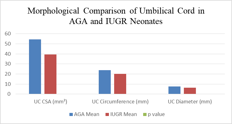
Figure:1 Comparison of umbilical cord cross-sectional area (CSA), circumference, and diameter between AGA and IUGR neonates. All parameters were significantly reduced in the IUGR group (p = 0.0001).
Ratio of total vessel area to lumen area (5.44±5.18 to 3.74±1.81, p=0.023) and vessel wall area to lumen area (4.44±5.18 to 2.74±1.81, p=0.023) were significantly increased in HUV of IUGR group compared to AGA group. In contrast, both ratios were increased in HUAs of IUGR but only HUA-2 increase was significant (p=0.018). No significant (p>0.05) differences in CSAs of umbilical vessels was observed between male and female in both groups.
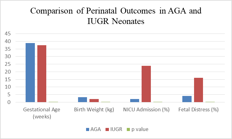
Figure:2 Comparison of gestational age, birth weight, NICU admission, and fetal distress between AGA and IUGR neonates. Birth weight was significantly lower, while NICU admission and fetal distress were significantly higher among IUGR neonates.
Perinatal outcomes of AGA and IUGR neonates are summarized in figure 2. Gestational age at delivery was significantly lower in the IUGR group compared with the AGA group (37.4 ± 1.1 weeks vs. 38.9 ± 1.2 weeks, p < 0.001). Similarly, birth weight was markedly reduced among IUGR neonates (1.98 ± 0.12 kg vs. 3.25 ± 0.50 kg, p < 0.001).
DISCUSSION :
The present study demonstrated significant reductions in total vessel area, vessel wall area, and luminal area of the human umbilical vein (HUV) in IUGR neonates. Similar findings were reported by Jakó M et al., who observed marked reductions in umbilical vein dimensions and vessel wall area in growth-restricted fetuses[1]. The authors suggested that vascular remodeling of the umbilical vein may occur as an adaptive response to chronic placental insufficiency.
Likewise, Visan V et al. demonstrated structural alterations of the umbilical vein in IUGR pregnancies, characterized by reduced vascular dimensions and impaired nitric oxide-mediated relaxation. These findings support the hypothesis that endothelial dysfunction contributes significantly to altered fetoplacental circulation[2].
In the present study, both umbilical arteries exhibited significantly smaller total vessel and vessel wall areas in IUGR neonates, although luminal areas did not differ significantly between groups. Similar observations have been reported in previous morphometric studies, indicating that arterial remodeling in IUGR predominantly affects vessel wall architecture rather than luminal dimensions [5].
The increased vessel wall-to-lumen ratio observed in the umbilical vein and arteries suggests structural vascular remodeling and increased vascular resistance. Such alterations may contribute to reduced fetoplacental blood flow and impaired nutrient transport, thereby exacerbating fetal growth restriction.
Several biological mechanisms may explain the histomorphometric alterations observed in the present study. Chronic placental hypoxia and oxidative stress are recognized contributors to endothelial dysfunction in IUGR [7,17,25]. Experimental studies have demonstrated reduced endothelial nitric oxide synthase (eNOS) activity and impaired nitric oxide bioavailability in placental and umbilical vessels from growth-restricted pregnancies.
Malhotra et al. reported that an imbalance between eNOS and arginase pathways contributes to endothelial dysfunction in IUGR, resulting in impaired vasodilation and increased vascular resistance. Similarly, Chillarkuraet al. demonstrated reduced placental eNOS expression in pregnancies complicated by fetal growth restriction [25].
In addition, alterations in growth-regulating hormones may influence umbilical cord development. Reduced concentrations of insulin-like growth factor-1 (IGF-1), IGF-binding proteins, and leptin have been documented in growth-restricted fetuses and may contribute to reduced Wharton's jelly deposition and diminished vascular growth. Verhaeghe et al. and Ong et al. reported significant associations between cord blood IGF-1, leptin concentrations, and birth weight.
The present study found significantly lower gestational age at delivery and birth weight among IUGR neonates. Furthermore, NICU admission and fetal distress were significantly more frequent in the IUGR group. These findings are consistent with previous studies demonstrating that reduced umbilical cord dimensions are associated with adverse neonatal outcomes.
Ratio et al. reported that fetuses with lean umbilical cords were more likely to be born small for gestational age and experience fetal distress during labor. Similarly, Ghezzi et al. observed significant associations between reduced umbilical vessel morphometry and adverse perinatal outcomes, including low birth weight and neonatal complications [27].
The increased frequency of NICU admission observed in the present study likely reflects the combined effects of prematurity, chronic hypoxia, and low birth weight. Baschat emphasized that abnormal fetoplacental circulation is a major determinant of neonatal morbidity and the need for intensive neonatal care in growth-restricted pregnancies.
The major strength of this study is the detailed histomorphometric evaluation of both the umbilical cord and individual umbilical vessels in a well-defined North Indian population. Furthermore, the correlation of structural alterations with clinical and perinatal outcomes provides valuable insight into the pathophysiology of fetal growth restriction.
However, several limitations should be acknowledged. The sample size was relatively modest, and the study was conducted at a single tertiary care center, which may limit generalizability. In addition, molecular markers of endothelial dysfunction, angiogenesis, and oxidative stress were not evaluated. Future multicenter studies incorporating morphometric, molecular, and Doppler parameters may provide a more comprehensive understanding of fetoplacental vascular remodeling in IUGR.
CONCLUSION :
The present study demonstrates that intrauterine growth restriction is associated with significant histomorphometric alterations of the umbilical cord and its vessels, characterized by reduced cord dimensions, decreased vessel area, and increased vessel wall-to-lumen ratios. These structural abnormalities are accompanied by adverse perinatal outcomes, including lower birth weight, increased NICU admissions, and fetal distress. Umbilical cord morphometry may therefore serve as a useful morphological marker of placental insufficiency and fetal compromise in growth-restricted pregnancies.
BIBLIOGRAPHY:
1. Jakó M, Surányi A, Kaizer L, Németh G, Bártfai G. Maternal hematological parameters and placental and umbilical cord histopathology in intrauterine growth restriction. Medical Principles and Practice. 2019 May 7;28(2):101-8.
2. Vişan V, Balan RA, Costea CF, Cărăuleanu A, Haba RM, Haba MŞ, Socolov G, Mogoş RA, Bogdănici CM, Nemescu D, Tănase DM. Morphological and histopathological changes in placentas of pregnancies with intrauterine growth restriction. Romanian Journal of Morphology and Embryology. 2020 Dec 21;61(2):477.
3. Almasry SM, Elfayomy AK. Morphometric analysis of terminal villi and gross morphological changes in the placentae of term idiopathic intrauterine growth restriction. Tissue and Cell. 2012 Aug 1;44(4):214-9.
4. Ghomian N, Amouian S, Tavassoli F, Arbabzadeh T. Comparison of placental morphology and histopathology of intrauterine growth restriction and normal infants.
5. Kumar D, Soni JS, Dave M. Histomorphometric Analysis of Umbilical Cord Blood Vessel Thickness amongst Normal and Intrauterine Growth-restricted Newborns. Advances in Human Biology. 2025 Jul 21:10-4103.
6. Güven D, Altunkaynak BZ, Altun G, Alkan I, Kocak I. Histomorphometric changes in the placenta and umbilical cord during complications of pregnancy. Biotechnic & Histochemistry. 2018 Apr 3;93(3):198-210.
7. Biswas S, Ghosh SK. Gross morphological changes of placentas associated with intrauterine growth restriction of fetuses: a case control study. Early human development. 2008 Jun 1;84(6):357-62.
8. Armengaud JB, Yzydorczyk C, Siddeek B, Peyter AC, Simeoni U. Intrauterine growth restriction: Clinical consequences on health and disease at adulthood. ReprodToxicol2021;99:168 76.
9. Martins JG, Biggio JR, Abuhamad A. Society for maternal fetal medicine consult series 52: Diagnosis and management of fetal growth restriction. Am J Obstet Gynecol 2020;223:B2 17.
10. Darby JR, Varcoe TJ, Orgeig S, Morrison JL. Cardiorespiratory consequences of intrauterine growth restriction: Influence of timing, severity and duration of hypoxaemia. Theriogenology 2020;150:84 95.
11. Sharma D, Shastri S, Sharma P. Intrauterine growth restriction: Antenatal and postnatal aspects. Clin Med Insights Pediatr2016;10:67 83.
12. Fang H, Li Q, Wang H, Ren Y, Zhang L, Yang L. Maternal nutrient metabolism in the liver during pregnancy. Front Endocrinol (Lausanne) 2024;15:1 12.
13. Roberts H, Woodman AG, Baines KJ, Jeyarajah MJ, Bourque SL, Renaud SJ. Maternal iron deficiency alters trophoblast differentiation and placental development in rat pregnancy. Endocrinology 2021;162:bqab215.
14. Tunster SJ, Watson ED, Fowden AL, Burton GJ. Placental glycogen stores and fetal growth: Insights from genetic mouse models. Reproduction 2020;159:R213 35.
15. Tsikouras P, Antsaklis P, Nikolettos K, Kotanidou S, Kritsotaki N, Bothou A, et al. Diagnosis, prevention, and management of fetal growth restriction (FGR). J Pers Med 2024;14:698.
16. Bendix I, Miller SL, Winterhager E. Editorial: Causes and consequences of intrauterine growth restriction. Front Endocrinol (Lausanne) 2020;11:205.
17. Malhotra A, Allison BJ, Castillo Melendez M, Jenkin G, Polglase GR, Miller SL. Neonatal morbidities of fetal growth restriction: Pathophysiology and impact. Front Endocrinol (Lausanne) 2019;10:55.
18. Kotrannavar SS, Shirol VS, Dhumale HA, Humbarwadi RS. Histomorphometry of umbilical vessels of intrauterine growth restricted neonates. Int J Reprod Contracept Obstet Gynecol2016;5:129-34.
19. Edwards AK, McKnight SM, Askelson K, McKnight JR, Dunlap KA, Satterfield MC. Adaptive responses to maternal nutrient restriction alter placental transport in ewes. Placenta 2020;96:1 9.
20. Gantenbein KV, Kanaka Gantenbein C. Highlighting the trajectory from intrauterine growth restriction to future obesity. Front Endocrinol (Lausanne) 2022;13:1 16.
21. Glorieux FH, Bonewald LF, Harvey NC, van der Meulen MC. Potential influences on optimizing long term musculoskeletal health in children and adolescents with X linked hypophosphatemia (XLH). Orphanet J Rare Dis 2022;17:30.
22. Ilias I, Milionis C, Zoumakis E. An overview of glucocorticoid induced osteoporosis. In: Feingold KR, Ahmed SF, Anawalt B, Blackman MR, Boyce A, Chrousos G, et al, editors. Endotext. [Internet]:MDText.com, Inc.; 2000. Available from: https://www.ncbi.nlm.nih.gov/books/ NBK278968/.[Last accessed on 2025 May 05].
23. Wardinger JE, Ambati S. Placental insufficiency. In: StatPearls. [Internet]: StatPearls Publishing; 2025. Available from: https://www.ncbi.nlm.nih. gov/books/NBK563171/[Last accessed on 2025 May 05].
24. 24.Santana EF, Castello RG, Rizzo G, Grisolia G, Araujo Júnior E, Werner H, et al. Placental and umbilical cord anomalies diagnosed by two and three dimensional ultrasound. Diagnostics (Basel) 2022;12:2810.
25. Chillakuru S, Velichety SD, Rajagopalan V. Human umbilical cord and its vessels: A histomorphometric study in difference severity of hypertensive disorders of pregnancy. Anat Cell Biol 2020;53:68 75.
26. Lan Y, Yang Z, Huang M, Cui Z, Qi Y, Niu H. Morphological and structural changes of umbilical veins and clinical significance in preeclampsia. Hypertens Pregnancy 2018;37:105 10.
27. Raio L, Ghezzi F, Di Naro E, Franchi M, Bolla D, Schneider H. Altered sonographic umbilical cord morphometry in early onset preeclampsia. Obstet Gynecol2002;100:311 6.