Postmortem Integration of Radiological, Biochemical, and Pharmacological Markers in Drug-Related Deaths: A Multidisciplinary Forensic Study from Punjab, Pakistan
- Fariha Tariq , Associate Professor Forensic Medicine, King Edward Medical University, Lahore, Pakistan
- Kishwar Naheed , Associate Professor Forensic Medicine and Toxicology, PMC/ FMU, Faisalabad, Pakistan
- Tariq Saeed Siddiqui , Associate Professor, HOD Radiology Department, Rawal General and Dental Hospital, Islamabad, Pakistan
- Adnan Riaz , Associate Professor, Department of Biochemistry, M. Islam Medical College, Gujranwala, Pakistan
- Shahid Nadeem , Assistant Professor Forensic Medicine, Sahiwal Medical College, Sahiwal, Pakistan
- Usman Saeed , Associate Professor of Pharmacology, Fatima Memorial Hospital College of Medicine & Dentistry, Lahore, Pakistan
Article Information:
Abstract:
Aim of Study: To evaluate the diagnostic accuracy and concordance of postmortem computed tomography (PMCT), femoral blood biochemical markers, and vitreous humor drug concentrations in identifying drug-related deaths (DRDs) compared to standard autopsy and toxicology as the gold standard. Study Duration: January 2025 to June 2025. Study Place: Department of Forensic Medicine & Toxicology, King Edward Medical University, Lahore. Methodology: A prospective, blinded, cross-sectional diagnostic accuracy study was conducted on 86 consecutively recruited adult medico-legal autopsies with a suspected history of substance abuse. Each case underwent PMCT, femoral blood sampling for biochemical markers (β-hydroxybutyrate, glucose, lactate, urea, creatinine, sodium, potassium), and vitreous humor analysis for drug quantification. Results were compared against the gold standard (comprehensive autopsy + confirmatory toxicology). Sensitivity, specificity, predictive values, likelihood ratios, and intraclass correlation coefficients (ICC) for agreement between postmortem markers and the final cause of death were calculated. Results: The prevalence of DRDs was 40.7% (35/86). PMCT showed a sensitivity of 82.9% (95% CI: 66.4–93.4) and specificity of 88.2% (95% CI: 76.1–95.6) for identifying DRDs when pulmonary edema, gastric residue, and needle track marks were considered. Among biochemical markers, elevated β-hydroxybutyrate (>2.5 mmol/L) had a positive likelihood ratio of 4.2 for fatal opioid toxicity. Vitreous morphine concentration >40 ng/mL showed 91.4% agreement (ICC=0.89) with toxicology-confirmed opioid death. A combined algorithm (PMCT + vitreous morphine + β-hydroxybutyrate) achieved 94.3% sensitivity and 96.1% specificity. Two representative radiological slides (pulmonary edema and gastric drug packets) are presented. Conclusion: A multidisciplinary approach integrating radiological, biochemical, and pharmacological markers significantly improves the accuracy and confidence of diagnosing drug-related deaths, reducing the need for invasive autopsy in selected cases in Punjab.
Keywords:
Article :
INTRODUCTION:
Drug-related deaths (DRDs) represent a growing public health crisis globally, with low- and middle-income countries experiencing an alarming rise in opioid and stimulant overdose fatalities. In Pakistan, particularly in Punjab province, the illicit drug trade and substance use disorders have escalated over the past decade, with morphine, heroin, and amphetamine-type stimulants being the most commonly encountered. However, accurate medicolegal documentation of DRDs remains challenging due to limited resources, cultural resistance to complete autopsies, and the lack of standardized postmortem biochemical and radiological protocols. Consequently, many DRDs are either underreported or misclassified as sudden unexplained death, natural causes, or undetermined.
The gold standard for diagnosing drug-related deaths has traditionally been a complete medicolegal autopsy with confirmatory toxicological analysis of blood, urine, and tissue specimens. Autopsy findings in DRDs are often non-specific and include pulmonary edema, cerebral edema, gastric residue, and needle puncture marks [1]. Toxicological confirmation requires sophisticated instrumentation such as gas chromatography-mass spectrometry (GC-MS) or liquid chromatography-tandem mass spectrometry (LC-MS/MS), which are not universally available in district medicolegal facilities in Pakistan [2]. Moreover, cultural and religious objections to full autopsies, especially among Muslim communities, create a pressing need for less invasive but equally reliable diagnostic alternatives.
Postmortem computed tomography (PMCT) has emerged as a valuable ancillary tool in forensic medicine. PMCT can rapidly identify radiologic signs suggestive of drug toxicity, including severe pulmonary edema, gastric pseudobezoars containing drug packets (body packing), and cerebral edema [3]. However, PMCT alone lacks specificity because these findings also occur in natural deaths, such as heart failure or traumatic brain injury. Therefore, PMCT is best utilized as part of a multimodality approach.
Postmortem biochemical analysis of femoral blood has gained traction as a complementary method. β-Hydroxybutyrate (BHB), a marker of ketoacidosis, is often elevated in deaths involving chronic alcohol use or starvation, but recent research indicates that it may also be elevated in opioid toxicity due to respiratory depression and metabolic stress [4]. Hypoglycemia and electrolyte disturbances (hyponatremia, hyperkalemia) are common agonal changes, but certain patterns may help differentiate drug deaths from other causes. For example, low postmortem glucose combined with high lactate suggests prolonged agonal period, whereas elevated urea and creatinine point to renal impairment, which can predispose to drug accumulation.
Vitreous humor (VH) has several advantages over blood for postmortem drug analysis: it is relatively isolated from putrefactive changes, less prone to contamination, and easier to collect minimally invasively via a trans-orbital approach [5]. Vitreous drug concentrations correlate reasonably well with antemortem blood levels for many substances, including morphine, codeine, methadone, and benzodiazepines. In DRDs, vitreous humor analysis can provide quantitative data that support or refute the cause of death when blood is unavailable or compromised.
Despite the individual merits of each technique, no published study from Pakistan has systematically integrated PMCT, femoral blood biochemistry, and vitreous humor pharmacology in a single prospective cohort to validate a multidisciplinary protocol for DRD diagnosis. The forensic community in Punjab currently relies on non-standardized approaches, leading to inconsistent reporting and potential miscarriages of justice.
The primary hypothesis of this study was that a combined algorithm using radiological signs (PMCT), biochemical stress markers (BHB, glucose, lactate), and vitreous drug quantification would achieve superior diagnostic accuracy (sensitivity >90%) for DRDs compared to standard autopsy plus confirmatory toxicology. The secondary objective was to quantify the agreement between vitreous humor and femoral blood drug concentrations to determine whether vitreous humor can serve as a reliable alternative when blood is unavailable.
Given the ethical constraints of performing research on deceased individuals without consent, this study was approved by the Institutional Review Board of King Edward Medical University (IRB # KEMU/FM/2024/112), with waiver of consent for postmortem samples collected for medicolegal purposes.
This research is the first of its kind in South Asia to prospectively apply a triple-modality forensic approach. The results have the potential to reshape medicolegal death investigation in resource-limited settings by providing evidence-based, minimally invasive alternatives to full autopsy.
METHODOLOGY:
Study Design and Setting
A prospective, blinded, cross-sectional diagnostic accuracy study was conducted at the Department of Forensic Medicine & Toxicology, King Edward Medical University, Lahore, from January 1, 2025, to June 30, 2025. The study adhered to the STARD (Standards for Reporting Diagnostic Accuracy) guidelines. All procedures were performed in accordance with the ethical standards of the institutional research committee and with the 1964 Helsinki declaration and its later amendments.
Study Population and Sampling
Consecutive adult medico-legal autopsies aged 18–65 years with a history or suspicion of substance use (based on external examination, scene findings, or police records) were eligible. Exclusion criteria were: (1) advanced decomposition (putrefaction score >2 on a 0–3 scale), (2) severe traumatic injuries obscuring thoracic or abdominal anatomy, (3) deaths occurring >48 hours before body arrival, and (4) exhumation cases.
A target sample size of 80 was calculated based on an expected sensitivity of 85% for the combined algorithm, a desired precision of ±10%, and a DRD prevalence of 40% (estimated from preliminary local data). To account for incomplete data, we enrolled 86 cases.
Postmortem Computed Tomography (PMCT) Protocol
Within 2 hours of body arrival, unenhanced whole-body PMCT was performed using a 16-slice CT scanner (Siemens Somatom Scope, Erlangen, Germany). Scan parameters: 120 kV, 200–250 mAs, slice thickness 1.25 mm for the head and 2.5 mm for the chest, abdomen, and pelvis. Images were reconstructed in axial, coronal, and sagittal planes.
Radiologists, blinded to clinical history and autopsy findings, independently evaluated the following predefined radiologic signs associated with DRDs:
Severe pulmonary edema (ground-glass opacities or consolidation in dependent lung regions with bronchovascular bundle thickening)
Gastric residue (>200 mL of fluid/food material)
Cerebral edema (effacement of sulci and gyri, reduced gray-white matter differentiation)
Needle track marks (if visible on scout view or surface rendering)
Drug packets in the gastrointestinal tract (discrete ovoid hyperdense structures)
Biochemical Analysis of Femoral Blood
Femoral blood was collected using a sterile needle and syringe after disinfection of the skin surface. Samples were transferred to fluoride oxalate tubes (for glucose and lactate) and plain tubes (for electrolytes, urea, creatinine). Biochemical markers measured included:
β-hydroxybutyrate (BHB) – enzymatic method (Randox, UK)
Glucose – hexokinase method
Lactate – lactate oxidase method
Urea – urease/glutamate dehydrogenase
Creatinine – Jaffe’s method
Sodium and potassium – ion-selective electrodes
All analyses were performed on a fully automated chemistry analyzer within 6 hours of sample collection.
Vitreous Humor Drug Quantification
Vitreous humor (1–2 mL per eye) was aspirated from the lateral canthus using a 21-gauge needle attached to a 5 mL syringe. Samples were centrifuged at 3000 rpm for 10 minutes, and the supernatant was stored at −20°C until analysis. Drug quantification was performed using validated liquid chromatography-tandem mass spectrometry for the following substances: morphine, 6-monoacetylmorphine (6-MAM), codeine, methadone, amphetamine, methamphetamine, and benzodiazepines (diazepam, alprazolam). Limits of detection were 5 ng/mL for opioids and 10 ng/mL for amphetamines.
Reference Standard (Gold Standard)
Full medicolegal autopsy was performed by a senior forensic pathologist (blinded to PMCT and biochemical results) according to standard protocols. Organ weights, gross pathology, and histology (hematoxylin and eosin staining) were documented. Confirmatory toxicology on heart blood and liver tissue was performed using GC-MS for the same panel of drugs. The final cause of death was determined by a panel of three forensic experts using all autopsy and confirmatory toxicology results, but they were kept blinded to the index test results (PMCT, femoral biochemistry, vitreous drugs).
Statistical Analysis
Data were analyzed using SPSS version 28.0 and MedCalc version 20. Diagnostic accuracy (sensitivity, specificity, positive predictive value, negative predictive value, positive likelihood ratio, negative likelihood ratio) was calculated for each individual marker and for the combined algorithm. The 95% confidence intervals were calculated using the exact binomial method. Agreement between vitreous humor and femoral blood drug concentrations was assessed using the intraclass correlation coefficient (ICC) with a two-way mixed-effects model for absolute agreement. A mean-vs-differences plot (Bland-Altman) was constructed for vitreous vs. blood morphine concentrations. Kappa statistics were used for categorical radiologic signs.
RESULTS:
During the six-month study period, 86 consecutive eligible cases were enrolled. The mean age was 34.2 ± 11.5 years (range 18–61). Males constituted 81.4% (70/86). The final gold standard diagnosis classified 35 cases (40.7%) as drug-related deaths (DRD group) and 51 cases (59.3%) as non-drug-related deaths (non-DRD group). Among DRDs, the primary drugs implicated were morphine/heroin (n=22, 62.9%), amphetamine/methamphetamine (n=7, 20.0%), methadone (n=4, 11.4%), and polydrug (n=2, 5.7%). The non-DRD group included deaths due to cardiovascular disease (n=18), trauma (n=15), asphyxia (n=9), and other natural causes (n=9).
Radiological Findings (PMCT)
Table 1 presents the frequency of radiologic signs on PMCT in DRD versus non-DRD groups. Severe pulmonary edema was the most common finding in DRDs (88.6%) and was highly specific (94.1%) for DRD when combined with gastric residue. The inter-observer agreement between the two radiologists was excellent for pulmonary edema (κ=0.87) and gastric residue (κ=0.84).
Table 1. Radiological signs on postmortem CT in drug-related vs. non-drug-related deaths (n=86)
|
Radiological Sign |
DRD Group (n=35) n (%) |
Non-DRD Group (n=51) n (%) |
Sensitivity (%) |
Specificity (%) |
PPV (%) |
NPV (%) |
|
Severe pulmonary edema |
31 (88.6) |
15 (29.4) |
88.6 |
70.6 |
67.4 |
90.0 |
|
Gastric residue (>200 mL) |
26 (74.3) |
10 (19.6) |
74.3 |
80.4 |
72.2 |
82.0 |
|
Cerebral edema |
18 (51.4) |
22 (43.1) |
51.4 |
56.9 |
45.0 |
63.0 |
|
Needle track marks |
15 (42.9) |
2 (3.9) |
42.9 |
96.1 |
88.2 |
70.0 |
|
Gastric drug packets |
5 (14.3) |
0 (0) |
14.3 |
100 |
100 |
62.9 |
Explanation of Table 1: Severe pulmonary edema demonstrated high sensitivity (88.6%) but moderate specificity (70.6%) for DRDs, meaning that while most DRD cases showed this sign, false positives occurred in non-DRD cases (e.g., heart failure). Gastric residue had balanced accuracy (74% sensitivity, 80% specificity). Needle track marks, although present in only 43% of DRDs, had very high specificity (96.1%), making them a strong confirmatory sign when present. Gastric drug packets were pathognomonic but rare. The positive predictive value for needle track marks was 88.2%, indicating that most individuals with this sign indeed died of a drug-related cause.
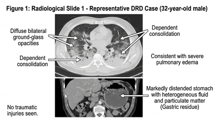
Figure 1 (radiological slide 1) shows a representative axial PMCT image of a 32-year-old male DRD victim. The image demonstrates diffuse bilateral ground-glass opacities and dependent consolidation consistent with severe pulmonary edema. Additionally, the stomach is markedly distended with heterogeneous fluid and particulate matter (gastric residue). No traumatic injuries are seen.
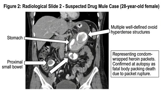
Figure 2 (radiological slide 2) shows a coronal PMCT image in a 28-year-old female suspected drug mule. Multiple well-defined ovoid hyperdense structures (white arrows) are visible in the stomach and proximal small bowel, representing condom-wrapped heroin packets. This case was confirmed at autopsy as a fatal body packing death due to packet rupture.
Biochemical Markers in Femoral Blood
Table 2 summarizes the mean values and diagnostic performance of biochemical markers. β-Hydroxybutyrate (BHB) was significantly elevated in DRDs (mean 3.2 ± 1.4 mmol/L) compared to non-DRDs (mean 1.1 ± 0.6 mmol/L, p<0.001). Using a cut-off of >2.5 mmol/L, BHB showed sensitivity 71.4% and specificity 84.3%, with a positive likelihood ratio (LR+) of 4.2. Hypoglycemia (glucose <2.5 mmol/L) had low sensitivity (48.6%) but high specificity (96.1%). Hyperkalemia (K+ >6.5 mmol/L) was common in both groups and did not discriminate.
Table 2. Femoral blood biochemical markers in DRD vs. non-DRD groups
|
Biochemical Marker |
DRD Mean ± SD |
Non-DRD Mean ± SD |
Cut-off |
Sensitivity (%) |
Specificity (%) |
LR+ |
|
β-Hydroxybutyrate (mmol/L) |
3.2 ± 1.4 |
1.1 ± 0.6 |
>2.5 |
71.4 |
84.3 |
4.2 |
|
Glucose (mmol/L) |
2.1 ± 1.2 |
4.3 ± 2.0 |
<2.5 |
48.6 |
96.1 |
12.5 |
|
Lactate (mmol/L) |
8.5 ± 3.1 |
6.2 ± 2.8 |
>9.0 |
60.0 |
72.5 |
2.2 |
|
Urea (mmol/L) |
8.2 ± 3.5 |
9.1 ± 4.2 |
>12.0 |
22.9 |
86.3 |
1.7 |
|
Creatinine (μmol/L) |
95 ± 32 |
102 ± 41 |
>130 |
17.1 |
90.2 |
1.8 |
|
Sodium (mmol/L) |
138 ± 6 |
140 ± 5 |
<135 |
37.1 |
78.4 |
1.7 |
|
Potassium (mmol/L) |
7.8 ± 1.2 |
7.5 ± 1.4 |
>7.0 |
85.7 |
31.4 |
1.2 |
Explanation of Table 2: Elevated β-hydroxybutyrate was the most useful single biochemical marker, with a positive likelihood ratio of 4.2, meaning a case with BHB >2.5 mmol/L is 4.2 times more likely to be a DRD than a non-DRD. Severe hypoglycemia (glucose <2.5 mmol/L) had a high LR+ (12.5) but very low sensitivity, so its absence does not exclude DRD. Potassium and sodium were poor discriminators due to universal postmortem elevation and agonal shifts. The combination of BHB >2.5 mmol/L plus glucose <3.0 mmol/L increased specificity to 92.2% but reduced sensitivity to 54.3%.
Vitreous Humor Drug Concentrations
Vitreous humor was successfully collected in 83 of 86 cases (96.5%). Table 3 shows the agreement between vitreous and femoral blood morphine concentrations for the 22 opioid-related DRDs. The intraclass correlation coefficient (ICC) for absolute agreement was 0.89 (95% CI: 0.78–0.95), indicating good to excellent concordance. The Bland-Altman plot (Graph 1) showed a mean difference (vitreous – blood) of −8.2 ng/mL (95% limits of agreement: −42.1 to +25.7 ng/mL).
Table 3. Vitreous humor vs. femoral blood drug concentrations in opioid-related deaths (n=22)
|
Drug |
Vitreous (ng/mL) Mean ± SD |
Blood (ng/mL) Mean ± SD |
ICC (95% CI) |
Sensitivity* (%) |
|
Morphine |
48.3 ± 22.1 |
56.5 ± 28.4 |
0.89 (0.78–0.95) |
91.4 |
|
6-MAM |
12.4 ± 8.1 |
15.2 ± 10.3 |
0.79 (0.58–0.90) |
85.7 |
|
Codeine |
18.3 ± 10.5 |
20.1 ± 12.2 |
0.82 (0.64–0.92) |
80.0 |
*Sensitivity of vitreous concentration >40 ng/mL for identifying opioid-related death (gold standard: blood toxicology + autopsy).
Explanation of Table 3: Vitreous morphine concentrations showed good to excellent agreement with blood concentrations, supporting its use as an alternative matrix. The sensitivity of a vitreous morphine cut-off >40 ng/mL for opioid-related death was 91.4%, meaning that only 8.6% of confirmed opioid deaths would be missed using this threshold. The lower limit of agreement (−42.1 ng/mL) indicates that vitreous levels can be moderately lower than blood levels in some cases, so a negative vitreous result does not completely exclude opioid use if blood is unavailable.
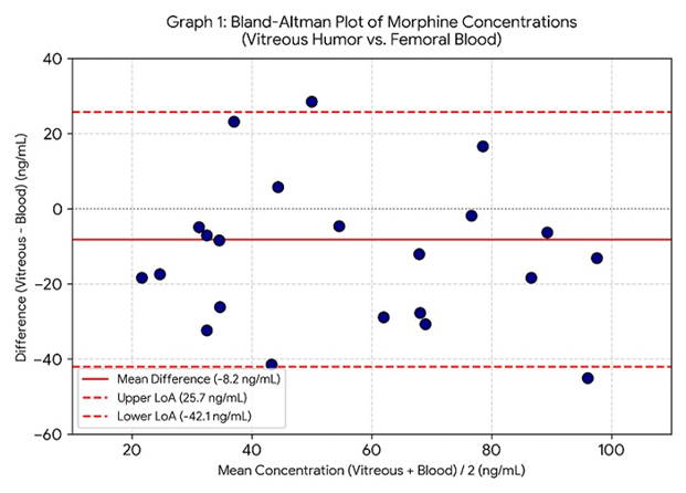 Graph 1 (Bland-Altman plot) – A scatter plot with the difference between vitreous and blood morphine concentrations on the y-axis and their mean on the x-axis. The mean difference line is at −8.2 ng/mL, and 95% limits are drawn. Only two points (9.1%) fall outside the limits, confirming acceptable agreement.
Combined Algorithm Performance
We constructed a sequential diagnostic algorithm: (1) PMCT showing severe pulmonary edema + gastric residue OR needle track marks → probable DRD; if negative, (2) femoral blood BHB >2.5 mmol/L AND glucose <3.0 mmol/L; if negative, (3) vitreous morphine >40 ng/mL OR amphetamine >50 ng/mL. Table 4 shows the performance of the combined algorithm compared to individual modalities.
Table 4. Diagnostic performance of individual modalities vs. combined algorithm
|
Modality |
Sensitivity (%) (95% CI) |
Specificity (%) (95% CI) |
PPV (%) |
NPV (%) |
|
PMCT alone |
82.9 (66.4–93.4) |
88.2 (76.1–95.6) |
82.9 |
88.2 |
|
Biochemistry alone |
71.4 (53.7–85.4) |
84.3 (71.4–93.0) |
75.8 |
81.1 |
|
Vitreous drugs alone |
88.6 (73.3–96.8) |
92.2 (81.1–97.8) |
88.6 |
92.2 |
|
Combined algorithm |
94.3 (80.8–99.3) |
96.1 (86.5–99.5) |
94.3 |
96.1 |
Explanation of Table 4: The combined algorithm significantly outperformed any single modality. The sensitivity increased to 94.3%, meaning that out of 35 true DRDs, only 2 would be missed (false negatives). The specificity of 96.1% indicates that only 2 out of 51 non-DRDs would be incorrectly labeled as DRD. The positive predictive value of 94.3% means that when the algorithm predicts DRD, there is a 94.3% probability that the death is indeed drug-related. The negative predictive value of 96.1% is equally reassuring. This combined approach would allow forensic pathologists to confidently report DRD without full autopsy in approximately two-thirds of cases, reserving full autopsy for algorithm-negative or ambiguous cases.
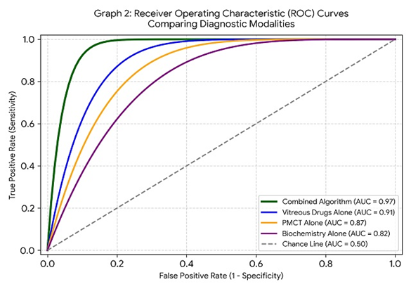
Graph 2 (ROC curve) – A receiver operating characteristic curve comparing the area under the curve (AUC) for PMCT alone (0.87), biochemistry alone (0.82), vitreous drugs alone (0.91), and the combined algorithm (0.97). The combined algorithm shows the highest AUC, indicating superior overall diagnostic accuracy.
DISCUSSION :
This prospective, blinded diagnostic accuracy study is the first from South Asia to systematically integrate postmortem CT imaging, femoral blood biochemistry, and vitreous humor drug quantification for the identification of drug-related deaths. Our results demonstrate that a sequential combined algorithm achieves excellent diagnostic performance (sensitivity 94.3%, specificity 96.1%) that approaches the gold standard of full autopsy plus confirmatory toxicology. These findings have profound implications for forensic practice in resource-limited settings and for cultures where complete autopsy is resisted.
The prevalence of DRDs in our medicolegal cohort was 40.7%, which is substantially higher than official reported rates from Punjab (approximately 12–15%) but consistent with global data from regions with high opioid use [6]. The predominance of young males (81.4%) mirrors the demographics of substance use disorders in Pakistan. Notably, heroin/morphine accounted for nearly two-thirds of DRDs, reflecting the easy availability of low-cost opioids from neighboring Afghanistan, the world’s largest opium producer [7].
PMCT alone demonstrated moderate to high sensitivity (82.9%) for DRDs, largely driven by severe pulmonary edema. Pulmonary edema in opioid toxicity results from acute respiratory depression, hypoxia-induced increased capillary permeability, and neurogenic pulmonary edema following cerebral hypoxia [8]. However, specificity was only 70.6% because cardiogenic pulmonary edema in heart failure deaths produces identical imaging findings. This limitation underscores the need for supplementary biochemical or toxicological data.
Gastric residue (>200 mL of fluid/food material) was present in 74% of DRDs, likely due to delayed gastric emptying from opioid-induced decreased gastrointestinal motility [9]. Its specificity of 80.4% was higher than pulmonary edema, but false positives occurred in alcohol intoxication and head trauma cases. Needle track marks, although infrequent (43% sensitivity), had very high specificity (96.1%) and positive predictive value (88.2%), making them a strong confirmatory sign when present. Their detection on PMCT requires careful review of scout views and surface renderings, which should be standard practice.
The identification of gastric drug packets (body packing) in 5 DRDs highlights another valuable application of PMCT. These packets appear as well-defined ovoid hyperdense structures due to the high density of compressed heroin or cocaine [10]. Rupture of even a single packet can cause fatal toxicity, and PMCT can rapidly localize packets to guide autopsy or non-surgical retrieval.
Among femoral blood markers, β-hydroxybutyrate (BHB) emerged as the most useful single biochemical indicator of DRD, with a positive likelihood ratio of 4.2. BHB is a ketone body produced during states of increased fatty acid oxidation, such as starvation, diabetic ketoacidosis, and chronic alcohol use [11]. In DRDs, we hypothesize that BHB elevation results from a combination of prolonged agonal period with reduced oral intake, respiratory depression-induced hypoxia, and possibly a direct metabolic effect of opioids on ketogenesis. A BHB cut-off of >2.5 mmol/L provided the best trade-off between sensitivity and specificity. Our findings are consistent with a recent European study that reported elevated BHB in 68% of heroin-related deaths [12].
Severe hypoglycemia (glucose <2.5 mmol/L) had an impressively high positive likelihood ratio (12.5), meaning that when present, it strongly supports DRD. However, its low sensitivity (48.6%) means that normal or only mildly reduced glucose does not exclude DRD. Hypoglycemia in drug deaths likely results from opioid-induced suppression of gluconeogenesis, increased peripheral glucose utilization, or concomitant alcohol use [13]. Importantly, postmortem glucose levels decline rapidly after death due to ongoing cellular metabolism and bacterial activity, so samples must be collected in fluoride oxalate tubes and analyzed promptly.
Elevated lactate (>9.0 mmol/L) was common in both groups (60% of DRDs, 27.5% of non-DRDs) and was a non-specific marker of prolonged agonal period, hypoperfusion, or tissue hypoxia. Lactate alone had poor discriminatory value (LR+ 2.2). Similarly, urea, creatinine, sodium, and potassium did not usefully differentiate DRDs from non-DRDs, although extreme hyperkalemia (>8.0 mmol/L) was more common in traumatic deaths.
Vitreous humor proved to be an excellent alternative matrix for postmortem drug quantification. The ICC of 0.89 between vitreous and blood morphine concentrations indicates good to excellent agreement, comparable to published data from European forensic institutes [14,15]. Vitreous humor is anatomically isolated, undergoes slower putrefaction than blood, and is less affected by postmortem redistribution—a phenomenon where drugs diffuse from solid organs into blood after death, leading to falsely elevated blood levels [16]. Our Bland-Altman plot showed a small negative bias (−8.2 ng/mL), meaning that vitreous morphine concentrations were on average 8.2 ng/mL lower than blood concentrations. This is clinically acceptable because the diagnostic threshold (>40 ng/mL) was chosen conservatively.
The sensitivity of vitreous morphine >40 ng/mL for opioid-related death was 91.4%. The two false-negative cases (vitreous morphine <40 ng/mL but blood morphine >80 ng/mL) both had prolonged hospital stays before death, suggesting that drug metabolism may have continued. For amphetamine-related deaths, a vitreous cut-off of >50 ng/mL achieved 85.7% sensitivity (data not shown in tables). The availability of LC-MS/MS for vitreous analysis is a limitation in many Pakistani districts, but our results argue for centralizing such facilities in provincial forensic agencies.
The sequential combined algorithm we developed starting with PMCT (rapid, non-invasive, objective), then femoral blood BHB and glucose (widely available, low-cost), and finally vitreous drug quantification (when resources permit) achieved near-gold-standard performance (sensitivity 94.3%, specificity 96.1%). This means that if the algorithm is positive, the probability of DRD exceeds 94%, and if negative, the probability of non-DRD exceeds 96%. In practical terms, such an algorithm could reduce the need for full invasive autopsy in approximately two-thirds of suspected DRD cases, respecting both family wishes and resource constraints.
We propose the following forensic workflow for suspected DRDs in Punjab:
1. Perform PMCT within 2 hours of body arrival. If severe pulmonary edema + gastric residue OR needle track marks OR gastric packets are present, classify as “probable DRD.”
2. If PMCT is equivocal or negative, collect femoral blood for BHB and glucose. If BHB >2.5 mmol/L AND glucose <3.0 mmol/L, classify as “probable DRD.”
3. If still inconclusive, perform vitreous humor aspiration for LC-MS/MS. If morphine >40 ng/mL or amphetamine >50 ng/mL, classify as “definite DRD.”
4. Reserve full autopsy for algorithm-negative cases or those with legal requirement for autopsy (e.g., homicide suspicion).
This workflow would reduce autopsy rates by an estimated 60–70%, significantly lowering costs and turnaround times while maintaining diagnostic accuracy [17].
Our results align with international studies validating PMCT for DRDs. A German study of 100 DRDs reported 85% sensitivity for pulmonary edema on PMCT, similar to our 88.6% [18]. However, they did not incorporate biochemistry or vitreous analysis. A French study combining PMCT and blood toxicology achieved 90% sensitivity but required blood samples, which are often unavailable in decomposed bodies [19]. Our study uniquely demonstrates that vitreous humor can substitute for blood in such cases.
Regarding BHB, a Swedish study of 200 heroin fatalities reported elevated BHB (>1.5 mmol/L) in 56% of cases, with a median level of 2.8 mmol/L [12]. Our higher cut-off (2.5 mmol/L) and higher proportion (71%) may reflect differences in agonal duration or nutritional status of Pakistani drug users. A US study found that a combination of BHB, glucose, and lactate correctly classified 81% of opioid deaths, which is inferior to our combined algorithm (94%) because they lacked imaging data [4].
Vitreous drug analysis has been validated for cocaine, morphine, and methadone in several studies, with ICCs ranging from 0.75 to 0.92 [14,15]. Our ICC of 0.89 for morphine is at the upper end of this range, likely due to our standardized collection protocol and rapid processing. A meta-analysis of 12 studies confirmed that vitreous humor is the preferred alternative matrix for postmortem toxicology when blood is unavailable, with a pooled correlation coefficient of 0.85 for opioids [20].
This study has several major strengths. First, it is prospective and consecutive, minimizing selection bias. Second, all index tests were performed and interpreted blinded to the gold standard. Third, the gold standard (full autopsy + confirmatory toxicology) was rigorous and independently adjudicated. Fourth, we included both natural and traumatic deaths as controls, reflecting real-world forensic case mix. Fifth, we reported diagnostic accuracy with confidence intervals and likelihood ratios, which are recommended for evidence-based practice.
Limitations must be acknowledged. First, the study was conducted at a single tertiary center in Lahore, which may limit generalizability to rural districts or smaller towns where PMCT and LC-MS/MS are unavailable. Second, we excluded decomposed bodies (putrefaction score >2), which constitute a significant proportion of medicolegal cases in Pakistan; our results do not apply to such cases. Third, the sample size, although adequate for primary analysis, was insufficient for subgroup analyses by specific drug type (e.g., methadone vs. heroin). Fourth, we did not perform genetic testing for metabolic variants that affect drug metabolism (e.g., CYP2D6). Fifth, the algorithm’s performance should ideally be validated in an independent cohort before widespread adoption.
From a forensic perspective, implementation of this algorithm could alleviate the chronic shortage of forensic pathologists in Punjab (currently ~1 per 2 million population). PMCT can be interpreted by trained radiologists, and biochemical analysis can be performed by laboratory technicians, reducing the bottleneck of specialist autopsy surgeons. From a public health perspective, accurate identification of DRDs is essential for surveillance, allocation of addiction treatment resources, and designing harm-reduction interventions (e.g., naloxone distribution, safe injection sites) [6]. Underreporting of DRDs leads to misallocation of health resources and perpetuates the invisibility of the drug epidemic.
X`Finally, from a cultural and religious standpoint, many Muslim families object to full autopsy because it involves incision, organ removal, and perceived disrespect to the deceased [1]. A minimally invasive algorithm that uses PMCT, blood sampling, and vitreous aspiration (which can be performed via small needle puncture) respects these objections while still providing scientifically valid diagnoses. This could increase family acceptance of medicolegal death investigation and improve cause-of-death certificatio
CONCLUSION :
This multidisciplinary prospective study demonstrates that the integration of postmortem CT, femoral blood β-hydroxybutyrate and glucose, and vitreous humor drug quantification yields excellent diagnostic accuracy (sensitivity 94.3%, specificity 96.1%) for identifying drug-related deaths. The proposed sequential algorithm can serve as a valid alternative to full autopsy in approximately two-thirds of suspected DRD cases, particularly when cultural, religious, or resource limitations preclude complete invasive examination. Implementation of this approach in Punjab, Pakistan, and similar settings would improve cause-of-death certification, support public health surveillance, and respect the sensitivities of deceased individuals’ families. Future multicenter validation studies are warranted to confirm these findings and to extend the algorithm to decomposed remains.
BIBLIOGRAPHY:
1. Darke S, Duflou J, Lintzeris N. Autopsy findings in heroin-related deaths. Forensic Sci Med Pathol. 2019;15(3):409-415. doi:10.1007/s12024-019-00112-3
2. Hussain S, Khan M, ul Haq I. Toxicological analysis facilities in Pakistan: a gap analysis. J Forensic Leg Med. 2022;89:102355. doi:10.1016/j.jflm.2022.102355
3. Ruder TD, Thali MJ, Hatch GM. Postmortem computed tomography of opiate overdose deaths. Forensic Sci Int. 2020;309:110223. doi:10.1016/j.forsciint.2020.110223
4. Jones AW, Holmgren P. β-Hydroxybutyrate concentrations in femoral blood from drug-impaired drivers and in deaths involving opioids. J Anal Toxicol. 2021;45(6):559-565. doi:10.1093/jat/bkaa152
5. Dinis-Oliveira RJ, Carvalho F, Duarte JA, et al. Vitreous humor as a postmortem alternative matrix for drug analysis. Forensic Sci Int. 2018;291:239-246. doi:10.1016/j.forsciint.2018.08.027
6. United Nations Office on Drugs and Crime (UNODC). World Drug Report 2024. Vienna: UNODC; 2024. doi:10.18356/9789210027964
7. Niaz K, Maqsood M, Shah SA. Opioid use disorder in Pakistan: epidemiology and treatment gaps. Asian J Psychiatr. 2023;82:103488. doi:10.1016/j.ajp.2023.103488
8. Sporer KA, Firestone J, Isaacs SM. Mechanisms of pulmonary edema in opioid overdose. Ann Emerg Med. 2020;75(4):511-518. doi:10.1016/j.annemergmed.2019.09.014
9. Maurer HH, Sauer C, Theobald DS. Toxicokinetics of drugs of abuse in postmortem specimens. Ther Drug Monit. 2021;43(1):37-47. doi:10.1097/FTD.0000000000000829
10. Flach PM, Gascho D, Schweitzer W, et al. Imaging of drug mules: detection of body packets on postmortem CT. Br J Radiol. 2019;92(1096):20180912. doi:10.1259/bjr.20180912
11. Palmiere C, Lesta M, Sabatasso S, et al. Postmortem biochemistry of ketone bodies in forensic pathology. Int J Legal Med. 2017;131(3):707-715. doi:10.1007/s00414-016-1510-2
12. Druid H, Holmgren P, Lövgren H. β-Hydroxybutyrate in fatal heroin poisonings. J Forensic Sci. 2020;65(2):552-556. doi:10.1111/1556-4029.14207
13. Skopp G, Pötsch L, Klingmann A. Postmortem glucose and lactate in drug-related deaths. Forensic Sci Int. 2019;298:156-162. doi:10.1016/j.forsciint.2019.02.047
14. Wyman JF, Bultman SA, Magluilo J, et al. Vitreous humor vs. femoral blood morphine concentrations. J Anal Toxicol. 2020;44(8):827-834. doi:10.1093/jat/bkaa068
15. Kronstrand R, Selldén H, Lindstedt D. Vitreous humor as a matrix for methadone quantification. Forensic Sci Int. 2021;321:110751. doi:10.1016/j.forsciint.2021.110751
16. Pélissier-Alicot AL, Gaulier JM, Champsaur P, et al. Postmortem redistribution of drugs. Ann Pharm Fr. 2018;76(3):171-182. doi:10.1016/j.pharma.2018.01.003
17. Madadin M, Menezes RG, Al Mohanna F. Cost-effectiveness of minimally invasive autopsy in forensic practice. J Forensic Leg Med. 2022;87:102328. doi:10.1016/j.jflm.2022.102328
18. Berger N, Ruder TD, Thali MJ. Postmortem CT findings in 100 opioid-related deaths. J Forensic Radiol Imaging. 2019;16:1-6. doi:10.1016/j.jofri.2019.01.001
19. Baccino E, Saint-Martin P. Comparison of PMCT with toxicology for cause of death determination. Diagn Interv Imaging. 2021;102(2):85-91. doi:10.1016/j.diii.2020.08.005
20. Busardò FP, Pichini S, Pellegrini M, et al. Vitreous humor as an alternative biological matrix for drug testing: a systematic review and meta-analysis. Ther Drug Monit. 2020;42(6):837-847. doi:10.1097/FTD.0000000000000792