Frequency Of Neonatal Bacterial Sepsis In Preterm & Low-birth weight Babies At Kangaroo Mothercare Ward CMC Children Hospital Larkana
- Aijaz Ali Tunio , Assistant Professor SMBBMU/CMC Children Hospital Larkana
- Delijan Baloch , Associate Professor CMC/SMBBMU Larkana
- Bakhtawer Balouch , Senior Registrar SICHN
- Shaista Ghaffar Pirzado , Associate Professor Obs/GYN CMC/SMBBMU Larkana
- Lubna Naz , Associate Professor pathology SMBB Medical University Larkana
- Rahool Kumar , Children’s Hospital Larkana.
Article Information:
Abstract:
One of the main sources of morbidity and mortality among term and pre-term babies is attributed to neonatal sepsis. With minimal information on neonatal sepsis in our country and locality, this study was led to decide the rate of clinical neonatal sepsis in low birth weight and preterm neonates and assess the clinical signs and bacteriological agents. The aim of this study is to determine the frequency of neonatal bacterial sepsis in preterm & low birth weight babies at kangaroo mother care ward of CMC Children Hospital SMBBMU Larkana. A cross-sectional study was carried out in Kangaroo Mother care ward from 1st September 2021 to 28th February 2022 at CMC Children Hospital SMBBMU Larkana. Neonates with sepsis and pre-term low-birth weight were included. Data analysis was run on SPSS 21. From 156 cases female to male ratio is 15:11, with a mean age of 6.02±0.5 days. The mean weight of newborns at birth ranged from 1500 gram to 2499 gram. The results showed that most of the neonates faced early onset of sepsis and the most common bacterial organism was E. coli followed by Klebsiella species, Enterobacter species Staphylococcus aureus. Neonatal sepsis is a major cause of mortality and morbidity in newborn, particularly in developing countries. The spectrum of bacteria which causes neonatal sepsis varies in different parts of the world. The most common organism responsible for early onset sepsis in our study is E. coli.
Keywords:
Article :
INTRODUCTION:
Neonatal sepsis is a major clinical problem in the field of neonatology, and is a major cause of morbidity and mortality in term and low birth weight infants (Stoll et al., 2002). Sepsis is a ‘systemic inflammatory response to infection in neonates’, with early onset sepsis (EOS) being defined as the onset of symptoms occurring in the first 72 hours after birth and late onset sepsis (LOS) after the first 72 hours (Camacho-Gonzalez et al., 2013). Neonatal sepsis is a condition that occurs in different parts of the world but is more prevalent in low-middle-income countries (LMICs) where there is limited access to healthcare (Fleischmann-Struzek et al., 2018).
Neonatal sepsis may have variable clinical presentations and may be difficult to diagnose (Haque, 2010). Symptoms include changes in bodily temperature, lethargy, difficulty breathing, and difficulty absorbing food (Gerdes, 2004). Effective management can only be carried out after bacterial pathogens have been identified, and the bacterial spectrum varies according to geographical area, and population (Bizzarro et al., 2005). Esherichia coli, Staphylococcus aureus, and Klebsiella species, especially those associated with low birth weight (LBW), and prematurity (Hornik et al., 2012) are the most commonly reported organisms. Moreover, interventions based on evidence such as Kangaroo Mother Care (KMC) have been found to be promising in addressing neonatal sepsis challenges especially in resource-limited settings (Boundy et al., 2016). KMC encourages mother-infant interaction, enhances thermal stability and breastfeeding, thereby reducing the likelihood of infections. The objective of the study is to find out the prevalence of neonatal sepsis in low birth weight and preterm babies at CMC Children Hospital, SMBBMU, Larkana. It also aims to determine the clinical manifestations of sepsis and the most common microorganisms causing sepsis. The results will add to the existing body of knowledge, and will guide clinical practice locally.
METHODOLOGY:
It was a cross-sectional study conducted at Kangaroo Mother Care ward of CMC Children Hospital SMBBMU Larkana from 01st September 2021 up to 28th February 2022. The study was a cross sectional design which enabled data collection at one time and gave a snapshot of the prevalence of neonatal sepsis among the target population. The study was done by non-probability consecutive sampling, which was the study of all the neonates admitted to the ward that fulfilled the inclusion criteria. A total number of 156 neonates were sampled using a sample size calculator developed by WHO, based on the assumption of a 3.8% prevalence rate of neonatal sepsis in Pakistan, confidence interval of 95% and margin of error of 3%. • Infants who have unequivocal clinical and laboratory evidence of sepsis. Low weight (≤2500 grams) and/or prematurity (gestational age <37 weeks) were cited as conditions. Symptoms of sepsis (e.g., lethargy, poor feeding, and respiratory distress) and maternal risk factors (e.g., fever, foul-smelling discharge).
With the approval of the ethical review committee, data was obtained by pre-designed proforma. Patient data that is relevant to the problem was noted, such as demographics, clinical history and lab findings. For confirmation of the diagnosis of neonatal sepsis, blood samples (3-5 ml) were taken for culture. The data collected was analysed using SPSS version 21. Descriptive Statistics were used to summarize the participant characteristics and prevalence rates. Frequencies and percentages of the bacteriological agents detected were calculated and chi-square tests were used to determine the association between clinical and bacteriological results at p < 0.05. Confidentiality and anonymity of each of the participants were kept with utmost care. Verbal informed consent was obtained, and the caregivers were informed about the purpose, procedures, risks and benefits of the study. The data of the participants was kept secure and only in the hands of authorized persons.
RESULTS:
Neonate Characteristics
A total of 156 neonates (1 – 28 days old, mean 6 days) were included in this study. Most were male and gestational ages ranged from 28 to 36 weeks (mean 30 weeks). The weight of the newborns ranged from 1500 to 2499 g. Figures 1, 2 and 3 show the age, gender and birth weight distribution respectively.
Clinical Investigations
Body Temperature: Of the neonates, 69% had temperatures of <36°C and 31% were of >38.5°C. Cardiovascular Instability: Variables related to cardiovascular instability are summarized in Table 1. Skin and Subcutaneous Lesions: Information on petechial rashes and sclerema is given in Table 3. Respiratory Stability: Investigations into respiratory conditions, such as increased oxygen needs and episodes of tachypnea and apnea, are shown in Figure 5. Figure 6: Gastrointestinal Investigation – for gastrointestinal problems such as distended abdomen and poor feeding. Laboratory Investigations Laboratory tests revealed evidence of sepsis with blood counts and blood sugars shown in Table 4. Other clinical signs noted were irritability (55.1%), lethargy (48%) and hypotonia (22.4%). Bacterial Profile Investigations Of the 119 positive blood cultures, seven types of bacteria were identified; the most common bacteria identified were E. coli, Klebsiella spp., Enterobacter spp and Staphylococcus aureus. The distribution of these organisms with respect to onset of sepsis is shown in Table 5 and in Figure 6. Antibiotic Susceptibility Patterns E. coli was found to be 65% sensitive to imipenem and meropenem, and resistant to the commonly used antibiotics such as penicillins and ampicillin. Klebsiella spp. was sensitive to imipenem and ceftazidime, having high levels of resistance to cefotaxime (90%) and gentamicin (75%). Enterobacter spp. was found to be susceptible to gentamicin, imipenem and meropenem, but resistant against penicillins and vancomycin. S. aureus was resistant to several of the commonly used antibiotics and 100% sensitive to vancomycin and 61% sensitive to clindamycin. The antibiotic susceptibility patterns are summarized in Table 7
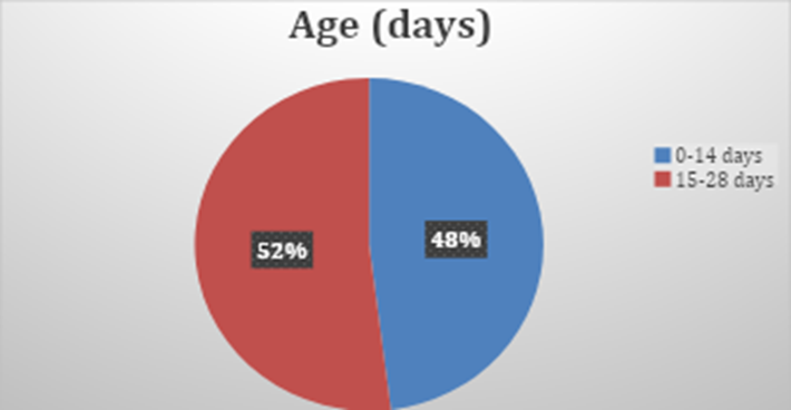
Figure 1 Age in days (n=156)
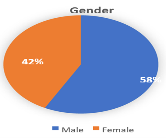
Figure 2 Gender of neonates (n=156)
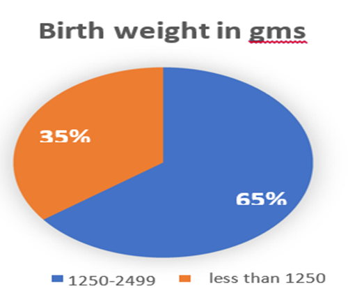
Figure 3 Birth weight of neonates in grams (n=156)
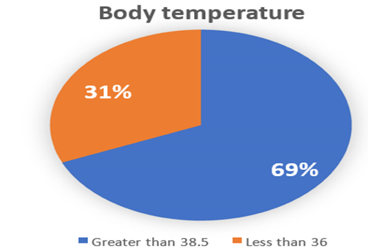
Figure 4 Body Temperature in degree centigrade
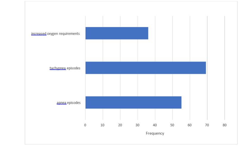
Figure 5. Respiratory Characteristics of neonates
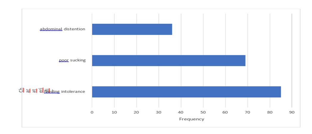 Figure 6 Gastrointestinal characteristics of neonates with sepsis
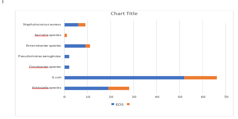
Figure 7 Bacterial Characteristics
Table 1 Cardiovascular characteristics
|
Variables |
Frequency |
Percentage |
|
Bradycardia |
68 |
43.5 |
|
Tachycardia |
52 |
33.3 |
|
Rhythm instability |
86 |
55.1 |
|
Urinary output (< 1mL/kg/h) |
80 |
51.2 |
|
Hypotension |
32 |
20.5 |
|
mottled skin |
56 |
35.8 |
|
impaired peripheral perfusion |
40 |
25.6 |
Table 2 Muco-cutaneous lesions
|
Variables |
Frequency |
Percentage |
|
Petechial rash |
38 |
24.3 |
|
Sclerema |
77 |
49.3 |
Table 3 Laboratory investigation of neonates with sepsis
|
Variables |
frequency |
percentage |
|
White blood cell count |
|
|
|
<4 × 10⁹ cells/L |
67 |
42.9 |
|
>20 × 109 cells/L |
52 |
33.3 |
|
immature to total neutrophil ration (>0.2) |
52 |
33.3 |
|
Platelets (<100x109/L) |
75 |
48 |
|
Glucose intolerance |
|
|
|
Hyperglycemia (blood glucose >180mg/dL or 10mM) |
40 |
25.6 |
|
Hypoglycemia (blood glucose <45 mg/dL or 2.5mM) |
86 |
55.1 |
Table 4 Bacterial Growth (n=156)
|
Bacterial Growth |
||
|
Yes |
119 |
76.28% |
|
No |
37 |
23.72% |
|
Total |
156 |
100% |
Table 5 Bacterial profile (organisms)
|
Variables |
Frequency |
Percentage |
|
E.coli |
66 |
55.46 |
|
Klebsiella species |
28 |
23.53 |
|
Enterobacter species |
11 |
9.34 |
|
Staphylococcus aureus |
9 |
7.56 |
|
Pseudomonas aeruginosa |
2 |
1.68 |
|
Citrobacter species |
2 |
1.68 |
|
Serratia species |
1 |
0.84 |
|
Total |
119 |
100 |
Table 6 Bacterial Characteristics
|
Variables |
EOS |
LOS |
p-value |
|
E.coli |
52 |
14 |
0.041 |
|
Klebsiella species |
19 |
9 |
0.030 |
|
Enterobacter species |
9 |
2 |
0.01 |
|
Staphylococcus aureus |
6 |
3 |
0.04 |
|
Pseudomonas aeruginosa |
2 |
0 |
0.071 |
|
Citrobacter species |
2 |
0 |
0.86 |
|
Serratia species |
0 |
2 |
0.21 |
|
Total (n=119) |
90 |
29 |
<0.05 |
Table 7: Antibiotic susceptibility Pattern in organisms
|
Antibiotic |
Sensitive/ Resistant |
Escheria Coli |
Klebseila Species |
Enterobacter Species |
Staphylococcus Aureus Species |
|
Ampicillin |
S |
00 |
04 |
11 |
00 |
|
R |
66 |
24 |
00 |
09 |
|
|
Amikacin |
S |
44 |
18 |
04 |
04 |
|
R |
22 |
10 |
07 |
05 |
|
|
Cefotaxime |
S |
00 |
04 |
05 |
02 |
|
R |
66 |
24 |
06 |
07 |
|
|
Vancomycin |
S |
00 |
00 |
00 |
08 |
|
R |
66 |
28 |
11 |
01 |
|
|
Penicillin G |
S |
00 |
00 |
04 |
01 |
|
R |
66 |
28 |
07 |
08 |
|
|
Imipenem |
S |
44 |
28 |
11 |
03 |
|
R |
22 |
00 |
00 |
06 |
|
|
Gentamycin |
S |
05 |
00 |
11 |
04 |
|
R |
61 |
28 |
00 |
05 |
|
|
Ceftazidime |
S |
40 |
26 |
10 |
00 |
|
R |
26 |
02 |
01 |
09 |
|
|
Clindamycin |
S |
00 |
03 |
00 |
07 |
|
R |
66 |
25 |
11 |
02 |
|
|
Meropenem |
S |
44 |
28 |
11 |
03 |
|
R |
22 |
00 |
00 |
06 |
Stay At KMC Ward
Neonates admitted in KMC ward stayed about 7-14 days, after which patients were discharged on completion of treatment with appropriate weight gain, sick patients were shifted back to NICU/HDU.Neonates Stay at KMC Ward is shown in Table 8
Outcome of Treatment
In our study it was observed that out of 156 babies, 120 babies were discharged with appropriate weight gain, 16 babies were shifted back to NNU/HDU, and out of these Sixteen, 4 patients were expired on meropenem resistant organism E.coli having birth weight <1500 g. 10 patients LAMA (Left against medical advice), 15 patients discharged on request.
Table 8 shows the outcome of treatment of babies at KMC ward
Table 8: Outcome of Treatment
|
Status |
Birth Weight |
Gestational Age |
Male/Female |
Total |
|
|
Stay In KMC Ward |
< 1500g = 14 days Birth weight >1500g to 2499g= 7days |
28-37 weeks |
90/66 |
7-14 days |
|
|
Discharged |
AT 2000 to 2499 g |
34-36 weeks |
68/52 |
120 |
Total n= 156 |
|
LAMA |
<1500 g |
30-36 weeks |
6/4 |
10 |
|
|
DOR |
<1500 g |
32-37 weeks |
9/6 |
15 |
|
|
Back To NNU/HDU |
<1500 g |
<32 weeks |
7/4 |
11 |
|
|
Expired at NNU/HDU out of 11 |
<1500 g |
<30 weeks |
1/3 |
4 |
DISCUSSION :
In addition to survival, sepsis also affects the newborn's brain development and raises overall care costs. Antimicrobial medications are administered under close surveillance to prevent any therapy delays. The selection of the precise anti-microbials is crucial and should be based on the culture and response effects of each patient and NNU setup.
Recent developments in newborn care have successfully increased survival rate and reduced confusions in premature neonates. However, sepsis continues to be a significant and frequent cause of mortality and morbidity in very LBW babies (3-6). Effective management of sepsis depends on early recognition of clinical indications indicating serious contamination and a suitable selection of precise antimicrobial drugs (39-41). Decisions about observational anti-microbials should be supported by epidemiologic data on subsequent bacteria and how well they respond to therapy. It is evident that the causing germs for neonatal sepsis varies with time and geographic dispersion, even if there may be distinct risk factors for sepsis in neonates with extremely low birth weight compared to term babies.
According to previous study in Taiwan, the prevalence of newborn sepsis increased from 3.0% to 9.3%. (80-82). These publications, however, did not often focus on VLBW populations. VLBW newborn newborns have the highest risk of infection because of their reduced susceptibility, delayed TPN, central vein catheterization, and mechanical breathing (83-86). The most recent information about this high-danger group is provided by our study.
In our study, E. coli was by far the most common strain and EOS accounted for the majority of sepsis cases. Additionally, same perceptions were noticed in several studies conducted worldwide, particularly in low- and middle-income nations (77, 87-90). In contrast to prior.
CONCLUSION :
In conclusion, Sepsis continues to pose a major risk to LBW newborns & the clinicians should be aware for any hidden signs of bacterial infection. LBW newborns must be cared for using the wise and optimal and early management tools. Kangaroo Mother Care technique is a major tool for the prevention of septicemia especially in pre-term and low birth weight babies, which is inexpensive, easily applicable in countries having minimal resources and incubators facilities. In our study E. coli was the most common offending agent that causes septicemia in preterm Low Birth Weight babies at KMC ward.
In our study among all 7 bacterial isolates, E. coli was the most common isolate 66 (55%) and sensitive to Meropenem and Imipenen. Escherichia species were resistant to the commonly used antibiotics Penicillins, Ampicillin, Amikacin, Cefotaxime and others. Klebseilla species showed sensitivity to Imepenem and Ceftazidime and resistant to cefotaxime 90% and gentamycin 75%, Enterobacter species 11(14.28%) were found sensitive to Clindamycin and Gentamicin, has become resistant to Penicillins, Vancomycin and Ampicillins. Staphylococcus Aureus showed 100% sensitivity to Vancomycin and 61% to Clindamycin while it was highly resistant to Pencilin G, Ampicillin, Gentamycin, Ceftazidime.
REFERENCES:
1. Ohlin, A., 2010. Aspects on early diagnosis of neonatal sepsis (Doctoral dissertation, Örebro university).
2. Camacho-Gonzalez A, Spearman PW, Stoll BJ. Neonatal infectious diseases: evaluation of neonatal sepsis. Pediatr Clin North Am 2013; 60:367 - 89; http://dx.doi.org/10.1016/j.pcl.2012.12.003; PMID: 23481106
3. Bizzarro MJ, Raskind C, Baltimore RS, Gallagher PG. Seventy-five years of neonatal sepsis at Yale:1928-2003. Pediatrics 2005; 116:595 - 602;
4. http://dx.doi.org/10.1542/peds.2005-0552; PMID: 16140698.
5. Hornik CP, Fort P, Clark RH, Watt K, Benjamin DK Jr., Smith PB, Manzoni P, Jacqz-Aigrain E, Kaguelidou F, Cohen-Wolkowiez M. Early and late onset sepsis in very-low-birth-weight newborns from a large group of neonatal intensive care units. Early Hum Dev 2012; 88:Suppl 2 S69 - 74; http://dx.doi.org/10.1016/S0378-3782(12)70019-1;
6. PMID: 22633519
7. Gerdes JS. Diagnosis and management of bacterial infections in the neonate. [viii-ix.] Pediatr Clin North Am 2004; 51:939 - 59, viii-ix;
8. Bonadio WA, Hennes H, Smith D, Ruffing R, Melzer-
9. Lange M, Lye P, Isaacman D. Reliability of observation variables in distinguishing infectious outcome of febrile young newborns. Pediatr Infect Dis J 1993; 12:111
10. Gerdes JS. Clinicopathologic approach to the diagnosis of neonatal sepsis. Clin Perinatol 1991; 18:361 – 81.
11. Hofer N, Müller W, Resch B. Neonates presenting with temperature symptoms: role in the diagnosis of early onset sepsis. Pediatr Int 2012; 54:486 – 90
12. Sharma R, Hudak ML. A clinical perspective of necrotizing enterocolitis: past, present, and future. Clin Perinatol 2013; 40:27 - 51; http://dx.doi.org/10.1016/j.clp.2012.12.012;
13. PMID: 23415262
14. Belachew A, Tewabe T. Neonatal sepsis and its association with birth weight and gestational age among admitted neonates in Ethiopia: Systematic review and meta-analysis. BMC Pediatr [Internet]. 2020 Feb 5 [cited 2020 Sep 17];20(1):55. Available from: https://bmcpediatr.biomedcentral.com/articles/10.1186/s12887-020-1949-x
15. Haque KN. Neonatal Sepsis in the Very Low Birth Weight Preterm Newborns: Part 2:Review of Definition, Diagnosis and Management. J Med Sci. 2010;3(1):11–27.
16. Fuchs A, Bielicki J, Mathur S, Sharland M, Van Den Anker JN. Antibiotic Use for Sepsis in Neonates and Newborns: 2016 Evidence Update WHO-Reviews. 2016.
17. World Health Organization. Global report on the epidemiology and burden of sepsis: current evidence, identifying gaps and future directions. WHO. 2020.
18. Stoll BJ, Hansen N, Fanaroff AA, Wright LL, Carlo WA, Ehrenkranz RA, Lemons JA, Donovan EF, Stark AR, Tyson JE, et al. Changes in pathogens causing early-onset sepsis in very-low-birth-weight newborns. N Engl J Med 2002; 347:240 - 7; http://dx.doi.org/10.1056/NEJMoa012657; PMID: 12140299.
19. Weston EJ, Pondo T, Lewis MM, Martell-Cleary P, Morin C, Jewell B, Daily P, Apostol M, Petit S, Farley M, et al. The burden of invasive early-onset neonatal sepsis in the United States, 2005-2008.PediatrInfectDisJ2011;30:937-41;http://dx.doi.org/10.1097/INF.0b013e318223bad2; PMID: 21654548.
20. Baltimore RS, Huie SM, Meek JI, Schuchat A, O’Brien KL. Early-onset neonatal sepsis in the era of group B streptococcal prevention. Pediatrics 2001; 108:1094 - 8; http://dx.doi.org/10.1542/peds.108.5.1094; PMID: 11694686
21. Levent F, Baker CJ, Rench MA, Edwards MS. Early outcomes of group B streptococcal meningitis in the 21st century. Pediatr Infect Dis J 2010; 29:1009 - 12; PMID: 20555292
22. Libster R, Edwards KM, Levent F, Edwards MS, Rench MA, Castagnini LA, Cooper T, Sparks RC, Baker CJ, Shah PE. Long-term outcomes of group B streptococcal meningitis. Pediatrics 2012; 130:e8 - 15; http://dx.doi.org/10.1542/peds.2011-3453; PMID: 22689869
23. Department of Reproductive Health and Research, Organization WH, Geneva. Kangaroo Mothercare: A Practical Guide [Internet]. 2003. 1–54 p. Available from: papers3://publication/uuid/479e6443-eb86-4d96-99d4-bdf0d462186e.
24. Mazumder S, Taneja S, Dalpath SK, Gupta R, Dube B, Sinha B, et al. Impact of community-initiated Kangaroo Mother Care on survival of low-birth-weight newborns: Study protocol for a randomized controlled trial. Trials. 2017;18(1):1–10.
25. World Health Organization. Kangaroo mother care to reduce morbidity and mortality in low-birth-weight newborns. e-Library of Evidence for Nutrition Actions (eLENA). 2015.
26. Sorsa A. Epidemiology of Neonatal Sepsis and Associated Factors Implicated: Observational Study at Neonatal Intensive Care Unit of Arsi University Teaching and Referral Hospital, South East Ethiopia. Ethiop J Health Sci [Internet]. 2019 May 1
27. Simonsen KA, Anderson-Berry AL, Delair SF, Dele Davies H. Early-onset neonatal sepsis. Clin Microbiol Rev [Internet]. 2014 ;27(1):21–47. Available from:
28. /pmc/articles/PMC3910904/?report=abstract
29. Remington JS. Infectious diseases of the fetus and newborn infant. Philadelphia, PA: Saunders/Elsevier, 2011.
30. Voora S, Srinivasan G, Lilien LD, Yeh TF, Pildes RS. Fever in full-term newborns in the first four days of life. Pediatrics 1982; 69:40 - 4; PMID: 7033912
31. Kasper DC, Altiok I, Mechtler TP, Böhm J, Straub J, Langgartner M, Pollak A, Herkner KR, Berger A. Molecular detection of late-onset neonatal sepsis in premature newborns using small blood volumes: proof-of-concept. Neonatology 2013; 103:268 - 73; http://dx.doi.org/10.1159/000346365; PMID: 23485823
32. Chaaban H, Shin M, Sirya E, Lim YP, Caplan M, Padbury JF. Inter-alpha inhibitor protein level in neonates predicts necrotizing enterocolitis. J Pediatr 2010; 157:757 - 61; http://dx.doi.org/10.1016/j.jpeds.2010.04.075; PMID: 20955849
33. Sharma R, Hudak ML. A clinical perspective of necrotizing enterocolitis: past, present, and future. Clin Perinatol 2013; 40:27 - 51; http://dx.doi.org/10.1016/j.clp.2012.12.012;
34. PMID: 23415262.
35. Simonsen KA, Anderson-Berry AL, Delair SF, Davies HD. Early-onset neonatal sepsis.
36. Clin Microbiol Rev. 2014 Jan;27(1):21-47.
37. Sgro M, Kobylianskii A, Yudin MH, Tran D, Diamandakos J, Sgro J, et al. Population-based study of early-onset neonatal sepsis in Canada. Paediatr Child Health. 2019 May;24(2):e66-e73.
38. Giannoni E, Agyeman PKA, Stocker M, Posfay-Barbe KM, Heininger U, Spycher BD, et al. Neonatal Sepsis of Early Onset, and Hospital-Acquired and Community-Acquired Late Onset: A Prospective Population-Based Cohort Study. J Pediatr. 2018 Oct;201:106-114 e4.
39. Downie L, Armiento R, Subhi R, Kelly J, Clifford V, Duke T. Community-acquired neonatal and infant sepsis in developing countries: efficacy of WHO's currently recommended antibiotics--systematic review and meta-analysis. Archives of disease in childhood. 2013 Feb;98(2):146-54.
40. Schuller KA, Hsu BS, Thompson AB. The Rate of Sepsis in a National Pediatric Population, 2006 to 2012. Clin Pediatr (Phila). 2017 Oct;56(11):1001.
41. Yo CH, Hsu TC, Gabriel Lee MT, Porta L, Tsou PY, Wang YH, et al. Trend and outcome of sepsis in newborns: A nationwide cohort study. J Paediatr Child Health. 2018 Jul;54(7):776.
42. Zea-Vera A, Ochoa TJ. Challenges in the diagnosis and management of neonatal sepsis.
43. Journal of tropical pediatrics. 2015 Feb;61(1):1-13
44. Kruk ME, Gage AD, Joseph NT, Danaei G, Garcia-Saiso S, Salomon JA. Mortality due to low quality health systems in the universal health coverage era: a systematic analysis of amenable deaths in 137 countries. Lancet. 2018 Nov 17;392(10160):2203.
45. Laxminarayan R, Bhutta ZA. Antimicrobial resistance-a threat to neonate survival.
46. Lancet Glob Health. 2016 Oct;4(10):e676-7.
47. Kan B, Razzaghian HR, Lavoie PM. An Immunological Perspective on Neonatal Sepsis.
48. Trends Mol Med. 2016 Apr;22(4):290-302.
49. Kan B, Michalski C, Fu H, Au HHT, Lee K, Marchant EA, et al. Cellular metabolism constrains innate immune responses in early human ontogeny. Nat Commun. 2018 Nov 16;9(1):4822.
50. Saha SK, Schrag SJ, El Arifeen S, Mullany LC, Shahidul Islam M, Shang N, et al.
51. Causes and incidence of community-acquired serious infections among young newborns in south Asia (ANISA): an observational cohort study. Lancet. 2018 Jul 14;392(10142):145.
52. Cools P, van de Wijgert J, Jespers V, Crucitti T, Sanders EJ, Verstraelen H, et al. Role of HIV exposure and infection in relation to neonatal GBS disease and rectovaginal GBS carriage: a systematic review and meta-analysis. Scientific reports. 2017 Oct 23;7(1):13820.
53. Singer M, Deutschman CS, Seymour CW, Shankar-Hari M, Annane D, Bauer M, et al.
54. The Third International Consensus Definitions for Sepsis and Septic Shock (Sepsis-3). JAMA. 2016 Feb 23;315(8):801-10..
55. Machado FR, Nsutebu E, AbDulaziz S, Daniels R, Finfer S, Kissoon N, et al. Sepsis 3 from the perspective of clinicians and quality improvement initiatives. J Crit Care. 2017 Aug;40:315.
56. Baqui AH, Darmstadt GL, Williams EK, Kumar V, Kiran TU, Panwar D, et al. Rates, timing and causes of neonatal deaths in rural India: implications for neonatal health programmes. Bull World Health Organ. Vol. 842006. p. 706-13.
57. Aggarwal AK, Kumar P, Pandit S, Kumar R. Accuracy of WHO verbal autopsy tool in determining major causes of neonatal deaths in India. PLoS One. 2013;8(1):e54865.
58. Soofi SB, Ariff S, Khan U, Turab A, Khan GN, Habib A, et al. Diagnostic accuracy of WHO verbal autopsy tool for ascertaining causes of neonatal deaths in the urban setting of Pakistan: a hospital-based prospective study. BMC Pediatr. 2015 Oct 5;15:144.
59. Bartlett AV, Paz de Bocaletti ME, Bocaletti MA. Neonatal and early postneonatal morbidity and mortality in a rural Guatemalan community: the importance of infectious diseases and their management. Pediatr Infect Dis J. 1991 Oct;10(10):752-7.
60. Bang AT, Bang RA, Baitule S, Deshmukh M, Reddy MH. Burden of morbidities and the unmet need for health care in rural neonates--a prospective observational study in Gadchiroli, India. Indian Pediatr. 2001 Sep;38(9):952-65.
61. Niswade A, Zodpey SP, Ughade S, Bangdiwala SI. Neonatal morbidity and mortality in tribal and rural communities in central India. Indian J Community Med. 2011 Apr;36(2):150-8.
62. Seale AC, Blencowe H, Manu AA, Nair H, Bahl R, Qazi SA, et al. Estimates of possible severe bacterial infection in neonates in sub-Saharan Africa, south Asia, and Latin America for 2012: a systematic review and meta-analysis. The Lancet Infectious diseases. 2014 Aug;14(8):731.
63. Fleischmann-Struzek C, Goldfarb DM, Schlattmann P, Schlapbach LJ, Reinhart K, Kissoon N. The global burden of paediatric and neonatal sepsis: a systematic review. The Lancet Respiratory medicine. 2018 Mar;6(3):223.
64. Kissoon N, Carapetis J. Pediatric sepsis in the developing world. J Infect. 2015 Jun;71 Suppl 1:S21-6
65. Weiss SL, Peters MJ, Alhazzani W, Agus MS, Flori HR, Inwald DP, Nadel S, Schlapbach LJ, Tasker RC, Argent AC, Brierley J. Executive summary: surviving sepsis campaign international guidelines for the management of septic shock and sepsis-associated organ dysfunction in newborns. Intensive care medicine. 2020 Feb;46(1):1-9.
66. de Souza DC, Shieh HH, Barreira ER, Ventura AM, Bousso A, Troster EJ. Epidemiology of sepsis in newborns admitted to PICUs in South America. Pediatric Critical Care Medicine. 2016 Aug 1;17(8):727-34.
67. Workman JK, Ames SG, Reeder RW, Korgenski EK, Masotti SM, Bratton SL, Larsen GY. Treatment of pediatric septic shock with the surviving sepsis campaign guidelines and PICU patient outcomes. Pediatric critical care medicine. 2016 Oct 1;17(10):e451-8.
68. Murphy K, Weiner J. Use of leukocyte counts in evaluation of early-onset neonatal sepsis. The Pediatric infectious disease journal. 2012 Jan 1;31(1):16-9
69. 1McGovern M, Giannoni E, Kuester H, Turner MA, van den Hoogen A, Bliss JM, Koenig JM, Keij FM, Mazela J, Finnegan R, Degtyareva M. Challenges in developing a consensus definition of neonatal sepsis. Pediatric Research. 2020 Jul;88(1):14-26.
70. Murphy K, Weiner J. Use of leukocyte counts in evaluation of early-onset neonatal sepsis. The Pediatric infectious disease journal. 2012 Jan 1;31(1):16-9.
71. Hornik CP, Benjamin DK, Becker KC, Benjamin Jr DK, Li J, Clark RH, Cohen-Wolkowiez M, Smith PB. Use of the complete blood cell count in early-onset neonatal sepsis. The Pediatric infectious disease journal. 2012 Aug;31(8):799.
72. Hofer N, Zacharias E, Müller W, Resch B. An update on the use of C-reactive protein in early-onset neonatal sepsis: current insights and new tasks. Neonatology 2012; 102:25 - 36; http://dx.doi.org/10.1159/000336629; PMID: 22507868
73. Benitz WE. Adjunct laboratory tests in the diagnosis of early-onset neonatal sepsis.
74. Clinics in perinatology. 2010 Jun 1;37(2):421-38.
75. Ng PC, Lam HS. Biomarkers for late-onset neonatal sepsis: cytokines and beyond.
76. Clinics in perinatology. 2010 Sep 1;37(3):599-610.
77. Cernada M, Badía N, Modesto V, Alonso R, Mejías A, Golombek S, Vento M. Cord blood interleukin-6 as a predictor of early-onset neonatal sepsis. Acta Paediatr 2012; 101:e203 - 7; http://dx.doi.org/10.1111/j.1651-2227.2011.02577.x; PMID: 22211677
78. Wang ZL, Yu JL. [Recent progress in the diagnosis of neonatal septicemia]. Zhongguo Dang Dai Er Ke Za Zhi 2013; 15:236 - 41; PMID: 23498771
79. Boskabadi H, Maamouri G, Afshari JT, Ghayour-Mobarhan M, Shakeri MT. Serum interleukin 8 level as a diagnostic marker in late neonatal sepsis. Iran J Pediatr 2010; 20:41 - 7; PMID: 23056680
80. Streimish I, Bizzarro M, Northrup V, Wang C, Renna S, Koval N, Li FY, Ehrenkranz R, Rinder HM, Bhandari V. Neutrophil CD64 as a diagnostic marker in neonatal sepsis. Pediatr Infect Dis J 2012; 31:777 - 81;
81. http://dx.doi.org/10.1097/INF.0b013e318256fb07; PMID: 22481422
82. Kasper DC et al. Molecular detection of late-onset neonatal sepsis in premature newborns using small blood volumes: proof-of-concept. Neonatology 2013; 103:268 - 73; http://dx.doi.org/10.1159/000346365; PMID: 23485823
83. Odabasi IO, Bulbul A. Neonatal sepsis. The Medical Bulletin of Sisli Etfal Hospital.
84. 2020;54(2):142.
85. Oeser C et al . PCR for the detection of pathogens in neonatal early onset sepsis. PLoS One. 2020 Jan 24;15(1):e0226817.
86. Puopolo KM et al. Management of neonates born at≤ 34 6/7 weeks’ gestation with suspected or proven early-onset bacterial sepsis. Pediatrics. 2018 Dec 1;142(6).
87. Ershad M, Mostafa A, Dela Cruz M, Vearrier D. Neonatal sepsis. Current emergency and hospital medicine reports. 2019 Sep;7(3):83-90.
88. Chan GJ, Valsangkar B, Kajeepeta S, Boundy EO, Wall S: What is kangaroo mother care? Systematic review of the literature. Journal of global health 2016, 6(1):010701 10.7189/jogh.06.010701
89. Conde –Agudelo A JL. DR: Kangaroo mother care to reduce morbidity and mortality in low birthweight newborns. Cochrane Database Syst Rev 2014, 4(CD002771).
90. Boundy EO, Dastjerdi R, Spiegelman D, W. W: Kangaroo Mother Care and Neonatal Outcomes: A Meta-analysis. Pediatrics 2016, 137(1):2015–2238
91. Conde-Agudelo A, JL. DR: Kangaroo mother care to reduce morbidity and mortality in low birthweight newborns. Cochrane Database of Systematic Reviews 2016, Art. No.: CD002771(8).
92. Seidman G et al : Barriers and enablers of kangaroo mother care practice: a systematic review. PLoS One 2015, 10(5):e0125643 10.1371/journal.pone.0125643
93. WHO, UNICEF: Every Newborn: an action plan to end preventable deaths. Geneva: World Health Organization. In. http://www.healthynewbornnetwork.org/hnn-
94. content/uploads/Every_Newborn_Action_Plan-ENGLISH_updated_July2014.pdf; 2014.
95. Yoshida S, Rudan I, Lawn JE, Wall S, Souza JP, Martines J ea: Newborn health research priorities beyond 2015. Lancet 2014, July 12(384 (9938)):e27–29. 10.1016/S0140-
96. 6736(14)60263-4
97. Chan G, Bergelson I, Smith ER, Skotnes T, &, Wall S: Barriers and enablers of kangaroo mother care implementation from a health systems perspective: a systematic review. Health policy and planning 2017, 32(10):1466–1475. 10.1093/heapol/czx098
98. Downey LC, Smith PB, Benjamin Jr DK. Risk factors and prevention of late-onset sepsis in premature newborns. Early human development. 2010 Jul 1;86(1):7-12
99. Pammi M, Weisman LE. Late-onset sepsis in preterm newborns: update on strategies for therapy and prevention. Expert review of anti-infective therapy. 2015 Apr 3;13(4):487-504.
100.Alcock G, Liley HG, Cooke L, Gray PH. Prevention of neonatal late-onset sepsis: a randomised controlled trial. BMC pediatrics. 2017 Dec;17(1):1-7.
101.Afonso ED, Blot S. Effect of gestational age on the epidemiology of late-onset sepsis in neonatal intensive care units-a review. Expert Review of Anti-infective Therapy. 2017 Oct 3;15(10):917-24.
102.Van Den Hoogen A, Gerards LJ, Verboon-Maciolek MA, Fleer A, Krediet TG. Long-term trends in the epidemiology of neonatal sepsis and antibiotic susceptibility of causative agents. Neonatology. 2010;97(1):22-8.
103.Bandyopadhyay T, Kumar A, Saili A, Randhawa VS. Distribution, antimicrobial resistance and predictors of mortality in neonatal sepsis. Journal of neonatal-perinatal medicine. 2018 Jan 1;11(2):145-53.
104.Ozkan H, Cetinkaya M, Koksal N, Celebi S, Hacımustafaoglu M. Culture-proven neonatal sepsis in preterm newborns in a neonatal intensive care unit over a 7 year period: Coagulase-negative S taphylococcus as the predominant pathogen. Pediatrics International. 2014 Feb;56(1):60-6.
105.Puopolo KM, Eichenwald EC. No change in the incidence of ampicillin-resistant, neonatal, early-onset sepsis over 18 years. Pediatrics. 2010 May;125(5):e1031-8.
106.Ma L et al. Late-onset sepsis in very low birth weight preterm newborns: 7 years' experience at a tertiary hospital in China. Pediatrics & Neonatology. 2021 Sep 1;62(5):529-35.
107.Jiang S et al. REIN-EPIQ Study Group. Epidemiology and microbiology of late-onset sepsis among preterm newborns in China, 2015–2018: a cohort study. International Journal of Infectious Diseases. 2020 Jul 1;96:1-9.
108.Zhao XP, Zhou W, Li XF, Song YY, Zhang TY, Liang H. Incidence of late-onset sepsis in very low birth weight and extremely low birth weight newborns and risk factors for late-onset sepsis. Zhongguo Dang dai er ke za zhi= Chinese Journal of Contemporary Pediatrics. 2017 Nov 1;19(11):1129-33.
109.Liu HQ, Tong XM. A clinical analysis of late-onset sepsis in very low birth weight and extremely low birth weight newborns. Zhongguo Dang dai er ke za zhi= Chinese Journal of Contemporary Pediatrics. 2019 Oct 1;21(10):1038-43.
110.Shah J, Jefferies AL, Yoon EW, Lee SK, Shah PS, Canadian Neonatal Network. Risk factors and outcomes of late-onset bacterial sepsis in preterm neonates born at< 32 weeks' gestation. American journal of perinatology. 2015 Jun;32(07):675-82.
111.El Hassani SE et al. Risk factors for late-onset sepsis in preterm newborns: a multicenter case-control study. Neonatology. 2019;116(1):42-51.
112.Tsai MH et al. Incidence, clinical characteristics and risk factors for adverse outcome in neonates with late-onset sepsis. The Pediatric infectious disease journal. 2014 Jan 1;33(1):e7-13
113.Caldas JP, Montera LC, Calil R, Marba ST. Temporal trend in early sepsis in a very low birth weight newborns' cohort: an opportunity for a rational antimicrobial use. Jornal de Pediatria. 2021 Aug 18;97:414-9.
114.El-Din S, Rabie EM, El-Sokkary MM, Bassiouny MR, Hassan R. Epidemiology of neonatal sepsis and implicated pathogens: A study from Egypt. BioMed research international 2015; Article ID 509484: 1-11.
115.Vergnano S, Sharland M, Kazembe P, Mwansambo C, Heath PT. Neonatal sepsis: An international perspective. Arch Dis Child Fetal Neonatal Ed 2005; 90(3): 220–4.
116.Malik FR, Amer K, Ullah M, Muhammad AS. Why our neonates are dying? Pattern and outcome of admissions to neonatal units of tertiary care hospitals in Peshawar from January, 2009 to December, 2011. J Pak Med Assoc 2016; 66(1): 40–4.
117.Mehar V, Agarwal S, Singh R, Agarwal A, Agrawal N, Majethia A, et al. Relationship between gestational age and mode of delivery with neonatal septicemia 2016; 3(3): 891–5.
118.Jain A, Roy I, Gupta MK, Kumar M, Agarwal SK. Prevalence of extended-spectrum β-lactamase-producing Gram-negative bacteria in septicaemic neonates in a tertiary care hospital. J Med Microbiol 2003; 52(5): 421–5. 8