Haematological Parameters Of Malaria Infection & Its Association With Various Haematological Parameters
- Dr. Seerat ul Urooj , Medicine Unit 2 Ghulam Muhammad Mahar Medical College and teaching Hospital Sukkur
- Dr. Saleh Muhammad Channa , Supervisor Professor Head of Department Medicine Unit 2 Ghulam Muhammad Mahar Medical College and Teaching Hospital Sukkur
Article Information:
Abstract:
Background: Malaria is a life-threatening protozoan infection caused by Plasmodium species and remains a major public health concern globally, particularly in tropical regions like Pakistan, with significant morbidity and mortality. Hematological abnormalities such as anemia and thrombocytopenia are common in malaria and may serve as useful indicators for early diagnosis and disease severity assessment. Material and Methods: This is a prospective, comparative cross-sectional study was carried out at the department “Medical Unit-2, Ghulam Muhammad Mahar Medical College and teaching Hospital Sukkur” from March to August, 2025. A total of 150 cases of malaria were included. Patients were identified according to the species of malarial parasites to compare the hematological parameters. Results: The mean age of the participants was 38.26 + 15.3 years. Males constituted 95 (63.3%) of the study population, while 55 (36.7%) were females. Plasmodium falciparum was identified as the causative species in 54 (36%) cases, whereas Plasmodium vivax accounted for the remaining 96 (64%) cases. A statistically significant difference was observed in several hematological parameters between infections caused by Plasmodium falciparum and Plasmodium vivax. Conclusion: The study's findings have shown relationship between certain hematological parameters and malarial parasite species. In suspected cases of malaria, routine hematological profiling may have significant diagnostic and prognostic significance.
Keywords:
Article :
INTRODUCTION:
Malaria remains a major global public health problem, particularly in tropical and subtropical regions, where it contributes significantly to morbidity and mortality. It is a protozoan disease caused by parasites of the genus Plasmodium and transmitted through the bite of infected female Anopheles mosquitoes. Five species infect humans, namely Plasmodium falciparum, Plasmodium vivax, Plasmodium ovale, Plasmodium malariae, and Plasmodium knowlesi, among which P. falciparum is the most virulent and responsible for the majority of severe disease and deaths.1,2
Although there have been advances in malaria control programs, this disease continues to affect millions of people worldwide, especially in low- and middle-income countries. P. vivax and P. falciparum are endemic in Pakistan with about 0.4-0.5 million confirmed cases reported annually. The continued transmission of malaria in the country has been attributed to environmental factors, poor diagnostic facilities and empirical treatment practices.3,4
The pathophysiology of malaria is primarily related to the asexual erythrocytic phase of the parasite life cycle. In this stage, parasites invade red blood cells (RBCs), multiply and cause hemolysis and release toxic by-products that elicit systemic inflammatory responses.⁵ In severe malaria (e.g. P. falciparum malaria), infected erythrocytes adhere to the vascular endothelium resulting in obstruction of the microvasculature and complications such as cerebral malaria, renal failure and multi-organ dysfunction.⁶
One of the most common manifestations of malaria is hematological abnormalities which play an important role in the disease severity and clinical presentations. These abnormalities include anemia, thrombocytopenia, leukopenia and changes in red cell indices that may be due to hemolysis, splenic sequestration, immune mediated destruction and bone marrow suppression.7 Thrombocytopenia is thought to be one of the most consistent hematological alterations among these and may be an important marker of malaria in endemic regions.⁸
Assessment of hematological parameters is particularly useful in resource-limited settings, as complete blood count testing is inexpensive, readily available and routinely performed in febrile patients. Such parameters could help clinicians in early suspicion of malaria, differentiation of Plasmodium species, assessment of severity of disease and early initiation of treatment.⁹
Microscopy, rapid diagnostic tests (RDTs) and polymerase chain reaction (PCR) are all available for the diagnosis of malaria but have limitations in sensitivity, specificity, cost and accessibility. Moreover, there is scarce local data on the association of hematological parameters with different Plasmodium species in Pakistani population.¹⁰
Despite high burden of malaria in Pakistan, there is paucity of recent local data on the association of hematological abnormalities with different Plasmodium species. Routine hematological parameters like anemia, thrombocytopenia and changes in leukocytes may be useful as inexpensive and easily available supportive markers for early diagnosis and clinical assessment especially in resource-poor settings where advanced diagnostic facilities are not easily available. Hence, the aim of this study was to determine hematological alterations in malaria patients and their association with Plasmodium species to generate updated evidence for the region to improve early clinical suspicion and patient management.
METHODOLOGY:
After receiving approval from the hospital's Ethical Review Committee [ERC/M. Unit-II/GMC TH/Suk/-037/2025 dated March, 2025], this comparative cross-sectional study was conducted over a six-month period from March to August, 2025, in the “Medical Unit-2 department of Ghulam Muhammad Mahar Medical College and teaching hospital Sukkur”. After obtaining written informed consent, a non-probability sample technique was utilised to include 150 cases of malaria, both male & female, with ages ranging from 18 to 70. The study excluded patients with immune-compromised conditions, other haematological problems, and those taking any medication, including antimalarial medicines. The sample size was determined utilising the OPENEPI sample size calculator. The mean platelets in falciparum were 88.0239 ± 1.17476 (x103 /µL)11 and in vivax were 98.1763 ± 0.9950211, with a power of study of 80% and a level of significance of 5%. However, because of their frequent turnover, we add 150 patients in order to improve the accuracy of the results. If a patient had at least three of the following clinical signs and symptoms—fever, abdominal discomfort, diarrhea, chills, nausea, headache, vomiting, and muscular or joint pain—a blood smear microscopy test was used to confirm the diagnosis of malaria parasite. Blood from a finger prick was utilised to create both thick and thin blood films for microscopy on a slide. Absolute methanol was used to fix the thin film. Experienced microscopists then used a 100× oil immersion objective to observe the blood film after staining it with 10% Giemsa stain for ten minutes. The gold standard for diagnosing malaria infection was also considered to be peripheral blood smear examination.
Two skilled microscopists separately counted the asexual stage Plasmodium parasite on a slide against 200 WBCs in thick blood film from each malaria case in order to calculate the density of the parasite. The parasite density was then calculated by averaging the two readers' responses.
Three milliliters of blood were extracted in an ethylene diamine-acetic acid (EDTA) tube after a sterile venepuncture using aseptic techniques. A Sysmex KX-21 automated haematology analyzer was used to create the CBC. Hemoglobin (g/dl), Red Blood Cell (x106/µL), Total Leukocyte Count (x103/µL), Monocyte (x103/µL), Lymphocyte (x103/µL), Eosinophil (x103/µL), Neutrophil (x103/µL), and Platelets (x103/µL) were all measured. To make thick smears for malarial parasites, a tiny drop of blood was deposited in the center of the slide and spread out using the corner of another slide to cover an area about four times its original size. The smears were allowed to dry at 37°C for at least 15 minutes prior to staining. As directed by Bain and Lewis, thick blood smears were made using Leishman's stain. After examining the slides, the pathologists found any malarial parasites.
SOPs were followed in all laboratory processes to ensure the quality of the data, and prior to beginning laboratory work, the primary investigator trained the laboratory assistants. Ten percent of the Plasmodium falciparum and Plasmodium vivax samples were reexamined by an independent laboratory technologist who was blind to the initial slide-reader's results in order to guarantee the correctness of malarial species determination. As advised by the manufacturer, a daily quality assurance check was carried out for an automated hematological analyzer. The use of suitable result formats and registrations ensured post-analytical quality assurance.
To ensure the secrecy of the results, each study participant received a corresponding code instead of their name. If hematological abnormalities were discovered, a doctor was consulted and the necessary therapies were administered. Antimalarial chemotherapy was administered to all malaria-positive individuals in accordance with normal protocols. Additionally, participants were guaranteed that the study's results would only be utilized for research.
The data was analyzed using Statistical Package for the Social Sciences (SPSS) version 27. Shapiro-wilk test was applied to check the normality of the data, as p-value was > 0.05, therefore, for quantitative data such as age, BMI, length of symptoms, Hb, RBC, TLC, monocyte, lymphocyte, eosinophil, neutrophil, and platelets, the mean and standard deviation were provided. For categorical characteristics including gender, smoking status, co-morbidities like diabetes and hypertension, and the kind of malarial parasite species, frequencies and percentages were reported. The hematological parameters of the several malarial parasite types were compared using an independent t-test. A p-value of less than or equal to 0.05 was considered significant
RESULTS:
A total of 150 patients were included in this study. The mean age of the patients was 39 + 15 years with mean BMI 27 + 3 kg/m2. 95 (63.3%) were male and 55 (36.6%) were females. 19 (13%) were smokers, 31 (20.6%) had diabetes and 33 (22%) had hypertension (Table # 1).
Plasmodium falciparum was identified as the causative species in 54 (36%) of the cases. However, remaining 96 (64%) cases were caused by Plasmodium vivax (Figure#1).
Mean hemoglobin, red blood cells, lymphocyte, eosinophil and platelets were found to be significantly lower in P.falciparum as compared to P.vivax (P<0.05). Conversely, the means of total leukocyte count, monocyte and neutrophil value were higher in the malaria P. falciparum patients than the P.vivax malarial patients in the study with statistically significant difference, p-value < 0.05 (Table # 2).
|
Baseline Data |
n(%)/ (mean + sd) |
|
Age (Years) |
38.26 + 15.3 |
|
BMI (kg/m2) |
27.12 + 3.0 |
|
Gender · Male · Female |
95 (63.3%) 55 (36.6%) |
|
Smoker · Yes · No |
19 (13%) 131 (87%) |
|
Diabetes · Yes · No |
31 (20.6%) 119 (79.3%) |
|
Hypertension · Yes · No |
33 (22%) 117 (78%) |
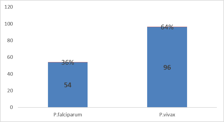
Figure#1: Type of Species of Malarial Parasites
Table#2 Haematological Parameters Of Malaria Infection (N=150)
|
Haematological Parameters |
P. falciparum (n=54) |
P. vivax (n=96) |
P-value |
|
Hemoglobin (g/dl) |
10.20 ± 0.16 |
11.36 ± 0.38 |
<0.001 |
|
Red blood cell (x106 /µL) |
4.1402 ± 0.11 |
4.4497 ± 0.11 |
<0.001 |
|
Total Leukocyte Count (x103 /µL) |
7.0212 ± 0.40 |
6.299 ± 0.11 |
<0.001 |
|
Monocyte (x103 /µL) |
6.9001 ± 0.19 |
5.9000 ± 1.24 |
<0.001 |
|
Lymphocyte (x103 /µL) |
23.3524 ± 0.63 |
26.1403 ± 0.46 |
<0.001 |
|
Eosinophil (x103 /µL) |
2.6104 ± 0.12 |
2.9800 ± 0.13 |
<0.001 |
|
Neutrophil (x103 /µL) |
70.0900 ± 1.51 |
65.3946 ± 0.34 |
<0.001 |
|
Platelets (x103 /µL) |
89.100 ± 1.20 |
99.177 ± 1.12 |
<0.001 |
DISCUSSION :
The present study identified Plasmodium vivax as the predominant malaria species (64%) compared to P. falciparum (36%). Significant hematological alterations were observed among malaria patients, with markedly lower hemoglobin levels, RBC counts, and platelet counts in P. falciparum infections compared to P. vivax. Thrombocytopenia and anemia were the most frequent hematological abnormalities, highlighting their potential role as supportive indicators in the diagnosis and clinical assessment of malaria.
The predominance of P. vivax observed in the present study is consistent with findings from Muhammad et al. in Khyber Agency, Pakistan, where P. vivax was the dominant species among symptomatic patients. However, no cases of P. ovale or P. malariae were detected. Similar patterns have also been reported in another Pakistani study, where P. vivax accounted for 97.3% of cases compared to 2.6% for P. falciparum.11,12 Comparable regional variations have also been reported internationally. Studies from India and Ethiopia have shown that P. vivax contributes approximately 40–60% of malaria cases in India, whereas Ethiopia demonstrates a contrasting pattern with P. falciparum predominance (approximately 60–66%) followed by P. vivax (approximately 34–40%).13,14
In the present study, malaria was more prevalent among males (63.3%) compared to females (36.6%). A similar trend has been reported from Swat, Pakistan, where male predominance (62.12%) was attributed to greater occupational and environmental exposure.15 The mean age of patients in the present study was 39 ± 15 years. Rehman et al. reported that 51% of malaria cases occurred in the 21–40-year age group, followed by 33.68% in the 1–20-year group.16
Malaria is commonly associated with multiple hematological abnormalities, which serve as important clinical and diagnostic indicators. In the present study, patients with P. falciparum had a lower mean hemoglobin level (10.20 ± 0.157) compared to those with P. vivax (11.36 ± 0.377), consistent with findings reported by Hyder A et al.12 Rehman S et al. reported a 100% positive predictive value (PPV) of anemia in P. falciparum infections.17 The multifactorial pathogenesis of malaria-associated anemia is primarily due to intravascular hemolysis of both parasitized and non-parasitized erythrocytes, leading to reduced hemoglobin levels.8,9 Secondary contributing factors include concomitant helminthic or protozoal infections and nutritional deficiencies such as iron, vitamin B12, and folate deficiency.10,11
Furthermore, the present study demonstrated lower RBC counts in P. falciparum infections compared to P. vivax, consistent with findings reported by Haroon A et al. and Kotepui et al.18,19 The parasite’s preference for erythrocyte age plays an important role in disease expression; while Plasmodium malariae tends to infect older erythrocytes, P. vivax and P. ovale preferentially invade reticulocytes.
Thrombocytopenia was another prominent finding in malaria patients. In the present study, platelet counts were significantly lower in P. falciparum infections compared to P. vivax. Similar hematological patterns have been reported globally. In Sudan, thrombocytopenia has been observed in nearly half of P. falciparum infections, while studies from Southeast Asia, including Thailand, have reported thrombocytopenia in up to 60–80% of falciparum malaria cases, significantly higher than in P. vivax infections.20,21
In contrast, some studies have reported a higher frequency of thrombocytopenia in P. vivax infections (71.4%) compared to mixed infections (66.7%) and P. falciparum (26%).19 Severe thrombocytopenia may therefore serve as an important diagnostic clue for malaria, particularly in endemic settings.22 The mean total leukocyte count in the present study was 6.66 ± 0.254 ×10³/μL, with relatively lower values observed in the P. falciparum group compared to P. vivax. This finding is consistent with Rasheed et al., who reported leukopenia in 20–22% of malaria cases, with higher frequency in P. falciparum infections.23
Differential leukocyte analysis showed that P. falciparum infections were associated with higher neutrophil response and lower eosinophil counts compared to P. vivax. This may be explained by neutrophil margination to inflammatory sites, splenic sequestration, lymphotoxic serum effects, and secondary bacterial infections.22
The present study demonstrated lower eosinophil counts in P. falciparum (2.61 ± 0.12/μL) compared to P. vivax (22.98 ± 0.13/μL). Previous studies have reported that P. falciparum infection may suppress pre-existing eosinophilia, whereas P. vivax has minimal impact on eosinophil levels.23
The present study provides clinically relevant region-specific evidence on hematological changes in malaria patients from an endemic setting in Pakistan, and highlights significant differences in hemoglobin, RBC, leukocyte and platelet parameters between Plasmodium falciparum and Plasmodium vivax infections. The use of routine hematological parameters highlights their potential utility as inexpensive and readily accessible ancillary diagnostic tools in resource-limited settings, and the species-specific comparison further confirms its clinical relevance. However, the interpretation of the findings should be made with some limitations such as the single center design, small sample size, short duration of the study, lack of molecular (PCR) confirmation of species and the failure to assess all potential confounding factors such as nutritional status, co-infections, and the presence of underlying hematological disorders which could affect the generalizability of the findings. Further multicenter studies with larger sample size and molecular diagnostic confirmation are recommended to validate these findings and explore the prognostic and diagnostic utility of hematological parameters in malaria.
CONCLUSION:
The study's findings have shown relationship between certain hematological parameters and malarial parasite species. In suspected cases of malaria, routine hematological profiling may have significant diagnostic and prognostic significance. By incorporating these results into national diagnostic recommendations, it may improve early detection, lower morbidity, and support larger initiatives to control and eventually eradicate malaria.
REFERENCES:
1. Song X, Wei W, Cheng W, Zhu H, Wang W, Dong H,et al. Cerebral malaria induced by plasmodium falciparum: clinical features, pathogenesis, diagnosis, and treatment. Front Cell Infect Microbiol. 2022 Jul 25;12:939532. DOI: https://doi.org/10.3389/fcimb.2022.939532
2. Khan MI, Qureshi H, Bae SJ, Khattak AA, Anwar MS, Ahmad S, Hassan F, Ahmad S. Malaria prevalence in Pakistan: A systematic review and meta-analysis (2006-2021). Heliyon. 2023 Apr 11;9(4):e15373. DOI: https://doi.org/10.1016/j.heliyon.2023.e15373.
3. Al-Saadi EA, Abdulnabi MA. Hematological changes associated with COVID-19 infection. J Clin Lab Anal.2022;36(1):e24064.
4. Asmerom H, Gemechu K, Sileshi B, Arkew M. Hematological Abnormalities Among Malaria Infected Adult Patients in Association with ABO Blood Groups at Jinella Health Center, Harar, Eastern Ethiopia. J Blood Med. 2023 Aug 21;14:463-476. doi: 10.2147/JBM.S419815.
5. Nadeem MF, Khattak AA, Zeeshan N. Molecular epidemiology of Plasmodium species in conflicted federally administered tribal area (FATA) Pakistan. Bra J Biol. 2021;83. DOI: https://doi.org/10.1590/1519-6984.247219.
6. Zafar S, Khan RSA., Jaffri AR, Zaidi SAR, Karim MN, Shaukat M. Clinical and hematological criteria for malaria diagnosis in endemic area with limited resources. Pak Armed Forces Med J.2025; 75(SUPPL-5):S701-5.
7. Ahmed Z, Ali B, Parveen K, Riaz Z, Abid W, Shah AH. Clinical presentations and hematological alterations in malaria patients and their association with Plasmodium species and disease severity at Ghambat Institute of Medical Sciences. Infect Dis J Pak. 2026;35(1):39–43.
8. https://doi.org/10.61529/idjp.v35i1.504
9. Habtemariam Y, Asnake M, Alemu M, Shash EP, Tessema TW, Tesso ZG. Plasmodium Vivax as a Causative Agent for Cerebral Malaria in a Group of Adults at Mizan Tepi Teaching Hospital: Case Series. Int Med Case Reports J.2024;17:161–66. DOI: https://doi.org/10.2147/IMCRJ.S440800.
10. Qaiser A, Ayub N, Ishtiaq Q, Tariq H, Amanyar H, Gulzar R, Gulzar S. Evaluation of hematological changes and types of anaemias in malaria. J Popul Ther Clin Pharmacol. 2025;32(4):466–471.
11. Ahad A, QadirS, Rashid AU, Swati AZ. Common Hematological Abnormalities in patients with Malaria presenting at Saidu Teaching Hospital, Swat Pakistan. Pak J Med Health Sci. 2022;16(4):298. https://doi.org/10.53350/pjmhs22164298
12. Khan T, Khan A, Khan A, Badshah F, Ibáñez-Arancibia E, De Los Ríos-Escalante PR, Maryam B, Noor N, Maria, Abdel-Maksoud MA, El-Tayeb MA, Hussain A. Assessment of hematological parameters in malarial suspected patients: Cross sectional study. Parasite Epidemiol Control. 2024 Jul 15;26:e00367. doi: 10.1016/j.parepi.2024.e00367.
13. Hyder A, Mahmood A, Mahmood R, Iqbal MI. Evaluation of haematological parameters in malaria infection and its association with different species of malarial parasite. Pak Armed Forces Med J 2020;70(5):1576-80.
14. Anvikar AR, Shah N, Dhariwal AC, Sonal GS, Pradhan MM, Ghosh SK, Valecha N. Epidemiology of Plasmodium vivax Malaria in India. Am J Trop Med Hyg. 2016 Dec 28;95(6 Suppl):108-120. doi: 10.4269/ajtmh.16-0163.
15. Deress T, Girma M. Plasmodium falciparum and Plasmodium vivax Prevalence in Ethiopia: A Systematic Review and Meta-Analysis. Malar Res Treat. 2019 Dec 3;2019:7065064. doi: 10.1155/2019/7065064.
16. Arévalo-Herrera M, Lopez-Perez M, Medina L, Moreno A, Gutierrez JB, Herrera S. Clinical profile of Plasmodium falciparum and Plasmodium vivax infections in low and unstable malaria transmission settings of Colombia. Malar J. 2015 Apr 11;14:154. doi: 10.1186/s12936-015-0678-3.
17. Naser RH, Rajaii T, Farash BR, Seyyedtabaei SJ, Hajali V, Sadabadi F, et al. Hematological changes due to malaria: an update. Mol Biochem Parasitol.2024;259:111635. doi: 10.1016/j.molbiopara.2024.111635.
18. Rahman S, Jalil F, Khan H, Jadoon MA, Ullah I, Rehman M, et al. Prevalence of malaria in district shangla, khyber Pakhtunkhwa, Pakistan. Jezs. 2017;5:678-82.
19. Haroon H, Fazel PA, Naeem M, Mobin A, Naqvi AH, Makki KJT. Hide and seek: hematological aspects of malaria a developing country perspective. J Infect Dev Ctries 2013;7(03):273-79. https://doi: 10.3855/jidc.2332.
20. Kotepui M, Piwkham D, Phun PB, Phiwklam N, Chupeerach C, Duangmano S. Effects of malaria parasite density on blood cell parameters. PLoS One 2015; 10(3): e0121057. https://doi: 10.1371/journal.pone.0121057.
21. Ebraheem AA, Abdelrahim YH, Gibreel AA, et al. Thrombocytopenia due to Plasmodium falciparum infection and its association with clinical symptoms. European Journal of Medical and Health Sciences. 2025;7(1):22–28.https://doi.org/10.24018/ejmed.2025.7.1.2229
22. Ali EA, Abdalla TM, Adam I. Platelet distribution width, mean platelet volume and haematological parameters in malaria. F1000Research. 2017;6:865. https://doi.org/10.12688/f1000research.11767.1
23. Haldar K, Mohandas NJAEPB. Malaria, erythrocytic infection, and anemia. Hematology 2009; 2009(1): 87-93. PMID: 20008186; PMCID: PMC2933134.
24. Mohtasim BM, Khan MT. Frequency & comparison of thrombocytopenia in patients suffering from P. Vivax and P. Falciparum 2018; 16(1):528. https://doi: 10.29309/TPMJ/2019.26.09.69