In-Vitro Comparative Evaluation of Cytocompatibility, Fibroblastic Activity, and Antioxidant Potential of Polycaprolactone–Polyvinylpyrrolidone (PCL–PVP) Scaffolds With and Without Pomegranate Peel Extract Using MTT and DPPH Assays for Periodontal Tissue Regeneration
- Mathivanan I , Ist year post graduate,RVS Dental College And Hospital, kumaran kottam,sulur,coimbatorte
- Reemitha P , IInd year post graduate ,RVS Dental College And Hospital, kumaran kottam,sulur,coimbatorte
- Gowri P , IInd year post graduate ,RVS Dental College And Hospital, kumaran kottam,sulur,coimbatorte
- Deepshika Saravanan , HOD ( head of the Department), RVS Dental College And Hospital, kumaran kottam,sulur,coimbatorte
- Senthil Kumar , professor, RVS Dental College And Hospital, kumaran kottam,sulur,coimbatorte
- Vijay , Director Cum Principal, RVS Dental College And Hospital, kumaran kottam,sulur, coimbatorte
Article Information:
Abstract:
Background: Periodontal soft-tissue regeneration requires biomaterials that are biocompatible, bioactive, and capable of supporting fibroblast adhesion and proliferation. Natural bioactive compounds derived from agricultural waste are gaining attention due to their sustainability and biological advantages. Pomegranate peel extract (PPE), rich in polyphenols and antioxidants, has shown promising regenerative potential. Aim: To evaluate the cytocompatibility, fibroblast activity, and antioxidant potential of an electrospun polycaprolactone–polyvinylpyrrolidone (PCL–PVP) scaffold incorporated with pomegranate peel extract. Materials and Methods: An in-vitro experimental study was conducted using L929 fibroblast cell lines. Electrospun nanofibrous scaffolds were fabricated using PCL–PVP (control) and PCL–PVP–PPE (test). Scaffold morphology was evaluated using scanning electron microscopy (SEM). Cytocompatibility was assessed by MTT assay, and antioxidant activity was evaluated using the DPPH free-radical scavenging assay. Statistical analysis was performed using Student’s t-test. Results: The PPE-incorporated scaffold demonstrated high fibroblast viability (~90%) with minimal cytotoxicity. SEM analysis revealed enhanced fiber distribution, surface porosity, and improved fibroblast adhesion with well-defined filopodial extensions. The DPPH assay showed approximately 71% free-radical scavenging activity.Conclusion: Pomegranate peel extract-incorporated electrospun PCL–PVP scaffolds exhibited excellent cytocompatibility and antioxidant potential, supporting their application in periodontal soft-tissue regeneration.
Keywords:
Article :
INTRODUCTION:
Periodontal disease leads to progressive destruction of periodontal tissues, including alveolar bone, periodontal ligament, and cementum, ultimately resulting in tooth loss if untreated. Conventional periodontal therapy primarily aims to arrest disease progression; however, regeneration of lost periodontal structures remains a major therapeutic challenge. The concept of periodontal regeneration involves restoration of the original architecture and function of periodontal tissues through biological and biomaterial-based approaches¹˒². Tissue engineering has emerged as a promising strategy to achieve periodontal regeneration by integrating scaffolds, cells, and signaling molecules to create an environment conducive to tissue repair³˒⁴. In this approach, biomaterial scaffolds play a critical role in providing a three-dimensional structure that mimics the extracellular matrix and supports cellular adhesion, proliferation, and differentiation⁵˒⁶. Among synthetic biodegradable polymers, polycaprolactone (PCL) has gained considerable attention in tissue engineering applications due to its excellent biocompatibility, biodegradability, and favorable mechanical properties⁷. PCL also exhibits good electrospinnability, allowing fabrication of nanofibrous scaffolds that closely resemble the architecture of natural extracellular matrix⁸˒⁹. However, the hydrophobic nature and slow degradation rate of PCL may limit its biological performance in certain applications. To overcome these limitations, PCL is often blended with hydrophilic polymers such as polyvinylpyrrolidone (PVP), which improves wettability, drug loading capacity, and cellular interactions¹⁰. The electrospinning technique allows the fabrication diffusion¹¹˒¹². In recent years, natural plant-derived bioactive compounds have gained attention in regenerative medicine due to their antioxidant, anti-inflammatory, and antimicrobial properties. Pomegranate (Punica granatum) peel extract contains high concentrations of polyphenols, flavonoids, and tannins that exhibit strong antioxidant and anti-inflammatory activities¹³˒¹⁴. These properties are particularly relevant in periodontal disease, where oxidative stress plays a significant role in tissue destruction¹⁵. Previous studies have demonstrated that incorporation of plant extracts into electrospun scaffolds can enhance cellular proliferation, improve antioxidant capacity, and promote tissue regeneration¹⁶. Pomegranate peel extract, specifically, has shown promising biological effects including enhanced cell proliferation, improved osteogenic activity, and inhibition of inflammatory responses. In addition, oxidative stress generated by reactive oxygen species (ROS) contributes significantly to periodontal tissue damage and inflammatory progression¹⁷. Antioxidant-loaded scaffolds may therefore help modulate oxidative stress and create a favorable microenvironment for periodontal regeneration. Based on these considerations, incorporation of pomegranate peel extract into electrospun PCL–PVP scaffolds may provide a bioactive platform capable of supporting fibroblast proliferation while simultaneously exhibiting antioxidant activity. Therefore, the present study aimed to evaluate and compare the cytocompatibility, fibroblastic activity, and antioxidant potential of electrospun PCL–PVP scaffolds with and without pomegranate peel extract using MTT and DPPH assays. The findings of this study may contribute to the development of novel biomaterial scaffolds for periodontal tissue regeneration.
MATERIALS AND METHODS:
Scaffold Preparation Electrospun scaffolds composed of polycaprolactone (PCL) and polyvinylpyrrolidone (PVP) were fabricated using the electrospinning technique. Electro spinning is a versatile method capable of producing nanofibers with high surface area-to-volume ratios that mimic extracellular matrix structures¹⁸˒¹⁹.
Two scaffold groups were prepared
Group 1: PCL–PVP scaffold (control)
Group 2: PCL–PVP scaffold incorporated with pomegranate peel extract (PPE)
The polymer solutions were electrospun under controlled conditions of voltage, flow rate, and collector distance to produce uniform nanofibrous membranes.
Cell Culture
L929 fibroblast cell lines were used to evaluate cytocompatibility and fibroblastic activity. Cells were cultured in Dulbecco’s Modified Eagle Medium supplemented with fetal bovine serum and antibiotics and maintained at 37°C in a humidified incubator with 5% CO₂.
MTT Assay for Cytocompatibility and Cell Viability
The cytocompatibility of scaffolds was evaluated using the MTT assay, which measures mitochondrial metabolic activity as an indicator of viable cells. Cells were seeded onto scaffold samples and incubated for a specified duration. Following incubation, MTT reagent was added and metabolized by viable cells to form insoluble formazan crystals, which were dissolved and quantified spectrophotometrically²⁰. Higher absorbance values indicated greater cell viability and proliferation.
DPPH Assay for Antioxidant Activity
The antioxidant potential of scaffolds was assessed using the DPPH radical scavenging assay. The DPPH radical is a stable free radical that changes color upon reduction by antioxidant compounds²¹. Scaffold extracts were mixed with DPPH solution and incubated in the dark, and absorbance was measured using a spectrophotometer. The percentage of radical scavenging activity was calculated using the standard formula: Antioxidant activity (%) = [(A₀ − A₁)/A₀] × 100 where A₀ represents control absorbance A₁ represents sample absorbance.
Statistical Analysis
Data were expressed as mean ± standard deviation. Statistical analysis was performed using analysis of variance (ANOVA) followed by appropriate post-hoc tests. A p-value less than 0.05 was considered statistically significant²².
RESULTS:
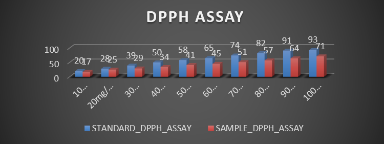
Antioxidant Activity (DPPH Assay) The antioxidant potential of the sample scaffold (PCL–PVP sheet incorporated with pomegranate peel extract) was evaluated using the DPPH radical scavenging assay and compared with the standard antioxidant (Vitamin C) across concentrations ranging from 10–100 mg/ml. The results demonstrated a concentration-dependent increase in radical scavenging activity for both the standard and the sample groups. However, the standard antioxidant consistently exhibited higher percentage inhibition compared to the scaffold sample at all concentrations. The highest antioxidant activity recorded for the standard group was 93% at 100 mg/ml, whereas the scaffold sample demonstrated 71% inhibition at the same concentration. The mean antioxidant activity of the standard group was 60 ± 25.66%, while the sample group showed a mean antioxidant activity of 43 ± 17.54%. Statistical analysis using a paired t-test revealed a highly significant difference between the groups (p < 0.001), indicating that although the scaffold exhibited antioxidant potential, its activity was significantly lower than the standard antioxidant. These findings indicate that the pomegranate peel extract incorporated within the PCL–PVP scaffold retained measurable antioxidant capacity and demonstrated dose-dependent radical scavenging activity.
Cytocompatibility and Cell Viability
The cytocompatibility of the scaffold material was evaluated using cell viability analysis on fibroblast cell lines. The results demonstrated 90% cell viability with only 10% cytotoxicity, indicating excellent cellular compatibility of the scaffold material. Based on standard cytotoxicity classification criteria, materials demonstrating greater than 70% cell viability are considered non-cytotoxic. Accordingly, the PCL–PVP scaffold incorporated with pomegranate peel extract was classified as non-cytotoxic, suggesting that the material is safe and suitable for biomedical applications involving cellular interaction. The high percentage of viable fibroblast cells suggests that the fabricated scaffold provides a favorable environment for cellular attachment and proliferation.
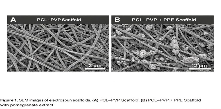
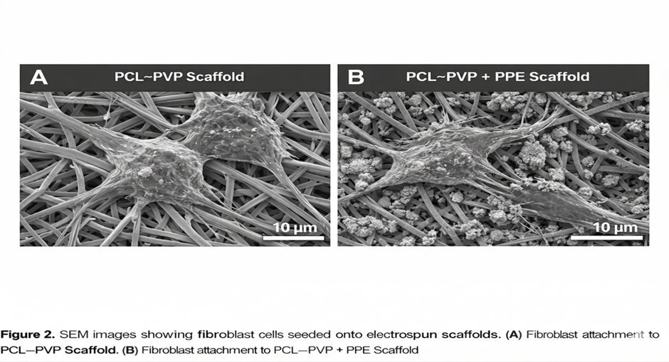
Table 1: Comparison of antioxidant activity (%) between Standard and Sample groups across concentrations using DPPH assay. Values represent percentage inhibition. Statistical significance was determined using a paired t-test.
|
|
STANDARD_DPPH_ASSAY |
SAMPLE_DPPH_ASSAY |
|
|
|
PERCENTAGE |
PERCENTAGE |
P VALUE |
||
|
CONCENTRATIONS |
10 mg/ml |
20 |
17 |
<0.001 |
|
20mg/ml |
28 |
25 |
||
|
30 mg/ml |
39 |
29 |
||
|
40 mg/ml |
50 |
34 |
||
|
50 mg/ml |
58 |
41 |
||
|
60 mg/ml |
65 |
45 |
||
|
70 mg/ml |
74 |
51 |
||
|
80 mg/ml |
82 |
57 |
||
|
90 mg/ml |
91 |
64 |
||
|
100 mg/ml |
93 |
71 |
||
|
|
Mean ± SD |
60 ± 25.656 |
43 ± 17.539 |
|
The antioxidant activity of the Sample (PCL with PVP sheet) was evaluated using the DPPH assay and compared against the Standard (Vitamin C) across concentrations ranging from 10 to 100 mg/ml. At each concentration, the Standard exhibited higher percentage inhibition than the Sample. The mean antioxidant activity of the Standard was 60 ± 25.66%, while the Sample showed 43 ± 17.54%. A paired t-test revealed a statistically significant difference between the two groups (p < 0.001), indicating that the Sample had significantly lower antioxidant activity compared to the Standard.
|
Test Metrics |
Sample |
|
Cytotoxicity (%) |
10% |
|
Cell Viability (%) |
90% |
|
Cytotoxic Reactivity |
Non-Cytotoxic |
This is a single sample tested for cytotoxicity, and the result is classified as Non-Cytotoxic based on established criteria
The sample (Pomegranate Peel Extract with PCL and PVP) showed 90% cell viability and 10% cytotoxicity, classifying it as non-cytotoxic according to standard biocompatibility threshold
DISCUSSION :
Periodontal regeneration requires biomaterials capable of supporting cellular proliferation while simultaneously controlling inflammation and oxidative stress within the periodontal microenvironment. The present in-vitro study evaluated the cytocompatibility and antioxidant activity of a PCL–PVP scaffold incorporated with pomegranate peel extract, with the objective of assessing its potential applicability in periodontal tissue engineering. Electrospun polymeric scaffolds have gained significant attention in regenerative medicine due to their ability to mimic the structural architecture of the extracellular matrix and facilitate cellular adhesion and proliferation. Nanofibrous scaffolds fabricated using electrospinning provide a highly porous structure with large surface area, enabling improved nutrient diffusion and cell–scaffold interactions¹³. Polycaprolactone (PCL) is widely used in tissue engineering applications because of its biocompatibility, mechanical stability, and slow degradation profile⁴. However, the hydrophobic nature of PCL may limit cellular attachment. Therefore, blending PCL with hydrophilic polymers such as polyvinylpyrrolidone (PVP) improves wettability and enhances biological interactions between cells and scaffold surfaces⁵. The combination of PCL and PVP has been reported to produce scaffolds with improved mechanical strength and enhanced cellular compatibility⁶. In the present study, cytocompatibility testing demonstrated 90% cell viability, indicating that the fabricated scaffold is non-cytotoxic and supports fibroblast survival. These findings are consistent with previous studies demonstrating excellent biocompatibility of PCL-based scaffolds in tissue engineering applications⁷. High fibroblast viability is particularly important in periodontal regeneration because fibroblasts play a key role in extracellular matrix synthesis, collagen production, and periodontal ligament repair⁸. Another important finding of the present study was the antioxidant activity demonstrated by the scaffold containing pomegranate peel extract. The DPPH assay results revealed that the scaffold exhibited dose-dependent radical scavenging activity, although the antioxidant effect was lower compared to the standard antioxidant (Vitamin C). Nevertheless, the presence of significant antioxidant activity indicates that the incorporated pomegranate peel extract retained its bioactive properties within the scaffold matrix. Oxidative stress plays a crucial role in the pathogenesis of periodontal disease. Excessive production of reactive oxygen species (ROS) contributes to tissue destruction, inflammatory cell activation, and alveolar bone resorption⁹. Therefore, biomaterials with antioxidant properties may help create a favorable microenvironment for periodontal regeneration by reducing oxidative stress and protecting cells from free radical damage¹⁰. Pomegranate peel extract is known to contain high concentrations of polyphenolic compounds such as ellagic acid, tannins, flavonoids, and anthocyanins, which exhibit potent antioxidant and anti-inflammatory activities¹¹–¹³. Several studies have reported that pomegranate-derived compounds can enhance cellular proliferation, promote osteoblastic activity, and inhibit inflammatory mediators involved in periodontal tissue destruction¹⁴. The incorporation of plant-derived bioactive compounds into polymeric scaffolds has been increasingly explored in regenerative medicine. These bioactive additives may improve biological performance by enhancing cellular responses, promoting angiogenesis, and modulating inflammatory pathways¹⁵. In particular, electrospun scaffolds loaded with natural extracts have been shown to significantly improve fibroblast attachment and proliferation due to the presence of bioactive phytochemicals¹⁶. In the context of periodontal regeneration, the use of antioxidant-loaded scaffolds may provide dual therapeutic benefits. First, the scaffold provides structural support for tissue regeneration, while second, the incorporated bioactive compounds help control oxidative stress and inflammation. This combination approach may enhance healing outcomes in periodontal defects. Although the antioxidant activity of the scaffold in the present study was lower than the standard antioxidant, the observed dose-dependent scavenging activity suggests that the incorporated extract remains biologically active within the polymeric matrix. The lower activity compared to Vitamin C may be attributed to the controlled release of the extract from the scaffold structure, which may actually be advantageous for sustained antioxidant effects in clinical applications. The cytocompatibility results further support the potential clinical applicability of the fabricated scaffold. Materials intended for tissue engineering applications must demonstrate minimal cytotoxicity and support cell survival. The high cell viability observed in the present study indicates that the scaffold does not release harmful degradation products and is suitable for cellular interaction. Another possible explanation for the favorable cytocompatibility observed may be the improved surface hydrophilicity provided by the PVP component of the scaffold. Hydrophilic surfaces are known to enhance protein adsorption and facilitate cell adhesion, which in turn promotes cellular proliferation and tissue integration¹⁷. Despite these promising findings, the present study has certain limitations. The evaluation was limited to in-vitro assays, and therefore the results may not fully represent the complex biological environment present in vivo. Factors such as immune response, vascularization, and mechanical forces may influence scaffold performance in clinical conditions. Future research should therefore include in-vivo studies using periodontal defect models to evaluate the regenerative potential of the scaffold in terms of new bone formation, periodontal ligament regeneration, and cementum deposition. Additionally, further studies investigating controlled release kinetics of the pomegranate extract and its anti-inflammatory effects may provide valuable insights into the therapeutic potential of this biomaterial system. Overall, the findings of this study suggest that PCL–PVP scaffolds incorporated with pomegranate peel extract exhibit favorable cytocompatibility and measurable antioxidant activity, indicating their potential application as bioactive scaffolds for periodontal tissue regeneration.
CONCLUSION :
Within the limitations of the present in-vitro study, the electrospun polycaprolactone–polyvinylpyrrolidone (PCL–PVP) scaffold incorporated with pomegranate peel extract demonstrated favorable cytocompatibility and measurable antioxidant activity. The results of the MTT assay revealed high fibroblast cell viability, indicating that the fabricated scaffold is biocompatible and non-cytotoxic, which is an essential requirement for biomaterials intended for periodontal tissue engineering applications¹˒⁷. The presence of approximately 90% viable cells suggests that the scaffold provides a supportive microenvironment for cellular attachment and proliferation, which may contribute to periodontal tissue regeneration. The DPPH assay demonstrated that the scaffold exhibited concentration-dependent radical scavenging activity, confirming the presence of antioxidant potential due to the incorporation of pomegranate peel extract. Although the antioxidant activity was lower compared with the standard antioxidant, the observed activity indicates that bioactive phytochemicals present in Punica granatum remain functionally active within the scaffold matrix¹³˒¹⁴. The antioxidant property of the scaffold may play a significant role in periodontal regeneration by reducing oxidative stress and protecting periodontal tissues from reactive oxygen species–mediated damage¹⁵˒¹⁷. The combination of biocompatible polymeric scaffolds and plant-derived bioactive compounds represents a promising strategy in regenerative medicine. Electrospun nanofibrous scaffolds provide structural support resembling the extracellular matrix, while natural antioxidants may enhance cellular responses and modulate inflammatory processes during tissue healing⁸˒¹¹˒¹⁶. Therefore, the findings of the present study suggest that PCL–PVP electrospun scaffolds incorporated with pomegranate peel extract may serve as a promising biomaterial for periodontal tissue engineering and regenerative applications. However, further in-vivo studies and clinical investigations are required to evaluate the long-term biological performance, degradation behavior, and regenerative potential of this scaffold in periodontal defects.
REFERENCES:
1. Nyman S, Lindhe J, Karring T, Rylander H. New attachment following surgical treatment of human periodontal disease. J Clin Periodontol. 1982;9(4):290–296.
2. Sculean A, Gruber R, Bosshardt DD. Soft tissue wound healing around teeth and dental implants. J Clin Periodontol. 2014;41(S15):S6–S22.
3. Giannobile WV. Periodontal tissue engineering by growth factors. Bone. 1996;19(1):23S–37S. Langer R, Vacanti JP. Tissue engineering. Science. 1993;260(5110):920–926.
4. Woodruff MA, Hutmacher DW. The return of a forgotten polymer—Polycaprolactone in tissue engineering. Prog Polym Sci. 2010;35(10):1217–1256.
5. Dash TK, Konkimalla VB. Poly-ε-caprolactone based formulations for drug delivery and tissue engineering. J Control Release. 2012;158(1):15–33.
6. Sill TJ, von Recum HA. Electrospinning: Applications in drug delivery and tissue engineering. Biomaterials. 2008;29(13):1989–2006.
7. Greiner A, Wendorff JH. Electrospinning: A fascinating method for the preparation of ultrathin fibers. Angew Chem Int Ed. 2007;46(30):5670–5703.
8. Li D, Xia Y. Electrospinning of nanofibers: Reinventing the wheel? Adv Mater. 2004;16(14):1151–1170.
9. Pham QP, Sharma U, Mikos AG. Electrospinning of polymeric nanofibers for tissue engineering applications. Tissue Eng. 2006;12(5):1197–1211.
10. Viuda-Martos M, Fernández-López J, Pérez-Álvarez JA. Pomegranate and its many functional components as related to human health. Compr Rev Food Sci Food Saf. 2010;9(6):635–654.
11. Ismail T, Sestili P, Akhtar S. Pomegranate peel and fruit extracts: A review of potential anti-inflammatory and anti-infective effects. J Ethnopharmacol. 2012;143(2):397–405.
12. Fischer UA, Carle R, Kammerer DR. Identification and quantification of phenolic compounds from pomegranate peel. Food Chem. 2011;127(2):807–813.
13. Oliveira LM, et al. Pomegranate extract promotes wound healing through antioxidant mechanisms. J Med Food. 2013;16(6):1–7.
14. Chapple ILC. Reactive oxygen species and antioxidants in inflammatory diseases. J Clin Periodontol. 1997;24(5):287–296.
15. Chapple ILC, Matthews JB. The role of reactive oxygen species and antioxidants in periodontal disease. J Clin Periodontol. 2007;34(5):412–418.
16. Battino M, Ferreiro MS, Gallardo I, Newman HN, Bullon P. The antioxidant capacity of saliva. Free Radic Biol Med. 2002;33(4):447–454.
17. Diomede F, et al. Pomegranate extract functionalized scaffolds enhance osteogenic differentiation of stem cells. Mater Sci Eng C. 2019;102:819–825.
18. Sadek H, Mamdouh W. Bioactive plant extract loaded nanofibrous scaffolds for tissue engineering applications. Int J Biol Macromol. 2018;118:1561–1568.
19. Hutmacher DW. Scaffolds in tissue engineering bone and cartilage. Biomaterials. 2000;21(24):2529–2543.
20. Place ES, George JH, Williams CK, Stevens MM. Synthetic polymer scaffolds for tissue engineering. Chem Soc Rev. 2009;38(4):1139–1151.
21. Huang ZM, Zhang YZ, Kotaki M, Ramakrishna S. A review on polymer nanofibers by electrospinning and their applications in nanocomposites. Compos Sci Technol. 2003;63(15):2223–2253.
22. Ma PX. Scaffolds for tissue fabrication. Mater Today. 2004;7(5):30–40.
23. O’Brien FJ. Biomaterials and scaffolds for tissue engineering. Mater Today. 2011;14(3):88–95.
24. Dalby MJ, Gadegaard N, Oreffo RO. Harnessing nanotopography and integrin-matrix interactions to influence stem cell fate. Nat Mater. 2014;13(6):558–569.
25. Geiger B, Spatz JP, Bershadsky AD. Environmental sensing through focal adhesions. Nat Rev Mol Cell Biol. 2009;10(1):21–33.
26. Humphrey JD, Dufresne ER, Schwartz MA. Mechanotransduction and extracellular matrix homeostasis. Nat Rev Mol Cell Biol. 2014;15(12):802–812.
27. Bacakova L, Filova E, Parizek M, Ruml T, Svorcik V. Modulation of cell adhesion and proliferation on biomaterial surfaces. Biotechnol Adv. 2011;29(6):739–767.
28. Keselowsky BG, Collard DM, García AJ. Integrin binding specificity regulates biomaterial surface chemistry effects on cell differentiation. PNAS. 2005;102(17):5953–5957.
29. Middleton JC, Tipton AJ. Synthetic biodegradable polymers as orthopedic devices. Biomaterials. 2000;21(23):2335–2346.
30. Puppi D, Chiellini F, Piras AM, Chiellini E. Polymeric materials for bone and cartilage repair. Prog Polym Sci. 2010;35(4):403–440.
31. Rezwan K, Chen QZ, Blaker JJ, Boccaccini AR. Biodegradable and bioactive porous polymer/inorganic composite scaffolds for bone tissue engineering. Biomaterials. 2006;27(18):3413–3431.
32. Venugopal J, Ramakrishna S. Applications of polymer nanofibers in biomedicine and biotechnology. Appl Biochem Biotechnol. 2005;125(3):147–157.
33. Bhardwaj N, Kundu SC. Electrospinning: A fascinating fiber fabrication technique. Biotechnol Adv. 2010;28(3):325–347.
34. Ramakrishna S, Fujihara K, Teo WE, Lim TC, Ma Z. An Introduction to Electrospinning and Nanofibers. Singapore: World Scientific; 2005.
35. Karabulut I, Topcu A, Duran A, Turan S, Ozturk B. Antioxidant activity of pomegranate peel extract. Food Chem. 2009;117(4):573–578.
36. Lansky EP, Newman RA. Punica granatum (pomegranate) and its potential for prevention and treatment of inflammation. J Ethnopharmacol. 2007;109(2):177–206.
37. Melcher AH. On the repair potential of periodontal tissues. J Periodontol. 1976;47(5):256–260.
38. Karring T, Nyman S, Lindhe J, Sirirat M. Potentials for root resorption during periodontal wound healing. J Clin Periodontol. 1984;11(1):41–52.
39. Polimeni G, Xiropaidis AV, Wikesjö UME. Biology and principles of periodontal wound healing/regeneration. Periodontol 2000. 2006;41:30–47.
40. Mirabella N, Castellani V, Sala S. Current options for the valorization of food manufacturing waste. J Clean Prod. 2014;65:28–41.
41. Gustavsson J, Cederberg C, Sonesson U. Global food losses and food waste. FAO Report. 2011.
42. Galanakis CM. Recovery of high value components from food waste. Food Res Int. 2012;44(8):2231–2238.
43. Kirchherr J, Reike D, Hekkert M. Conceptualizing the circular economy. Resour Conserv Recycl. 2017;127:221–232.
44. Ellen MacArthur Foundation. Towards the Circular Economy. 2013. Bartold PM, McCulloch CA, Narayanan AS, Pitaru S. Tissue engineering: A new paradigm for periodontal regeneration. J Periodontal Res. 2000;35(2):69–77.
45. Ivanovski S. Periodontal regeneration. Aust Dent J. 2009;54(S1):S118–S128.
46. Giannobile WV, Somerman MJ. Growth and amelogenin-like factors in periodontal regeneration. Periodontol 2000. 2003;19:59–75.