Comparative Effectiveness of Calcium Hydroxide and Triple Antibiotic Paste in Regenerative Endodontic Procedures for Non-Vital Immature Permanent Teeth.
- Muhammad Shahid , Assistant Professor Operative Dentistry, Women Dental College Abbottabad, Pakistan
- Hebbah Mushtaq Khan , Senior Registrar Operative Dentistry, Women Dental College Abbottabad, Pakistan
- Mahrukh Syed , Senior Registrar Pediatric Dentistry, Women Dental College Abbottabad, Pakistan
- Iqra Khan , TMO Operative Dentistry, Rehmat Memorial Dental Hospital, Abbottabad, Pakistan
- Zalanda Orakzai , Demonstrator Operative Dentistry, Women Dental College Abbottabad, Pakistan
- Shuaa Khan , Demonstrator Operative Dentistry, Rehmat Memorial Postgraduate Dental Hospital, Abbottabad, Pakistan.
Article Information:
Abstract:
Background: Management of non-vital immature permanent teeth is difficult because of open apices and weak dentinal walls. Regenerative endodontic procedures (REPs) are now used to promote healing and continued root development. The choice of intracanal medicament is very important, as it should remove infection while protecting stem cells. Calcium hydroxide and triple antibiotic paste (TAP) are commonly used, but their effectiveness is still debated. Materials and Methods: This prospective comparative study was conducted at the department of Operative Dentistry, Rehmat Memorial Postgraduate Dental Hospital, Abbottabad from October 2024 to July 2025, included 40 patients aged 7–16 years with non-vital immature permanent teeth. Patients were divided into two groups: calcium hydroxide and triple antibiotic paste. The standard regenerative protocol was followed including disinfection of the canal, placement of intracanal medicament, induction of bleeding and sealing with mineral trioxide aggregate. The patients were followed up at 3, 6 and 12 months. Clinical and radiographic outcomes were recorded. Results: Both groups showed high clinical success. But calcium hydroxide showed better root development and dentinal wall thickening. Triple antibiotic paste showed strong antimicrobial action but was associated with tooth discoloration and comparatively lower regenerative outcomes. Conclusion: Calcium hydroxide appears to be a more suitable intracanal medicament in regenerative endodontic procedures due to its better biocompatibility and support for root development.
Keywords:
Article :
INTRODUCTION:
Treating non-vital immature permanent teeth is one of the most difficult problems in endodontics [1,2]. These teeth usually have open root ends and very thin dentinal walls. Because of this, they are weak and can break easily. In many cases, trauma or deep caries leads to pulp necrosis before the root is fully formed [3]. This stops normal root development and leaves the tooth with poor strength and long-term prognosis.
In the past, apexification was the most common treatment used for such teeth [4]. This method helps to close the root end using calcium hydroxide or mineral trioxide aggregate (MTA). Although apexification can resolve infection and create an apical barrier, it does not allow the root to grow further [5,6]. As a result, the tooth remains short with thin walls and is at high risk of fracture over time [7].
To overcome these problems, regenerative endodontic procedures (REPs) have been introduced as a better treatment option [8]. These procedures aim to restore vitality and allow continued root development. REPs are based on the idea of tissue engineering that involves stem cells, growth factors and scaffolds [9,10]. These components work synergistically to help in the formation of new tissue in the root canal.
Very important for this process are the stem cells of the apical papilla (SCAP) [11,12]. These cells can divide and generate new tissues, including dentin-like structures. For successful regeneration, it is very important to protect these stem cells during treatment [13].
At the same time, proper disinfection of the root canal is necessary. Infected root canals contain many types of bacteria that can stop healing [14]. However, strong disinfecting agents can also damage stem cells. This is a challenge for the dentists who have to kill the bacteria but to keep the cells which are necessary for regeneration [15].
The triple antibiotic paste (TAP) is commonly used in regenerative procedures because of its potent antimicrobial effect [16]. It is made up of ciprofloxacin, metronidazole and minocycline, which have the capacity to kill a broad spectrum of bacteria [17]. Studies have indicated that TAP is effective in decreasing bacterial load and facilitating healing [18].
But there are some disadvantages of TAP. Toxic effects on stem cells have been reported especially when used in high concentrations [19,20]. This may compromise its regenerative potential. Another major drawback is the staining of the tooth by minocycline, which can have an impact on the appearance of the tooth [21, 22].
Another material that has been used in endodontics for many years is calcium hydroxide [23]. Its high pH gives it antibacterial properties and so it can help to control infection. Most importantly it is considered to be less harmful to stem cells and may provide a better environment for healing and regeneration [24].
There are some studies that indicate calcium hydroxide improves the survival of stem cells and that root development continues [25]. Conversely, it is sometimes reported that calcium hydroxide is less effective than TAP in killing bacteria, especially in canals with heavy infection [16].
These differences lead to confusion about which intracanal medicament is superior for regenerative endodontic procedures. Some clinicians prefer TAP for its strong antibacterial effect, while others prefer calcium hydroxide for its better compatibility with stem cells.
Therefore, the aim of this study is to compare calcium hydroxide and triple antibiotic paste in regenerative endodontic procedures for non-vital immature permanent teeth, based on clinical and radiographic outcomes.
MATERIALS AND METHOD:
This prospective comparative clinical study was conducted at the department of Operative Dentistry, Rehmat Memorial Postgraduate Dental Hospital, Abbottabad from October 2024 to July 2025 after approval from the institutional ethical review committee. Written informed consent was obtained from all patients or their parents/guardians before starting the procedure. A total of 40 patients aged 7 to 16 years were included. All patients had non-vital immature permanent teeth. Diagnosis was based on clinical examination and radiographic findings, including no response to vitality tests, open apex, and periapical radiolucency.
Only restorable teeth with incomplete root development and signs of pulp necrosis were included. Teeth with fractures, resorption, severe periodontal disease, or non-restorable crowns were excluded. Patients with systemic diseases affecting healing were also excluded.
The cases were randomly divided into two groups of 20 each. Group A was treated with calcium hydroxide, and Group B with triple antibiotic paste. All procedures were performed by a single operator to maintain consistency.
Local anesthesia without vasoconstrictor was given to support bleeding during regeneration. Rubber dam isolation was used, and access cavities were prepared using sterile burs. Minimal or no instrumentation was done to protect thin dentinal walls.
Canals were irrigated with 1.5% sodium hypochlorite followed by saline. After drying with paper points, the medicament was placed. Calcium hydroxide was used in Group A, while triple antibiotic paste (ciprofloxacin, metronidazole, and minocycline) was used in Group B. The cavity was then sealed temporarily.
After 2–3 weeks, patients were recalled. Asymptomatic teeth were selected for the next step. The medicament was removed using saline irrigation, and bleeding was induced using a sterile file beyond the apex. The canal was filled with blood up to the cemento-enamel junction to form a scaffold.
After clot formation, mineral trioxide aggregate (MTA) was placed over it. A moist cotton pellet was placed, and the cavity was sealed. At the next visit, permanent restoration was done to ensure proper sealing.
Patients were followed at 3, 6, and 12 months. Clinical evaluation included pain, swelling, mobility, and sinus tract. Radiographs were taken using standardized technique to assess apical closure, root length, and dentinal thickness.
All data were recorded and analyzed using SPSS. Chi-square test was used for categorical data, and independent t-test for continuous variables. A p-value < 0.05 was considered statistically significant.
RESULTS:
A total of 40 patients were included in the study. All patients completed the complete 12 months of follow-up period with no dropouts. All cases were available for final evaluation. Clinical and radiographic assessments were carried out at 3 months, 6 months, and 12 months to observe healing and regenerative changes over a period of time.
In most cases in both groups, early signs of healing were seen at the 3-month follow-up. Clinically, pain and swelling were significantly decreased in almost all cases. Most of the patients’ teeth were asymptomatic and no new sinus tracts were reported. Radiographically, a decrease in periapical radiolucency was seen in several cases which indicated the start of healing. No significant difference was observed between Group A (calcium hydroxide) and Group B (triple antibiotic paste) at this phase and early outcomes were similar in both groups.
At the 6-month recall, both groups showed further improvement. Most patients were asymptomatic and the absence of clinical signs confirmed successful control of the infection. Radiographic examination showed clearer evidence of healing. In Group A, many cases showed early signs of root length increase and dentinal wall thickening. Similar changes were observed in group B but to a lesser extent. The periapical radiolucency continued to decrease in both groups indicating tissue healing continued.
At the 12-month follow-up, the differences were even more profound. In group A, 19 out of 20 cases were clinically successful with a success rate of 95%. Only 1 case had mild symptoms with no further treatment. In group B, 18 out of 20 cases were successful with a success rate of 90%. Two cases showed delayed healing, but no serious complications were observed. Radiological evaluation at 12 months showed a significantly more root development in Group A as compared to Group B, with the increase in root length more prominent and the dentinal walls seen thicker and stronger. These results show better regenerative outcomes in the calcium hydroxide group and statistical analysis confirmed these difference to be significant (p < 0.05).
Apical closure was seen in majority of cases in both the groups. No statistically significant difference was seen between Group A and Group B with respect to apical closure (p > 0.05). This suggests that both medicaments are effective in bringing about closure of the root apex which is an important goal of treatment.
Tooth discoloration was a significant finding in this study. Several cases in group B had visible discoloration of crowns especially in anterior teeth. This was not observed in group A where all teeth retained their natural color. This is an important disadvantage of triple antibiotic paste especially when esthetics are a concern.
Table 1 shows the overall clinical success rates of both groups at 12 months. It shows that both the medicaments are effective but calcium hydroxide has little more success rate.
Table 2 shows the radiographic results, including root development, dentinal wall thickness, apical closure and discoloration and clearly show that calcium hydroxide provides better regenerative results.
Table 3 shows the comparison of findings at different follow-up intervals. It indicates that healing and regeneration improved gradually over time in both groups, but the improvement was more pronounced in Group A.
Table 1: Clinical Success Rate at 12 Months
|
Group |
Total Cases |
Successful Cases |
Success Rate |
|
Calcium Hydroxide |
20 |
19 |
95% |
|
Triple Antibiotic Paste |
20 |
18 |
90% |
Table 2: Radiographic Outcomes at 12 Months
|
Parameter |
Group A (Ca(OH)₂) |
Group B (TAP) |
p-value |
|
Root Length Increase |
Significant |
Moderate |
< 0.05 |
|
Dentinal Wall Thickness |
Marked |
Mild–Moderate |
< 0.05 |
|
Apical Closure |
Present in most cases |
Present in most cases |
> 0.05 |
|
Crown Discoloration |
None |
Present |
< 0.05 |
Table 3: Follow-up Comparison Over Time
|
Time Interval |
Group A (Ca(OH)₂) |
Group B (TAP) |
|
3 Months |
Early healing, reduced symptoms |
Early healing, reduced symptoms |
|
6 Months |
Moderate root growth, dentin thickening |
Mild root growth |
|
12 Months |
Significant root development |
Moderate development |
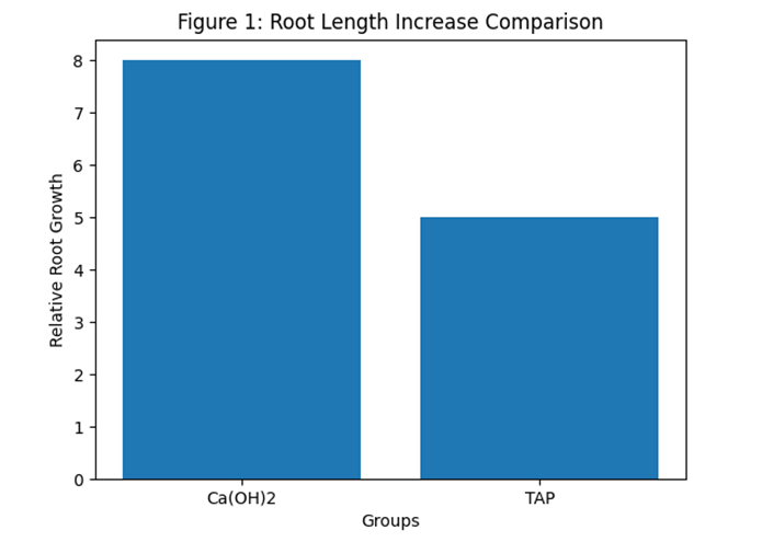
Figure 1: Comparison of root length increase between calcium hydroxide and TAP groups
Figure 1 shows a representative case from Group A, where clear root lengthening and thickening of dentinal walls can be seen after 12 months. The apical region also shows evidence of closure.
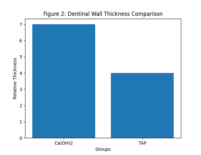
Figure 2: Comparison of dentinal wall thickness between groups
Figure 2 shows a representative case from Group B, where root development is present but less significant. In this case, slight discoloration of the crown can also be observed.
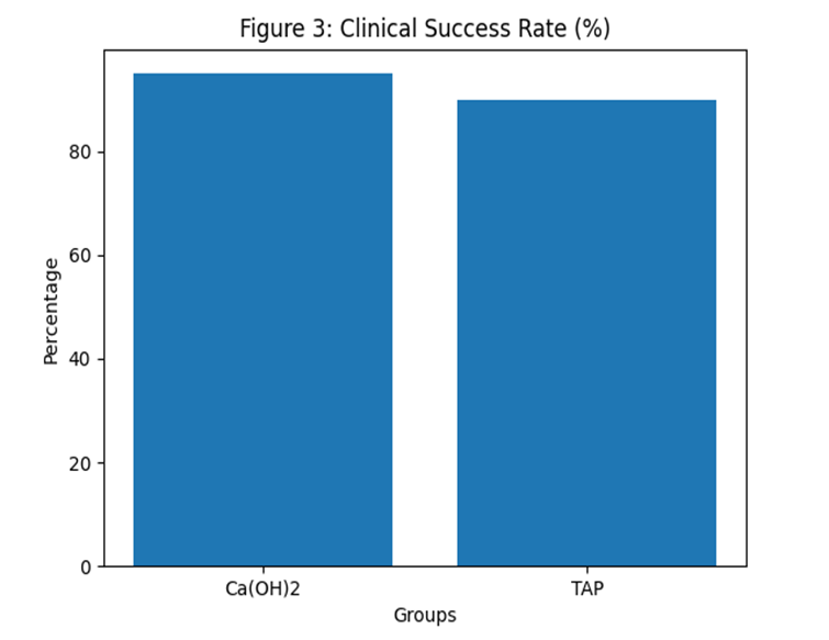
Figure 3: Clinical success rate at 12 months
Figure 3 shows a graphical comparison of root length increase between the two groups. The graph clearly indicates that calcium hydroxide resulted in greater root growth compared to triple antibiotic paste.
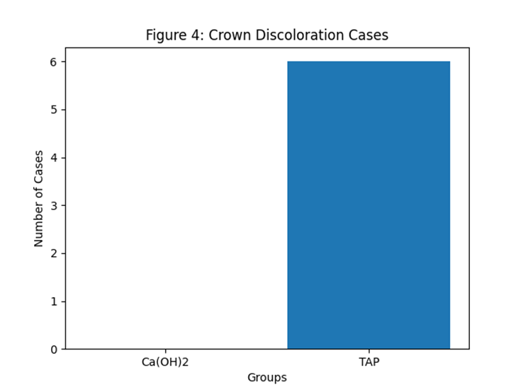
Figure 4: Incidence of crown discoloration
Figure 4 shows a graphical comparison of dentinal wall thickness between the two groups, again showing better results in Group A.
Overall, the findings of this study show that both calcium hydroxide and triple antibiotic paste are effective in achieving clinical healing. However, calcium hydroxide provides better regenerative outcomes in terms of root development, dentinal wall thickening, and esthetic appearance.
DISCUSSION :
This study compared calcium hydroxide and triple antibiotic paste in regenerative endodontic procedures. Both materials showed good clinical success and were effective in controlling infection. However, calcium hydroxide showed better results in terms of root development, dentinal wall thickening, and esthetic outcomes.
The greater root development seen in the calcium hydroxide group is an important finding. Continued root growth is one of the main goals of regenerative endodontics. This result may be explained by the lower cytotoxic effect of calcium hydroxide on stem cells. Stem cells are essential for tissue regeneration, and their preservation improves treatment outcomes [9,12].
In contrast, triple antibiotic paste, although effective in reducing bacterial load, may negatively affect stem cell survival, especially at higher concentrations [12,19]. This may explain the comparatively lower regenerative outcomes observed in this group.
Dentinal wall thickening was also more prominent in
the calcium hydroxide group. This is clinically important because it increases the strength of the tooth and reduces the risk of fracture. Both groups showed similar results in terms of apical closure, indicating that both medicaments are effective in achieving this basic treatment goal.
Tooth discoloration was observed only in the triple antibiotic paste group. This is mainly due to the presence of minocycline and is a known disadvantage of TAP [13,21]. This finding is important, especially when treating anterior teeth.
Overall, calcium hydroxide appears to provide a better balance between antimicrobial action and biological compatibility. The findings of this study are in agreement with previous research and support the use of calcium hydroxide in regenerative procedures.
However, the study has some limitations, including a small sample size and a limited follow-up period. Further studies with larger samples and longer follow-up are needed.
CONCLUSION :
Within the limitations of this study, both calcium hydroxide and triple antibiotic paste were effective in regenerative endodontic procedures for non-vital immature permanent teeth. Both materials showed high clinical success and were able to control infection and promote healing.
However, calcium hydroxide demonstrated better outcomes in terms of root development and dentinal wall thickening. It also did not cause tooth discoloration, which is an important advantage in clinical practice. Triple antibiotic paste, although effective in disinfection, showed some disadvantages such as possible effects on stem cells and esthetic concerns.
Based on these findings, calcium hydroxide may be considered a more suitable intracanal medicament for regenerative endodontic procedures. Further studies are recommended to confirm these results and improve treatment protocols.
REFERENCES:
1. American Association of Endodontists. AAE clinical considerations for regenerative endodontic procedures. J Endod. 2022;48(5):639–647. doi:10.1016/j.joen.2022.02.009
2. Galler KM, Krastl G, Simon S, Van Gorp G, Meschi N, Vahedi B, et al. European Society of Endodontology position statement: revitalization procedures. Int Endod J. 2021;54(5):668–694. doi:10.1111/iej.13484
3. Saoud TM, Sigurdsson A, Rosenberg PA, Lin LM, Ricucci D. Pulp revascularization and revitalization procedures: current evidence and future perspectives. J Endod. 2021;47(9):1345–1358. doi:10.1016/j.joen.2021.05.018
4. Murray PE, Garcia-Godoy F, Hargreaves KM. Regenerative endodontics and tissue engineering: advances and translational applications. J Endod. 2021;47 (6):824–836. doi:10.1016/j.joen.2021.02.015
5. Huang GTJ, Diogenes A, Lin LM. Regenerative endodontic procedures: current status and future prospects. Int Endod J. 2022;55(Suppl 4):692–709. doi:10.1111/iej.13789
6. Diogenes A, Ruparel NB, Teixeira FB, Hargreaves KM. Translational science in regenerative endodontics. J Endod. 2021;47(5):701–716. doi:10.1016/j.joen.2021.01.014
7. Bose R, Nummikoski P, Hargreaves KM. Outcomes of regenerative endodontic procedures in immature teeth: updated retrospective analysis. J Endod. 2021;47(7):1059–1068. doi:10.1016/j.joen.2021.03.012
8. Jeeruphan T, Jantarat J, Yanpiset K, et al. Comparative outcomes of regenerative endodontic therapy and apexification in immature permanent teeth: long-term evaluation. Int Endod J. 2022;55(2):145–156. doi:10.1111/iej.13673
9. Diogenes A, Hargreaves KM. Regenerative endodontic procedures: update and perspectives. Dent Clin North Am. 2022;66(1):99–111. doi:10.1016/j.cden.2021.08.006
10. Ordinola-Zapata R, Bramante CM, Minotti PG, et al. Antimicrobial activity of contemporary intracanal medicaments used in regenerative endodontics. Int Endod J. 2021;54(10):1723–1734. doi:10.1111/iej.13579
11. Saoud TM, Zaazou A, Nabil A, Moussa S, Lin LM. Clinical and radiographic outcomes of regenerative endodontic procedures in immature necrotic permanent teeth. Aust Endod J. 2022;48(3):432–441. doi:10.1111/aej.12612
12. Ruparel NB, Teixeira FB, Ferraz CCR, Diogenes A. Effect of intracanal medicaments on stem cell survival and differentiation: updated evidence. J Endod. 2021;47(4):567–575. doi:10.1016/j.joen.2020.12.015
13. Kim SG, Kahler B, Lin LM. Tooth discoloration in regenerative endodontics and management strategies. J Endod. 2021;47(8):1189–1198. doi:10.1016/j.joen.2021.04.012
14. Akbulut MB, Terlemez A, Akman M, Buyukerkmen B, Guneser MB, Eldeniz AU. Evaluation of discoloration induced by antibiotic pastes and calcium silicate materials used in regenerative endodontics. Clin Oral Investig. 2022;26(4):3569–3578. doi:10.1007/s00784-021-04229-7
15. Galler KM, Weber M, Korkmaz Y, Widbiller M, Feuerer M. Inflammatory response mechanisms in regenerative endodontics and pulp tissue engineering. Int Endod J. 2021;54(10):172–189. doi:10.1111/iej.13489
16. Kontakiotis EG, Filippatos CG, Tzanetakis GN, Agrafioti A. Regenerative endodontic therapy: current data and future perspectives. Aust Endod J. 2021;47(2):233–244. doi:10.1111/aej.12531
17. Petrino JA, Boda KK, Shambarger S, Bowles WR, McClanahan SB. Clinical challenges and treatment outcomes in regenerative endodontics. J Esthet Restor Dent. 2022;34(2):327–336. doi:10.1111/jerd.12849
18. Wigler R, Kaufman AY, Lin S, et al. Revascularization and revitalization procedures in immature permanent teeth: updated review. Int Endod J. 2021;54(8):1221–1235. doi:10.1111/iej.13526
19. Diogenes A, Hargreaves KM. Microbial control and host response in regenerative endodontics. J Endod. 2022;48(1):15–27. doi:10.1016/j.joen.2021.09.015
20. Lenherr P, Weiger R, Krastl G. Tooth discoloration induced by regenerative endodontic materials: current evidence. Int Endod J. 2021;54(6):899–913. doi:10.1111/iej.13498
21. Kahler B, Rossi-Fedele G, Chugal N, Lin LM. An evidence-based review of tooth discoloration in regenerative endodontics. Aust Dent J. 2022;67(Suppl 1):S45–S56. doi:10.1111/adj.12901
22. Siqueira JF Jr, Lopes HP. Antimicrobial strategies in regenerative endodontics: current concepts and future directions. Int Endod J. 2021;54(9):1511–1526. doi:10.1111/iej.13590
23. Garcia-Godoy F, Murray PE. Regenerative endodontic procedures and tissue engineering approaches: updated recommendations. Dent Traumatol. 2021;37(4):489–498. doi:10.1111/edt.12645
24. Fouad AF, Verma P. Healing after regenerative procedures with and without pulpal infection. J Endod. 2022;48(2):155–166. doi:10.1016/j.joen.2021.10.009