When Cure Triggers Crisis: Paradoxical Anti-Tubercular Therapy in Abdominal Tuberculosis
- Prof. Richa Jaiman , M.S. Professor, Department of Surgery
- Prof. Puneet Srivastava , M.S., M.Ch Professor, Department of Surgery
- Bhavana Verma , M.S. Assistant Professor, Department of Surgery
- Abhishek Pankaj , Resident, Department of Surgery
- Shubhanshu Agarwal , Resident, Department of General Surgery
- Sarthak Sirohi , Resident, Department of General Surgery
Article Information:
Abstract:
The phenomenon of Paradoxical Reaction (PR) in abdominal tuberculosis (ATB) represents a significant and under-recognized clinical challenge, occurring in an estimated 10–25% of patients who exhibit worsening symptoms or new lesions despite receiving appropriate and effective antitubercular therapy (ATT). While pulmonary PR has been well-characterized in both HIV-infected and immunocompetent populations [1,2], intestinal manifestations often present acutely as mechanical bowel obstruction, creating a complex diagnostic dilemma between treatment failure, drug resistance, and immune reconstitution inflammatory syndrome (IRIS). This comprehensive review synthesizes the current literature on the immunopathogenesis of PR within the IRIS paradigm, detailing the clinical timeline, radiological hallmarks (the 'fat halo' sign, comb sign, lymph node necrosis), and the critical role of inflammatory biomarkers such as C-reactive protein (CRP) and erythrocyte sedimentation rate (ESR) [3]. The review further evaluates the management dichotomy—systemic corticosteroids versus surgical intervention—providing an updated evidence-based algorithm for clinicians navigating the paradox of 'cure triggering crisis' [4,5].
Keywords:
Article :
INTRODUCTION:
Tuberculosis (TB) remains one of the most consequential infectious diseases threatening global public health in the modern era. According to the World Health Organization (WHO) Global Tuberculosis Report, an estimated 10.6 million people fell ill with tuberculosis in 2022, with the overwhelming burden concentrated in low- and middle-income countries of Southeast Asia, Sub-Saharan Africa, and the Western Pacific [1]. India alone accounts for approximately 26% of the world's TB burden, making contextual expertise in this condition indispensable for clinicians practicing in endemic zones.
While pulmonary TB constitutes the most prevalent manifestation, extrapulmonary tuberculosis (EPTB) accounts for approximately 15–20% of all TB cases in immunocompetent individuals and escalates dramatically to 40–50% in patients co-infected with HIV [2]. EPTB poses a far greater diagnostic challenge than its pulmonary counterpart, owing to its protean clinical manifestations and the inherent difficulty in procuring adequate tissue samples for histological and microbiological confirmation. Within the EPTB spectrum, abdominal tuberculosis (ATB) represents the sixth most frequent site of involvement globally [3], encompassing the gastrointestinal (GI) tract (with the terminal ileum and ileocecal junction being most vulnerable due to abundant lymphoid tissue), the peritoneum, mesenteric lymph nodes, and solid visceral organs including the liver, spleen, and pancreas.
Antitubercular therapy (ATT)—comprising the standard four-drug regimen of Isoniazid (H), Rifampicin (R), Pyrazinamide (Z), and Ethambutol (E)—is universally the cornerstone of ATB management [4]. The expectation shared by both clinician and patient is an unambiguous and progressive clinical improvement following initiation of bactericidal agents. However, in a clinically significant and often alarming proportion of patients (estimated at 10–25%), a paradoxical and counterintuitive phenomenon occurs: there is a transient but severe clinical, radiological, or histological deterioration despite appropriate, sensitive, and effective antimicrobial therapy. This is termed a Paradoxical Reaction (PR) [1,5].
When PR occurs in the gastrointestinal tract, it frequently and dramatically manifests as Paradoxical Intestinal Obstruction (PIO). The rapid, immune-mediated enlargement of mesenteric lymph nodes, intense perilesional edema infiltrating all layers of the bowel wall, and a fibrotic healing response can synergistically convert asymptomatic or subclinical strictures into complete mechanical obstructions within days to weeks. This precipitates an acute surgical emergency that challenges even the most seasoned clinicians. The fundamental clinical dilemma—Is the deterioration due to multidrug-resistant TB (MDR-TB), poor drug compliance, secondary bacterial superinfection, or a paradoxically hyperactive host immune response?—has profound implications for patient management, as the treatment strategies for these conditions are radically different and, in some cases, diametrically opposed [6].
The consequences of misdiagnosis are severe. Incorrectly attributing PIO to MDR-TB may lead to the unnecessary escalation to second-line ATT drugs, which carry a substantially higher burden of adverse effects and cost. Conversely, failing to recognize a true pharmacological failure or drug resistance may result in continued ineffective therapy, progressive disease, and high mortality. This review provides a comprehensive analysis of the etiology, immunopathogenesis, clinical features, diagnostic approach, and evidence-based management of PIO, aiming to equip clinicians with the knowledge to navigate this formidable diagnostic and therapeutic challenge.
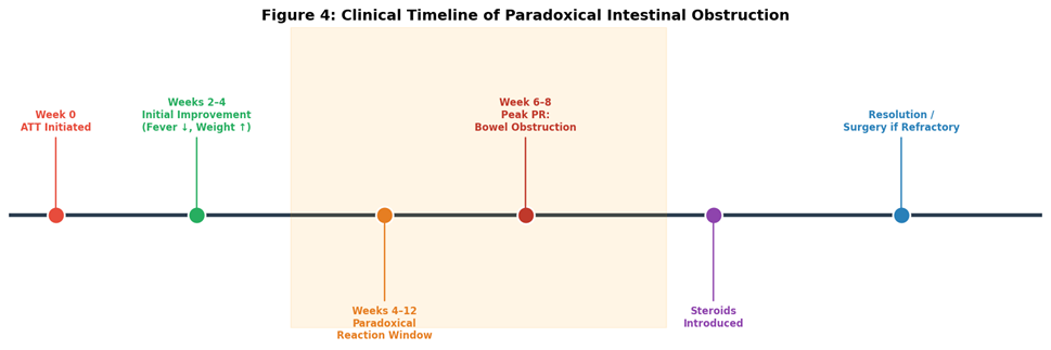
Figure 4: Clinical Timeline of Paradoxical Intestinal Obstruction – from ATT initiation through the paradoxical reaction window (weeks 4–12) to resolution or surgical intervention.
2. Epidemiology and Incidence of Paradoxical Reactions
The reported incidence of paradoxical reactions varies considerably across published series, reflecting heterogeneity in patient populations, diagnostic criteria, and the site of TB involvement. For pulmonary TB, PR has been reported in 2–23% of cases [1]. For extrapulmonary TB, including lymph node and abdominal disease, the rates are substantially higher, ranging from 15–25% [5]. A landmark multicenter study by Cheng et al. [1] documented PR in 11 of 49 (22.4%) non-HIV-infected patients with EPTB, establishing that this phenomenon is not limited to the immunocompromised.
Several patient-level risk factors predispose to PR. These include a younger age at disease onset, a larger pre-treatment disease burden as quantified by extent of lymphadenopathy or bowel wall thickening on cross-sectional imaging, low pre-treatment body mass index (BMI), and an aggressive baseline inflammatory response (high baseline CRP and ESR). Interestingly, paradoxical reactions appear more common in patients who achieve early and rapid bacteriological sterilization, suggesting that the immunological competence of the host—its ability to mount and sustain a Th1 response—is the critical determinant [7]. HIV co-infection, paradoxically, has a bidirectional relationship: while IRIS is classically described with HAART initiation in HIV-TB co-infected patients, the magnitude of PR may be attenuated in profoundly immunocompromised individuals (CD4 count < 50 cells/μL) who lack the T-cell infrastructure to mount the overwhelming inflammatory response [8].
2.1 Risk Factor Profile for PIO
Table 1: Risk Factors Associated with Paradoxical Intestinal Obstruction
|
Risk Category |
Specific Factor |
Clinical Implication |
|
Patient Demographics |
Age < 40 years; Female sex |
Higher Th1 immune reactivity |
|
Disease Burden |
Bulky lymphadenopathy (>2 cm nodes); Extensive bowel involvement |
Greater antigen load on bacterial lysis |
|
Nutritional Status |
Low BMI (<18 kg/m²) pre-treatment |
Paradoxical immune recovery on nutrition support |
|
Immunological Markers |
High baseline IFN-γ; Low IL-10 |
Pro-inflammatory cytokine dominance |
|
Bacteriological |
Rapid early mycobacterial clearance |
Sudden antigen surge; IRIS trigger |
3. Immunopathogenesis: The IRIS Framework
The pathogenesis of PR is fundamentally an immunological paradox rooted in the dynamic interplay between mycobacterial antigen burden and host immune competence. It is best understood through the Immune Reconstitution Inflammatory Syndrome (IRIS) framework—a phenomenon classically described in HIV patients commencing highly active antiretroviral therapy (HAART) but equally operant in HIV-negative individuals initiating ATT [8]. IRIS represents the "unmasking" or "paradoxical" worsening of an underlying infection as a consequence of a robust restoration of pathogen-specific immunity.
3.1 Phase I: The Pre-ATT Immune Landscape
Prior to ATT initiation, Mycobacterium tuberculosis employs multiple and sophisticated immune evasion strategies to establish chronic infection. The bacilli impair phagolysosome fusion within alveolar and gut-associated macrophages, preventing their degradation. They modulate antigen presentation by downregulating MHC class II molecules on macrophages, thereby diminishing CD4+ T-cell activation. Additionally, mycobacteria drive the production of anti-inflammatory cytokines such as Interleukin-10 (IL-10) and Transforming Growth Factor-beta (TGF-β), actively suppressing the Th1 inflammatory response. The net result is a state of controlled immune tolerance in which the host is unable to eradicate the pathogen but successfully contains it within granulomata—an immunological stalemate [8].
3.2 Phase II: The Antigenic Surge upon ATT Initiation
Upon the introduction of bactericidal agents—principally Isoniazid (which disrupts mycolic acid synthesis, a critical component of the mycobacterial cell wall) and Rifampicin (which inhibits bacterial RNA polymerase)—there is a rapid and massive destruction of viable mycobacteria within the bowel wall, mesenteric lymph nodes, and peritoneal granulomata [4]. This large-scale lysis releases an enormous deluge of mycobacterial antigens into the local tissue microenvironment and systemic circulation. These antigens include tuberculoproteins (particularly the 38-kDa and 16-kDa antigens), lipopolysaccharide components of the cell wall (lipoarabinomannan), and heat shock proteins [7]. This constitutes the central antigenic "trigger" of the paradoxical cascade.
3.3 Phase III: The Cytokine Storm and Granulomatous Expansion
The sudden availability of an immense antigen load triggers a delayed-type hypersensitivity (DTH) reaction of extraordinary magnitude. Professional antigen-presenting cells (APCs)—particularly dendritic cells (DCs) that had previously been held in an immunotolerogenic state by mycobacterial products—are now rapidly activated. They process and present mycobacterial peptides on MHC class II molecules to naive and memory CD4+ Th1 lymphocytes, which undergo clonal expansion [7,8]. These reinvigorated Th1 cells release a coordinated and overwhelming surge of pro-inflammatory cytokines, most notably:
• Interferon-gamma (IFN-γ): The signature Th1 cytokine; activates macrophages to a bactericidal, M1-polarized phenotype.
• Tumor Necrosis Factor-alpha (TNF-α): Drives granuloma formation, maintains granuloma integrity, and induces local tissue inflammation and edema.
• Interleukin-2 (IL-2): Potent T-cell proliferation factor; amplifies and sustains the immune response.
• Interleukin-17 (IL-17): Recruits neutrophils and amplifies the local inflammatory milieu.
Macrophages responding to this cytokine milieu are hyper-activated, differentiating into epithelioid cells and fusing to form multinucleated Langhans giant cells. While these are the physiological effectors of mycobacterial containment, the sheer velocity and magnitude of this response during PR leads to the rapid, uncontrolled expansion of existing granulomata within the bowel wall and mesentery [3].
3.4 Phse IV: Fibrostenosis and Luminal Compromise
Concurrent with the acute inflammatory response, healing in ATB is intrinsically and aggressively fibrotic. TGF-β—released both by macrophages and by damaged epithelial cells—is the master regulator of fibrogenesis [3]. It drives the activation and proliferation of myofibroblasts, which deposit large quantities of Type I and Type III collagen in the submucosal and serosal layers of the bowel. In the bowel, where luminal diameter is finite, this combination of acute inflammatory edema (causing mucosal and submucosal thickening) and progressive collagen deposition (causing transmural fibrosis and loss of bowel wall compliance) creates an inevitable and progressive luminal narrowing. In PIO, these processes are accelerated and amplified compared to uncomplicated ATB, converting a partial stricture—which may have been clinically quiescent for months—into a complete, functional, and painful mechanical obstruction within days [5,6].
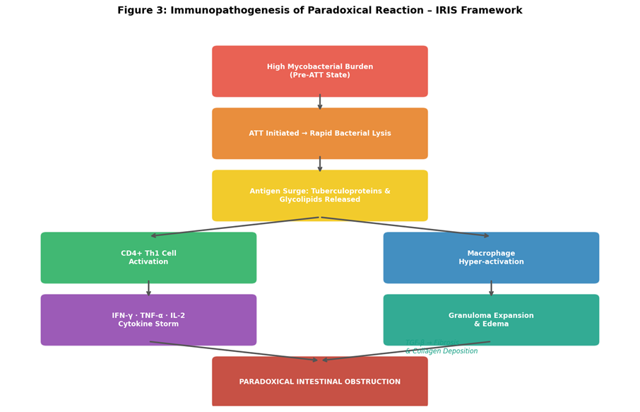
Figure 3: Immunopathogenesis of Paradoxical Intestinal Obstruction – the IRIS cascade from mycobacterial antigen release through the cytokine storm to mechanical bowel obstruction.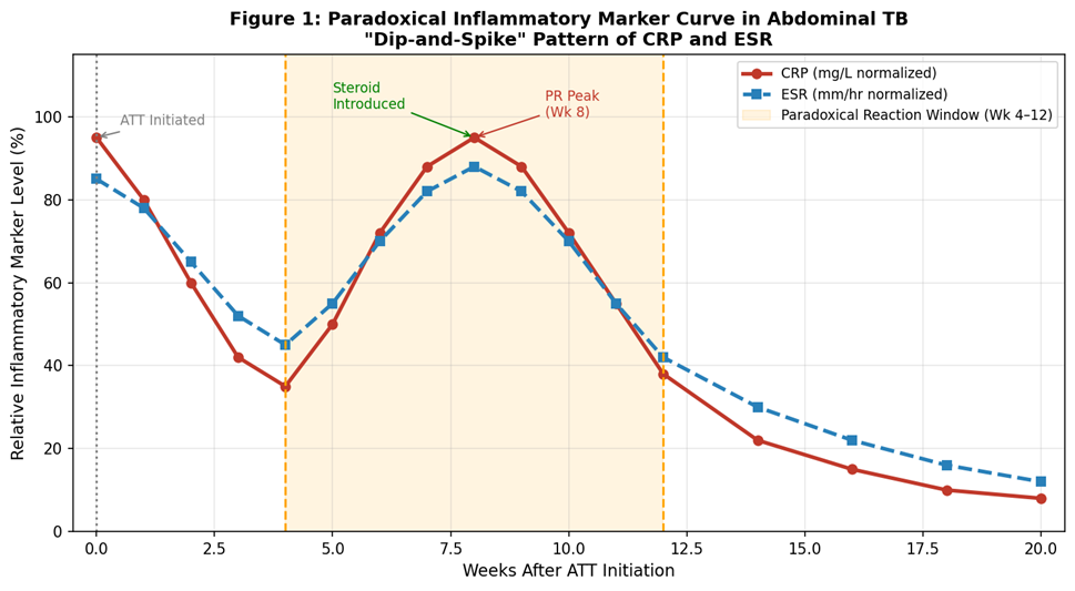
Figure 1: The 'Dip-and-Spike' pattern of inflammatory biomarkers (CRP and ESR) in Paradoxical Intestinal Obstruction. Note the initial decline after ATT initiation followed by a sharp rebound peak at weeks 6–10.
intervention.
4. Clinical Presentation and Temporal Characteristics
The clinical presentation of PIO is characterized as much by its chronology as by its symptomatology. By definition, the patient must have demonstrated unambiguous initial clinical improvement during the first 2–4 weeks of ATT—typically manifesting as resolution of fever (defervescence), improved appetite, early weight gain, and subjective well-being [1,5]. This initial improvement is critical to distinguishing PIO from primary ATT failure, where no improvement occurs. The paradoxical deterioration typically manifests within a characteristic window of 4 to 12 weeks post-initiation, with a peak incidence between weeks 6 and 10 [1,5]. Late-onset paradoxical reactions, occurring months after ATT initiation, have been documented but are significantly less common.
4.1 Cardinal Symptoms of Acute Obstruction
Patients with PIO typically present to the emergency setting with the full constellation of mechanical small bowel obstruction:
• Colicky Abdominal Pain: The hallmark symptom, characterized by a distinct crescendo-decrescendo pattern. Periumbilical or central in origin, reflecting hyperperistalsis of the small bowel against a fixed mechanical obstruction at the terminal ileum or ileocecal junction. Pain episodes may last 2–4 minutes, separated by pain-free intervals [3].
• Vomiting: May be bilious in proximal obstructions or feculent in distal small bowel obstructions. Progressive and frequent, leading to significant fluid and electrolyte derangements, particularly hypokalemic, hypochloremic metabolic alkalosis.
• Absolute Obstipation: The inability to pass flatus or stool, indicating complete luminal occlusion. This distinguishes complete from partial obstruction and has significant management implications.
• Abdominal Distension: Progressive, tympanic distension reflecting gas accumulation in obstructed loops proximal to the stricture. Auscultation reveals hyperactive, high-pitched 'tinkling' bowel sounds initially; sounds may become absent in later stages suggesting ileus or vascular compromise.
• Dehydration and Hemodynamic Compromise: Due to third-space fluid losses into obstructed bowel loops and persistent vomiting, patients may present with tachycardia, hypotension, and oliguria.
A clinically intriguing and paradoxical observation in PIO is the relative absence of systemic inflammatory signs. Unlike a pyogenic bowel obstruction, patients with PIO are frequently afebrile or only low-grade febrile. Moreover, these patients may appear nutritionally improved compared to their pre-treatment baseline—a reflection of improved absorption following mycobacterial eradication—emphasizing that PIO is a disease of immune hyper-competence rather than systemic immune failure [1,5].
4.2 Differential Diagnosis
The differential diagnosis of bowel obstruction in a patient on ATT must be approached systematically. Beyond PIO, the clinician must consider: (1) Mechanical obstruction due to adhesions from prior surgery or peritoneal TB; (2) MDR-TB with progressive bowel involvement; (3) Non-compliance resulting in incomplete bacterial suppression and ongoing inflammatory destruction; (4) Secondary bacterial enteritis superimposed on TB; (5) Crohn's disease—which can mimic ATB radiologically, histologically, and clinically, and may coexist in endemic regions [6]; (6) Bowel malignancy, including intestinal lymphoma, which must be excluded particularly in older patients or those with unusual radiological findings.
5. Diagnostic Evaluation: Differentiating PR from Treatment Failure
The diagnostic imperative in suspected PIO is the exclusion of drug resistance and treatment failure before labeling the deterioration as paradoxical. This requires a systematic, multimodal diagnostic approach integrating clinical history, laboratory biomarkers, microbiological testing, and advanced cross-sectional imaging.
5.1 Clinical Criteria
The clinical diagnosis of PR requires fulfillment of all three of the following criteria, as proposed by established consensus guidelines [1,5]: (1) Unambiguous clinical improvement during the first 2–4 weeks of ATT (the 'honeymoon period'); (2) Subsequent clinical, radiological, or laboratory deterioration occurring during the 4–12-week window; (3) Exclusion of alternative diagnoses including drug resistance, non-compliance, secondary infection, and drug toxicity.
5.2 Laboratory Biomarkers
Routine hemograms characteristically show a normalized or near-normal total white blood cell (WBC) count, contrasting with the leukocytosis seen in bacterial superinfection [3]. The pathognomonic laboratory signature of PIO is the characteristic 'dip-and-spike' pattern of acute-phase reactants: CRP typically normalizes within the first 3–4 weeks of ATT (corresponding to the 'honeymoon period'), and then surges dramatically—often to levels exceeding 150 mg/L—during the paradoxical phase [3,5]. ESR shows a similar but more attenuated and delayed pattern. Fecal calprotectin, a validated marker of intestinal neutrophilic inflammation, is markedly elevated in PIO, reflecting the intense mucosal and submucosal inflammatory infiltrate.
Crucially, microbiological testing is negative for viable organisms. AFB smear and culture from any tissue or fluid sampled (peritoneal fluid, lymph node aspirate) are negative, confirming sterile inflammation. GeneXpert MTB/RIF (Xpert)—the WHO-endorsed molecular diagnostic tool—shows no evidence of viable, replicating mycobacterial DNA in the vast majority of PIO cases, and critically, demonstrates no rifampicin resistance mutations, effectively ruling out MDR-TB [4]. This combination of negative microbiological data with florid clinical and inflammatory deterioration is the cornerstone of the PR diagnosis.
5.3 Radiological Signatures on CT Enterography
Computed Tomography (CT) enterography—performed with oral and intravenous contrast—is the imaging modality of choice for evaluating PIO [3,6]. It provides detailed information on the degree of luminal narrowing, bowel wall morphology, mesenteric changes, and complications. The key CT findings specific to PIO include:
• The 'Fat Halo' (Target) Sign: Pathognomonic of acute, intense inflammation. Submucosal edema appears as a low-attenuation (hypodense) layer between the brightly contrast-enhancing mucosa and the muscularis propria, creating a trilaminar or 'target' pattern on axial sections. Its presence in a patient on ATT strongly suggests PIO over fibrotic stricture alone [3].
• Comb Sign: Engorgement of the vasa recta supplying the inflamed bowel loop, creating a 'comb-like' pattern in the mesentery. Indicates active hyperemia and is a sensitive marker of acute inflammation.
• Reactive Mesenteric Lymphadenopathy: Rapid enlargement of mesenteric nodes (>1.5 cm in short axis), often with central low-attenuation necrosis and a peripheral rim of enhancement on contrast imaging—the 'central necrosis' sign. In PIO, multiple enlarged nodes may conglomerate into a mass-like configuration ('nodal conglomerate') causing extrinsic luminal compression of the ileum or colon [6].
• Sclerosing Encapsulating Peritonitis (Abdominal Cocoon): In severe peritoneal TB, PR can manifest as intense fibrous encapsulation of the entire small bowel, which becomes encased in a thick fibrotic membrane—the 'cocoon.' This catastrophic complication results in a functional closed-loop obstruction and typically requires surgical intervention.
Table 2: Differential Diagnosis – Paradoxical Reaction vs. Treatment Failure
|
Diagnostic Feature |
Paradoxical Reaction (PIO) |
Treatment Failure / MDR-TB |
|
Onset Timeline |
4–12 weeks after documented initial improvement |
Continuous worsening or no initial response to ATT |
|
Constitutional Fever |
Usually absent or low-grade |
Persistent high-grade fever |
|
Mycobacterial Culture |
Negative (sterile inflammation) |
Positive for M. tuberculosis |
|
GeneXpert MTB/RIF |
Negative; No rifampicin resistance |
Positive; May show rpoB mutation |
|
CRP / ESR |
Classic 'dip-and-spike' pattern |
Persistently or progressively elevated |
|
Radiological Pattern |
Fat halo sign, massive lymphadenopathy, new strictures |
Progressive disseminated or cavitatory disease |
|
Nutritional Status |
Improved or stable |
Progressive decline, cachexia |
|
Response to Steroids |
Dramatic improvement within 48–72 hours |
No response; may worsen |
6. Management Strategies: The 'Steroid vs. Steel' Debate
The management of PIO demands a multidisciplinary team approach—encompassing the gastroenterologist or internist directing ATT, the infectious disease specialist providing expert guidance on drug resistance and drug interactions, and the abdominal surgeon available for timely operative intervention when medical therapy fails. The overarching and non-negotiable principle is that ATT must not be discontinued, modified, or 'de-escalated' when a diagnosis of PR is established and drug resistance has been rigorously excluded [4,5]. Stopping ATT risks mycobacterial rebound, disease progression, and the development of acquired drug resistance.
6.1 Supportive and Resuscitative Measures
Initial management of the acute presentation follows standard surgical principles for bowel obstruction. Intravenous fluid resuscitation—using balanced crystalloids to correct dehydration and electrolyte imbalances (particularly hypokalemia and hyponatremia)—is paramount. A nasogastric (NG) tube is placed for gastric decompression, which reduces the volume of proximal bowel secretions and alleviates the distending pressure on the obstructed loop. Serial abdominal examinations and plain radiographs are performed to monitor for signs of bowel ischemia or perforation [6].
6.2 Pharmacological Immunomodulation: Systemic Corticosteroids
Given that PIO is driven by an orchestrated immune overreaction—specifically a TNF-α and IFN-γ dominated cytokine storm—targeted immunomodulation is the logical and evidence-supported first-line pharmacological intervention [4,5]. The therapeutic objective is to suppress the exuberant Th1 immune response, reduce capillary permeability and perilesional edema, and thus convert a functionally complete obstruction to a partial one through which luminal contents can traverse, allowing conservative management.
Prednisolone (oral) or Methylprednisolone (intravenous in patients unable to tolerate oral intake) is the mainstay of corticosteroid therapy in PIO. The recommended regimen is Prednisolone 0.5 to 1.0 mg/kg/day (maximum 40–60 mg/day) administered for 2 to 4 weeks, followed by a gradual, systematic taper at a rate of 5–10 mg per week over a further 6 to 8 weeks [4,5]. The clinical response to corticosteroids in established PIO, when correctly diagnosed, is often dramatic and therapeutically confirmatory: resolution of obstructive symptoms—including the resumption of flatus passage, reduction in vomiting, and symptomatic pain relief—frequently occurs within 48 to 72 hours of steroid initiation. This rapid response itself serves as a form of 'therapeutic diagnostic confirmation,' reinforcing the PR diagnosis.
It is imperative that corticosteroids are administered under the 'cover' of adequate, sensitive ATT to prevent the reactivation of latent, as yet unstabilized, mycobacterial foci. Prophylaxis against steroid-related complications—including Pneumocystis jirovecii pneumonia (PJP prophylaxis with co-trimoxazole in immunocompromised patients), osteoporosis, hyperglycemia, and gastric ulceration—must be concurrent with steroid therapy [4]
6.3 Biologic and Targeted Immunotherapy
In rare cases where patients have absolute contraindications to systemic corticosteroids (active peptic ulceration with hemorrhage, uncontrolled diabetes mellitus, severe psychiatric disorders worsened by steroids) or demonstrate steroid-refractory PIO after 5–7 days of adequate therapy, targeted biologic agents represent an emerging therapeutic frontier [5]. Infliximab, a chimeric monoclonal antibody that specifically neutralizes TNF-α, has been successfully employed in isolated case reports and small case series to terminate the paradoxical inflammatory cascade in steroid-refractory IRIS. The theoretical basis is compelling—TNF-α is a central mediator of the cytokine storm—and the precedent for anti-TNF therapy in refractory IRIS following HAART initiation is well-established [8]. However, the potential risk of reactivating residual mycobacterial infection by blocking TNF-α—a cytokine indispensable for granuloma maintenance and integrity—necessitates extreme caution, close microbiological monitoring, and currently limits this approach to tertiary centers with specialist expertise.
6.4 Surgical Intervention: Indications, Principles, and Techniques
Conservative pharmacological management is successful in resolving PIO in approximately 75–80% of cases [5,6]. However, surgery remains an indispensable component of the therapeutic armamentarium for refractory or complicated cases. The critical skill lies in identifying the precise indications for operative intervention and timing surgery to minimize morbidity while preventing catastrophic complications:
• Absolute Indications: Bowel perforation resulting in generalized purulent or fecal peritonitis; Gangrenous bowel with transmural ischemic necrosis (an immediate life-threatening emergency).
• Relative/Elective Indications: Failure of conservative management—defined as persistence of complete, absolute obstruction beyond 72–96 hours of maximal medical therapy (IV steroids, NG decompression, IV fluids); Clinically significant partial obstruction unresponsive to steroids after 2 weeks; Abdominal cocoon not amenable to conservative management.
The surgical philosophy in abdominal tuberculosis has fundamentally evolved over the past two decades. The historical approach of aggressive resection has been largely abandoned in favor of a bowel-conserving strategy, driven by the recognition that the bowel in ATB is friable, edematous, and poorly vascularized, making anastomoses at high risk of leakage, fistula formation, and dehiscence [3,6]. The two principal bowel-conserving techniques are:
• Stricturoplasty: The procedure of choice for short (< 5 cm: Heineke-Mikulicz stricturoplasty) or medium-length (5–15 cm: Finney stricturoplasty) strictures. The bowel is opened longitudinally along the antimesenteric border and closed transversely, widening the luminal diameter without sacrificing any bowel. This is particularly valuable in patients with multiple strictures, where sequential bowel resections would risk creating short bowel syndrome [3].
• Segmental Bowel Resection: Reserved for gangrenous bowel, long strictures (>15 cm) that are not amenable to stricturoplasty, or persistent fistula or perforation sites. Primary anastomosis, if performed, carries a high leakage risk in the setting of active ATB; a temporary diverting ileostomy followed by delayed anastomotic reversal is often the safer approach in contaminated fields [6].
• Peritonectomy for Abdominal Cocoon: In cases of sclerosing encapsulating peritonitis, careful, meticulous adhesiolysis and excision of the thickened fibrotic peritoneal membrane encasing the small bowel loops—often performed by experienced laparoscopic or open surgeons—is required. Inadvertent enterotomy during lysis, with subsequent fistula formation, is the most feared complication [3].
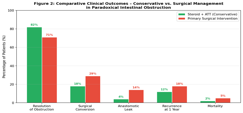
Figure 2: Comparative Clinical Outcomes in Paradoxical Intestinal Obstruction – conservative corticosteroid management versus primary surgical intervention across five key endpoints.
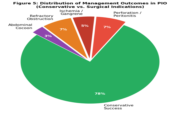
Figure 5: Distribution of management approaches in PIO — the majority (78%) resolve with conservative therapy; surgery is required in approximately 22% for specific indications.
6.5 Proposed Management Algorithm
Table 3: Step-by-Step Management Algorithm for Suspected Paradoxical Intestinal Obstruction
|
Step |
Action |
Detail / Rationale |
|
1 |
Confirm initial ATT response |
Document 2–4 weeks of clinical improvement; if absent, consider MDR-TB or non-compliance first |
|
2 |
Exclude drug resistance |
GeneXpert MTB/RIF on any accessible specimen; sputum if pulmonary co-disease; culture and DST |
|
3 |
Resuscitate & Decompress |
IV fluids, electrolyte correction, NGT, urinary catheter, strict I/O monitoring |
|
4 |
Continue ATT unchanged |
Never stop or modify ATT if PR is suspected; modification risks resistance development |
|
5 |
Initiate corticosteroids |
Prednisolone 0.5–1.0 mg/kg/day; monitor for resolution within 48–72 hrs; protect with PJP prophylaxis |
|
6 |
Serial clinical review |
Repeat abdominal XR at 24–48 hrs; clinical examination for peritonism; WBC, CRP trend |
|
7 |
Surgical review at 72–96 hrs |
If complete obstruction persists → OR; if partial obstruction with clinical improvement → continue conservative management; taper steroids over 8 weeks |
DISCUSSION :
The paradigm of PIO fundamentally and permanently reshapes how clinicians must conceptualize disease progression and therapeutic response in TB. The historical, intuitive, and deeply ingrained clinical assumption that 'worsening symptoms = treatment failure' is directly challenged by the IRIS framework. This conceptual reorientation has profound implications for clinical practice: it prevents the unnecessary, harmful, and sometimes iatrogenic escalation of ATT to second-line, nephrotoxic, and ototoxic drugs (aminoglycosides such as kanamycin; cycloserine; ethionamide) which carry a substantially greater adverse effect burden and which would be entirely inappropriate for a patient whose bacteria are in fact being effectively eradicated [4,8].
The diagnostic challenge is compounded by the significant clinical and radiological overlap between PIO, Crohn's disease (CD), intestinal malignancy, and complicated ATB. Crohn's disease, in particular, can mimic ATB in virtually every dimension: the clinical presentation, the predilection for the terminal ileum and ileocecal junction, the histological finding of non-caseating granulomata (though caseating granulomata favor TB), and the radiological appearance of mural thickening, strictures, and fistulae [6]. In endemic TB zones, misdiagnosis of Crohn's disease as ATB—or vice versa—is unfortunately common. The implications of this misdiagnosis are severe, as the treatment of CD with immunosuppressants (azathioprine, biological agents) in a patient with undiagnosed active TB is catastrophic [3].
The psychological dimension of PIO deserves dedicated and emphatic attention in clinical practice. A patient who has witnessed their own clinical improvement—the resolution of fever, the return of appetite, the ability to eat—only to be suddenly and painfully readmitted with bowel obstruction, often experiences profound demoralization, loss of trust in the therapeutic regimen, and anxiety about the future. The clinician's ability to explain PIO in accessible terms—that this painful crisis is, in fact, evidence of a robust and functionally recovering immune system successfully eliminating the bacteria—is both therapeutically supportive and crucial for maintaining ATT adherence during the subsequent weeks of corticosteroid therapy [1].
Looking forward, the future of PIO management lies in predictive medicine and personalized immunomodulation. Emerging research in transcriptomics and proteomics suggests that specific gene expression signatures at ATT initiation—particularly patterns in Type I interferon signaling and TNF superfamily gene clusters—may be capable of identifying patients at high risk of developing PR before any clinical symptoms manifest [7,8]. If validated prospectively, this could enable a prophylactic strategy: the co-administration of low-dose corticosteroids from ATT day one in high-risk patients, blunting the immune over-reaction before it translates into clinical crisis. Similarly, specific HLA haplotypes (particularly HLA-DR and HLA-DQ alleles involved in antigen presentation) are being investigated as genetic predispositions to PR. Pharmacogenomic profiling may ultimately allow a truly personalized ATT regimen that pre-empts immunological complications rather than reacting to them.
CONCLUSION :
Paradoxical Intestinal Obstruction is a severe, clinically deceptive, and potentially life-threatening immune-mediated complication of otherwise successful antitubercular therapy in abdominal tuberculosis. Driven by a massive release of mycobacterial antigens upon bacterial lysis and a subsequent, immune-reconstitution mediated TNF-α and IFN-γ cytokine storm, the resulting accelerated fibrotic and edematous changes within the bowel wall and mesentery precipitate mechanical obstruction in a subset of patients who are, paradoxically, responding to treatment.
Accurate and timely diagnosis rests on three pillars: a high and maintained index of clinical suspicion in any ATB patient deteriorating during the 4–12 week window; characteristic radiological findings on CT enterography (fat halo sign, lymph node necrosis, new strictures); and the rigorous exclusion of drug resistance via GeneXpert and culture. The cornerstone of management is the uninterrupted and unmodified continuation of sensitive ATT, coupled with systemic corticosteroids to suppress the immune over-reaction, with surgical intervention reserved strictly for refractory absolute obstructions, bowel ischemia, and perforation.
The recognition that 'cure can trigger crisis' is not merely an academic curiosity—it is a clinical imperative that, when understood and appropriately applied, prevents diagnostic misclassification, inappropriate ATT modification, unnecessary surgical morbidity, and patient demoralization. As the global TB burden remains immense and ATB continues to be encountered with significant frequency in endemic regions, a clear, evidence-based framework for navigating PIO is indispensable for every practitioner involved in the care of TB patients.
BIBLIOGRAPHY:
1. Cheng, V.C., Ho, P.L., Lee, R.A., et al. (2002). Clinical spectrum of paradoxical deterioration during antituberculosis therapy in non-HIV-infected patients. European Journal of Clinical Microbiology & Infectious Diseases, 21(11), 803–809.
2. Breen, R.A., Smith, C.J., Bettinson, H., et al. (2004). Paradoxical reactions during tuberculosis treatment in patients with and without HIV co-infection. Thorax, 59(8), 704–707.
3. Kapoor, V.K. (2014). Abdominal tuberculosis. Postgraduate Medical Journal, 74(874), 459–467.
4. World Health Organization. (2022). Global Tuberculosis Report 2022. WHO Press, Geneva. ISBN 978-92-4-006172-9.
5. Cho, O.H., Park, K.H., Kim, T., et al. (2009). Paradoxical responses in non-HIV-infected patients with peripheral lymph node tuberculosis. Journal of Infection, 59(1), 56–61.
6. Uzunkoy, A., Harma, M., & Harma, M. (2004). Diagnosis of abdominal tuberculosis: experience from 11 cases and review of the literature. World Journal of Gastroenterology, 10(24), 3647–3649.
7. Bell, L.C., & Noursadeghi, M. (2018). Pathogenesis of HIV-1 and Mycobacterium tuberculosis co-infection. Nature Reviews Microbiology, 16(2), 80–90.
8. Mehdorn, H.M., & Schirmer, M. (2019). Immune reconstitution inflammatory syndrome (IRIS) in tuberculosis. The Lancet Infectious Diseases, 19(5), 459–460.
9. Sharma, S.K., & Mohan, A. (2004). Extrapulmonary tuberculosis. Indian Journal of Medical Research, 120(4), 316–353.
10. Singh, B., & Gupta, R. (2020). Paradoxical reactions to anti-tuberculosis therapy: A comprehensive review. Journal of Infection and Chemotherapy, 26(9), 883–891.