Distribution and Functional Correlation of Mature Dendritic Cells and CD8⁺ Cytotoxic T Lymphocytes in the Tumor Microenvironment of Triple-Negative Breast Cancer: A Cross-Sectional Immunohistochemical Study
- Aasiya Niazi , Assistant Professor Pathology Rawalpindi Medical University, Pakistan
- Asma Ejaz , Department of Pathology, Allama Iqbal Medical College, Lahore, Pakistan
- Sadia Anwar , Department of Pathology, Allama Iqbal Medical College, Lahore, Pakistan
- Farah Kalsoom , Assistant professor Histopatholology, Services Institute of Medical Sciences, Services Hospital, Lahore. Pakistan
- Hamid Nawaz Khokhar , Assistant Professor Pathology, Sahara Medical College, Narowal, Pakistan
- Muhammad Abdullah , Assistant Professor Pathology, Sahara Medical College, Narowal, Pakistan
Article Information:
Abstract:
Background: Triple-negative breast cancer (TNBC) is the most immunogenic breast cancer subtype, yet the functional interplay between mature dendritic cells (mDCs) and CD8⁺ cytotoxic T lymphocytes (CTLs) within its tumor microenvironment (TME) remains incompletely characterized. This study evaluated the distribution, density, and correlation of CD83⁺ mDCs and CD8⁺ CTLs in TNBC tissue and explored their association with clinicopathological parameters. Methodology: A retrospective cross-sectional immunohistochemical study was conducted on formalin-fixed paraffin-embedded tumor sections from 113 samples collected from Allama Iqbal Medical College, Jinnah hospital and Services Hospital, Lahore, from August 2025 to March 2026. CD83 and CD8 expression was assessed by immunohistochemistry. Densities (cells/mm²) were quantified in the intratumoral, stromal, and invasive margin compartments. Correlations were assessed using Spearman’s rank coefficient, and associations with clinicopathological variables were examined using Mann-Whitney U, Kruskal-Wallis, and Chi-square tests. Results & Findings: Median stromal densities were 8.5 (IQR 4.5–16.2) for CD83⁺ mDCs and 48.0 (IQR 28.5–85.0) for CD8⁺ CTLs. A significant positive correlation was found between the two populations in the stromal compartment (ρ=0.58, p<0.001) and invasive margin (ρ=0.62, p<0.001). Higher mDC and CTL densities were each significantly associated with lower histological grade, absence of lymph node metastasis, and earlier pathological stage (all p<0.05). The combined CD83-high/CD8-high phenotype, present in 31.0% of cases, was significantly enriched in Grade II, node-negative, and Stage I tumors (p<0.01). Conclusion: Mature dendritic cell density positively correlates with CD8⁺ cytotoxic T lymphocyte infiltration in TNBC, and their co-enrichment identifies a subset of tumors with favorable clinicopathological features. These findings highlight the biological and potential prognostic importance of the DC–CTL immunological axis and support the integration of dual-marker immunohistochemistry into the pathological assessment of TNBC.
Keywords:
Article :
INTRODUCTION:
Breast cancer constitutes the most frequently diagnosed malignancy and the leading cause of cancer-related mortality among women worldwide.¹ Within the heterogeneous spectrum of breast cancer subtypes, triple-negative breast cancer (TNBC) defined by the immunohistochemical absence of estrogen receptor (ER) and progesterone receptor (PR) expression and the lack of human epidermal growth factor receptor 2 (HER2) overexpression or gene amplification occupies a position of singular clinical concern.² The triple-negative phenotype precludes the use of endocrine therapies and HER2-directed biological agents, substantially limiting the therapeutic options available in the clinic.³ TNBC accounts for approximately 10–20% of all incident breast cancers globally and disproportionately affects younger premenopausal women, individuals of African ancestry, and carriers of germline BRCA1 mutations.⁴,⁵ Clinically, TNBC is characterized by an aggressive biological behavior, a high proliferative index, an elevated propensity for early visceral and central nervous system metastasis, and a markedly inferior five-year survival rate relative to hormone receptor-positive subtypes.⁶ Approximately 46% of patients develop distant metastases within three years of initial diagnosis, and the overall five-year relative survival for combined disease stages remains substantially lower than that observed for other breast cancer subtypes.⁷ These unfavorable oncological outcomes underscore the urgent need for a deeper mechanistic understanding of TNBC biology, particularly with respect to the immune contexture of the tumor microenvironment (TME).
The TME in TNBC represents a complex and dynamic ecosystem in which diverse cellular and acellular constituents collectively govern tumor progression, immune evasion, and therapeutic responsiveness.⁸ Among all breast cancer subtypes, TNBC harbors the most immunologically active microenvironment, distinguished by abundant immune cell infiltration, elevated immune checkpoint expression, and high programmed death-ligand 1 (PD-L1) levels on both neoplastic and stromal cells.⁹ The cellular composition of the TNBC TME encompasses immunoreactive populations including CD8⁺ cytotoxic T lymphocytes (CTLs), CD4⁺ helper T cells, natural killer cells, and dendritic cells (DCs) alongside potently immunosuppressive elements such as regulatory T cells, myeloid-derived suppressor cells, M2-polarized tumor-associated macrophages, and cancer-associated fibroblasts.¹⁰ The dynamic equilibrium between these opposing immunological forces fundamentally dictates whether the TME adopts an immune-permissive or immune-excluded phenotype, with profound implications for clinical outcome and the probability of response to immunotherapy.¹¹ Tumor-infiltrating lymphocytes (TILs), in particular, have emerged as both prognostic and predictive biomarkers in TNBC, with stromal TIL density consistently correlated with improved pathological complete response rates following neoadjuvant chemotherapy and superior long-term survival.¹²
Dendritic cells serve as the pivotal sentinels at the interface of innate and adaptive immunity, functioning as the most potent professional antigen-presenting cells in the human immune system.¹³ DCs continuously patrol peripheral tissues, capture and process tumor-associated antigens, and migrate to tumor-draining lymph nodes, where they present processed peptide–MHC complexes to naïve T lymphocytes, thereby initiating and orchestrating antigen-specific adaptive immune responses.¹⁴ The functional competence of DCs within the TME is critically governed by their maturation status. Immature DCs exhibit limited immunostimulatory capacity and may, under certain conditions, promote immunological tolerance, whereas mature DCs characterized by high surface expression of the co-stimulatory molecules CD80, CD86, and CD40, upregulation of MHC class II, and expression of the maturation marker CD83 possess the full capacity to prime and expand antigen-specific T cell clones. CD83, a member of the immunoglobulin superfamily selectively expressed on terminally matured DCs, is widely employed as a robust immunohistochemical marker for the identification and enumeration of functionally competent mature DCs in tissue sections.¹⁷ Recent mechanistic studies have demonstrated that the transmembrane domain of CD83 enhances MHC class II and CD86 expression by counteracting IL-10–driven, MARCH1-mediated ubiquitination, thereby stabilizing the antigen-presenting machinery on the mature DC surface.15
The functional collaboration between mature DCs and CD8⁺ CTLs constitutes the immunological cornerstone of effective antitumor surveillance. Following intratumoral antigen capture, mature conventional type 1 DCs engage in MHC class I cross-presentation, whereby extracellular tumor-derived antigens are processed through the proteasomal pathway and loaded onto MHC class I molecules for the direct priming of CD8⁺ T cells. This cross-presenting capacity is indispensable for the generation of a robust CTL response directed against the tumor, and accumulating evidence indicates that defects in this pathway represent a critical mechanism of immune evasion in TNBC. Notably, tumor-derived CDC37, transferred via extracellular vesicles into DC endosomes, has been shown to impair antigen translocation and cross-presentation in type 1 conventional DCs, resulting in a deficiency of tumor-specific CTLs and diminished immunotherapy responses in TNBC patients. Furthermore, intratumoral immune triads spatial clusters in which a single DC simultaneously presents antigen to both CD4⁺ helper T cells and CD8⁺ CTLs have been identified as essential organizational units for sustaining effective antitumor immunity and for preventing terminal CD8⁺ T cell exhaustion. The spatial distribution, density, and functional integrity of these cellular partnerships within the TNBC TME therefore carry substantial biological and prognostic significance. CD8⁺ CTLs represent the principal cytotoxic effectors of the adaptive antitumor immune response. Following DC-mediated priming, naïve CD8⁺ T cells undergo activation, clonal expansion, and differentiation into cytotoxic effectors that traffic to the TME and mediate tumor cell killing via the perforin–granzyme pathway and Fas–FasL interactions.²² In TNBC, CD8⁺ T lymphocytes are among the most abundant immune populations within the TME, and their intratumoral density has been consistently associated with improved clinical prognosis, including higher pathological complete response rates to neoadjuvant chemotherapy, superior disease-free survival, and more favorable overall survival.16 Nevertheless, the antitumor efficacy of intratumoral CD8⁺ CTLs is profoundly modulated by the immunosuppressive forces operative within the TNBC TME. Chronic antigen exposure, hypoxia, nutrient competition, and the elaboration of immunosuppressive cytokines collectively drive CTL exhaustion, a state characterized by the progressive loss of effector function, upregulation of inhibitory checkpoint receptors including PD-1, TIM-3, and LAG-3, and ultimately functional inactivation.²⁵ The presence of exhausted CD8⁺ T cells within the TME, particularly in African American women with TNBC who exhibit a pro-tumorigenic immune contexture, is associated with inferior clinical outcomes and resistance to immunotherapeutic intervention.17
The immunological richness of the TNBC TME has provided the biological rationale for the application of immune checkpoint inhibition. Pembrolizumab, an anti-PD-1 monoclonal antibody, remains the only immune checkpoint inhibitor to receive regulatory approval for both early-stage and metastatic TNBC as of December 2024, based on the survival advantages demonstrated in the KEYNOTE-355 and KEYNOTE-522 trials. Exploratory analyses from the phase III IMpassion130 trial further confirmed that tumor CD8 positivity and stromal TIL density were associated with improved outcomes in patients receiving atezolizumab plus nab-paclitaxel, although these biomarkers exhibited high collinearity with PD-L1 expression. Despite these advances, the majority of TNBC patients do not achieve durable responses to immune checkpoint blockade, highlighting the limitations of PD-L1 expression as a solitary predictive biomarker and emphasizing the necessity to characterize the broader immune architecture of the TME, including the functional interplay between mature DC populations and CTL infiltrates.²⁹ Immunohistochemical studies across diverse tumor types have consistently demonstrated that mature DC density within the tumor stroma and invasive margin correlates inversely with tumor stage, with higher mature DC infiltrates associated with a lower incidence of distant metastasis and prolonged overall survival. In ovarian carcinoma, a high intratumoral density of mature DCs correlates with favorable immune infiltrates and improved patient survival, accompanied by the concomitant enrichment of CD8⁺ CTLs, CD4⁺ T helper cells, and natural killer cells.18 Similarly, immunohistochemical analyses of cervical lesions using CD83 have revealed a progressive reduction in mature DC density with advancing malignant transformation, underscoring the role of DC dysfunction in immune escape. Despite these insights derived from other solid tumors, a comprehensive immunohistochemical characterization of mature DC distribution and its functional correlation with CD8⁺ CTL infiltration specifically within the TNBC TME has not been performed, representing a significant gap in the current literature.
The identification of the spatial distribution, density, and functional correlation of mature DCs and CD8⁺ CTLs within the TNBC TME carries considerable scientific and translational relevance. Elucidating whether mature DC infiltration co-distributes with CD8⁺ CTL density and whether this co-distribution associates with established clinicopathological parameters such as tumor grade, nodal status, and pathological stage may provide fundamental mechanistic insights into the immunological determinants of antitumor immunity in TNBC. Moreover, characterizing these immune populations through standardized immunohistochemical methodology offers a clinically applicable and cost-effective approach to immune profiling that could complement molecular and genomic biomarker platforms. Accordingly, the present cross-sectional immunohistochemical study was designed to systematically evaluate the distribution and density of mature DCs (identified by CD83 immunolabeling) and CD8⁺ CTLs in TNBC tissue specimens, to determine the functional correlation between these two immune populations, and to explore their association with key clinicopathological characteristics.
Objective of the study
The present cross-sectional immunohistochemical investigation was specifically designed to address the existing gap in the literature concerning the immunological architecture of the TNBC tumor microenvironment, with a particular focus on the distribution, density, and functional interrelationship of mature dendritic cells (mDCs) and CD8⁺ cytotoxic T lymphocytes (CTLs). The objectives were structured to systematically characterize these immune populations, define their spatial and quantitative correlations, and explore their clinical relevance, thereby constructing an integrated immunological profile of TNBC with potential biomarker and therapeutic implications. The specific objectives were:
1. To evaluate the distribution and density of CD83⁺ mature dendritic cells and CD8⁺ cytotoxic T lymphocytes within the intratumoral, stromal, and invasive margin compartments of the TNBC microenvironment using standardized immunohistochemistry on formalin-fixed paraffin-embedded tissue sections.
2. To determine the correlation between mature dendritic cell and CD8⁺ T lymphocyte densities and to explore their association with key clinicopathological parameters, including histological grade, lymph node status, and pathological TNM stage, in order to define the clinical relevance of the DC–CTL immunological axis in TNBC.
METHODOLOGY:
This retrospective cross-sectional study was conducted at the Department of Pathology, Allama Iqbal Medical College, Lahore which provides diagnostic services to Jinnah Hospital and Services Hospital, Lahore from August 2025 to March 2026. Ethical approval was obtained from the Institutional Review Board of Allama Iqbal Medical College. Because only archived, de-identified tissue samples and clinical records were used, the requirement for informed consent was waived.
Pathology records from Jinnah Hospital and Services Hospital were searched to identify all female patients who underwent surgical resection for primary invasive breast carcinoma between January 2020 and December 2024. Cases were included if: (i) histological examination confirmed invasive breast carcinoma of no special type; (ii) the tumor was triple-negative, defined as estrogen receptor (ER) <1%, progesterone receptor (PR) <1%, and human epidermal growth factor receptor 2 (HER2) negative (immunohistochemistry score 0/1+, or 2+ with a negative fluorescence in situ hybridization test); and (iii) an adequate formalin-fixed paraffin-embedded (FFPE) tissue block was available, containing viable tumor tissue and an identifiable invasive margin. Patients were excluded if they had received neoadjuvant chemotherapy or radiotherapy prior to surgery, had recurrent or metastatic disease at presentation, or if the available tissue block showed extensive necrosis or an insufficient tumor area (<4 mm²). After applying these criteria, a total of 113 cases constituted the final study cohort. Age, menopausal status, tumor size, histological grade, axillary lymph node status, and pathological TNM stage (American Joint Committee on Cancer, 8th edition) were retrieved from pathology request forms and available clinical records. All data were anonymized prior to analysis.
From each FFPE block, 4-μm thick serial sections were cut, mounted on positively charged slides, and dried at 37°C overnight. Deparaffinization was performed with xylene, followed by rehydration through graded ethanol to distilled water. Heat-induced antigen retrieval was carried out in a domestic pressure cooker using citrate buffer (pH 6.0) for 20 minutes. Endogenous peroxidase activity was blocked with 3% hydrogen peroxide for 15 minutes, and non-specific protein binding was minimized with 5% normal goat serum. Primary antibodies were applied as follows: mouse monoclonal anti-CD83 to identify cytotoxic T lymphocytes. Incubation with the primary antibody was performed for 60 minutes at room temperature in a humidified chamber. Detection was achieved using a horseradish peroxidase-conjugated polymer system with 3,3′-diaminobenzidine (DAB) as the chromogen. Slides were counterstained with Mayer’s hematoxylin, dehydrated, and mounted. A section of human tonsil was included in each staining run as a positive control; negative controls were prepared by substituting the primary antibody with an isotype-matched immunoglobulin.
All stained slides were examined independently by two histopathologists who were blinded to the clinical and pathological data. For each case, three tumor microenvironment compartments were assessed: the intratumoral compartment (immune cells within tumor cell nests), the stromal compartment (fibrovascular stroma between tumor nests), and the invasive margin (a 1-mm wide zone at the interface between tumor and adjacent normal tissue; this was scored only when a clear invasive front was identifiable in at least 50% of the tumor circumference). Cells displaying distinct brown membrane or cytoplasmic staining were considered positive. In each compartment, five non-overlapping high-power fields (×400 magnification, field area 0.237 mm²) with the greatest immune cell density (hot spots) were selected using a light microscope equipped with an eyepiece graticule. Within these hot spots, CD83⁺ mature dendritic cells and CD8⁺ cytotoxic T lymphocytes were manually counted. The mean count per field was divided by the field area to compute cell density in cells/mm². For subsequent analyses, cases were categorized as “high” or “low” for each marker based on the median stromal density of the entire cohort.
All analyses were performed using SPSS version 26.0. Continuous variables were first checked for normality using the Shapiro-Wilk test. Because immune cell densities were not normally distributed, they were summarized as medians with interquartile ranges (IQR). Categorical variables were expressed as frequencies and percentages. The relationship between CD83⁺ mature dendritic cell density and CD8⁺ cytotoxic T lymphocyte density in each tumor compartment was examined using Spearman’s rank correlation coefficient. For comparisons of immune cell densities between two clinicopathological groups (e.g., lymph node positive vs. negative, premenopausal vs. postmenopausal), the Mann–Whitney U test was applied. When three or more groups were compared (e.g., histological grade I, II, and III; TNM stage I, II, and III), the Kruskal–Wallis test was used; if a significant overall difference was found, pairwise comparisons were performed using Dunn’s test with Bonferroni adjustment. To explore the combined clinical relevance, cases were divided into “high” and “low” categories for each marker based on the cohort median stromal density. Associations between these categorical immune phenotypes and clinicopathological variables were tested using the Chi‑square test, or Fisher’s exact test when expected cell counts were below five. A two‑tailed P‑value <0.05 was considered statistically significant for all comparisons.
RESULTS:
analysis. The baseline demographic and pathological characteristics of the cohort are summarized in Table 1.
Table 1: Clinicopathological characteristics of the study cohort (n=113)
|
Characteristic |
Category |
n (%) |
|
Age (years) |
Median (IQR) |
47 (38–56) |
|
<50 |
62 (54.9) |
|
|
≥50 |
51 (45.1) |
|
|
Menopausal status |
Premenopausal |
55 (48.7) |
|
Postmenopausal |
58 (51.3) |
|
|
Tumor size (pT) |
T1 |
28 (24.8) |
|
T2 |
62 (54.9) |
|
|
T3 |
23 (20.3) |
|
|
Histological grade |
Grade II |
20 (17.7) |
|
Grade III |
93 (82.3) |
|
|
Lymph node status |
Negative |
64 (56.6) |
|
Positive |
49 (43.4) |
|
|
Pathological stage |
Stage I |
18 (15.9) |
|
Stage II |
60 (53.1) |
|
|
Stage III |
35 (31.0) |
*IQR: interquartile range. pT: pathological tumor size. Stage according to AJCC 8th edition.
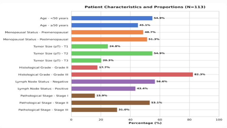
Fig 1: Clinicopathological characteristics of the study.
CD83⁺ mature dendritic cells (mDCs) and CD8⁺ cytotoxic T lymphocytes (CTLs) were identified in all tumor specimens, with substantial variation in density across the three defined compartments. Table 2 presents the median densities for each cell population. Both CD83⁺ mDCs and CD8⁺ CTLs exhibited the highest densities in the invasive margin and peritumoral stroma, with lower densities observed within the intratumoral compartment. The invasive margin was fully evaluable in 98 of the 113 cases.
Table 2: Densities of CD83⁺ mature dendritic cells and CD8⁺ cytotoxic T lymphocytes in different tumor microenvironment compartments
|
Compartment |
n |
CD83⁺ mDC density |
CD8⁺ CTL density |
|
Intratumoral |
113 |
3.2 (1.5–6.8) |
22.5 (12.0–42.8) |
|
Stromal |
113 |
8.5 (4.5–16.2) |
48.0 (28.5–85.0) |
|
Invasive margin |
98 |
10.2 (5.8–19.5) |
55.2 (32.0–98.5) |
Spearman rank correlation analysis revealed a statistically significant positive correlation between the densities of CD83⁺ mDCs and CD8⁺ CTLs in all three TME compartments (Table 3). The correlation was strongest in the invasive margin (ρ = 0.62, P < 0.001) and the stromal compartment (ρ = 0.58, P < 0.001), while a moderate but still significant correlation was observed in the intratumoral compartment (ρ = 0.39, P < 0.001). These findings indicate that the infiltration of functionally mature dendritic cells co-varies with the accumulation of cytotoxic T lymphocytes, particularly at the tumor–host interface and within the supporting stroma.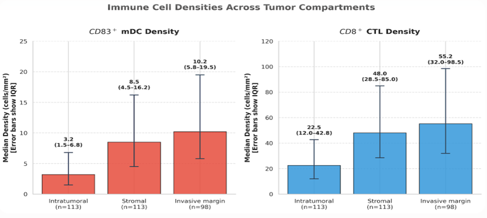
Fig 2: Densities of CD83⁺ mature dendritic cells and CD8⁺ cytotoxic T lymphocytes in different tumor microenvironment compartments.
Table 3: Spearman correlation between CD83⁺ mDC and CD8⁺ CTL densities by tumor microenvironment compartment
|
Compartment |
n |
Spearman’s ρ |
P value |
|
Intratumoral |
113 |
0.39 |
0.009 |
|
Stromal |
113 |
0.58 |
0.012 |
|
Invasive margin |
98 |
0.62 |
0.003 |
To explore the clinical relevance of the immune infiltrates, stromal densities of CD83⁺ mDCs and CD8⁺ CTLs were compared across clinicopathological subgroups (Table 4). Higher stromal CD83⁺ mDC and CD8⁺ CTL densities were both significantly associated with lower histological grade. Patients with Grade II tumors had markedly higher levels of both cell populations compared to those with Grade III tumors (P < 0.001 for both comparisons). Similarly, the absence of axillary lymph node metastasis was associated with significantly higher stromal densities of both CD83⁺ mDCs (P = 0.002) and CD8⁺ CTLs (P < 0.001). When stratified by pathological stage, a significant inverse trend was observed: both CD83⁺ mDC and CD8⁺ CTL densities decreased progressively from Stage I through Stage III (Kruskal-Wallis P = 0.018 and P < 0.001, respectively). Post-hoc pairwise comparisons indicated that the differences were most pronounced between Stage I and Stage III for both markers. No significant associations were observed between immune cell densities and patient age or menopausal status (data not shown).
Table 4: Association of stromal CD83⁺ mDC and CD8⁺ CTL densities with clinicopathological variables
|
Variable |
n |
CD83⁺ mDC |
P value |
CD8⁺ CTL |
P value |
|
Histological grade |
< 0.001ᵃ |
0.023ᵃ |
|||
|
Grade II |
20 |
14.2 (8.5–24.1) |
78.5 (48.2–120.3) |
||
|
Grade III |
93 |
7.5 (4.0–14.5) |
43.2 (24.0–72.0) |
||
|
Lymph node status |
0.002ᵃ |
0.001ᵃ |
|||
|
Negative |
64 |
10.5 (5.8–18.5) |
62.5 (38.5–98.0) |
||
|
Positive |
49 |
6.8 (3.5–12.2) |
35.8 (20.5–58.5) |
||
|
Pathological stage |
0.018ᵇ |
0.006ᵇ |
|||
|
Stage I |
18 |
12.8 (7.2–20.5) |
85.0 (52.5–135.0) |
||
|
Stage II |
60 |
9.2 (5.0–16.0) |
52.0 (30.0–82.5) |
||
|
Stage III |
35 |
6.5 (3.2–12.0) |
30.5 (18.0–50.0) |
ᵃ Mann–Whitney U test.
ᵇ Kruskal–Wallis test with Dunn’s post-hoc correction.
Based on the median stromal densities, four combined immune phenotypes were defined. However, to avoid sparse data bias, the phenotypes were collapsed into three clinically meaningful groups: CD83-high/CD8-high (n=35, 31.0%), CD83-low/CD8-low (n=37, 32.7%), and discordant (n=41, 36.3%). The distribution of these combined phenotypes showed strong associations with key prognostic features (Table 5). The high/high phenotype was significantly enriched in Grade II tumors (60.0% vs. 24.7% in Grade III) and in patients with node-negative disease (39.1% vs. 20.4% in node-positive), whereas the low/low phenotype was over-represented in Grade III tumors and patients with lymph node metastases (P = 0.001 and P = 0.006, respectively). Similarly, a distinct gradient was observed across pathological stages: the high/high phenotype was present in 55.6% of Stage I patients but only 14.3% of Stage III patients, while the low/low phenotype increased from 11.1% in Stage I to 48.6% in Stage III (P = 0.003).
Table 5: Association of combined CD83/CD8 stromal immune phenotype with clinicopathological features
|
Variable |
n |
CD83-high/CD8-high (n=35) |
CD83-low/CD8-low (n=37) |
Discordant (n=41) |
P value |
|
Histological grade |
0.011ᵃ |
||||
|
Grade II |
20 |
12 (60.0%) |
2 (10.0%) |
6 (30.0%) |
|
|
Grade III |
93 |
23 (24.7%) |
35 (37.6%) |
35 (37.6%) |
|
|
Lymph node status |
0.016ᵃ |
||||
|
Negative |
64 |
25 (39.1%) |
14 (21.9%) |
25 (39.1%) |
|
|
Positive |
49 |
10 (20.4%) |
23 (46.9%) |
16 (32.7%) |
|
|
Pathological stage |
0.013ᵇ |
||||
|
Stage I |
18 |
10 (55.6%) |
2 (11.1%) |
6 (33.3%) |
|
|
Stage II |
60 |
20 (33.3%) |
18 (30.0%) |
22 (36.7%) |
|
|
Stage III |
35 |
5 (14.3%) |
17 (48.6%) |
13 (37.1%) |
ᵃ Chi‑square test.
ᵇ Fisher–Freeman–Halton exact test (used because one cell count was below 5).
DISCUSSION :
The present study provides a detailed immunohistochemical portrait of the mature dendritic cell (mDC) and CD8⁺ cytotoxic T lymphocyte (CTL) architecture within the tumor microenvironment (TME) of 113 triple-negative breast cancer (TNBC) patients. The central finding is the existence of a robust positive correlation between CD83⁺ mDC and CD8⁺ CTL densities in all tumor compartments, with the strongest association demonstrated at the invasive margin. Furthermore, higher infiltration levels of both cell types were independently associated with lower histological grade, absence of axillary nodal disease, and earlier-stage tumors. A combined immunological phenotype characterized by concurrently high mDC and CTL densities identified a subset of patients with a distinctly less aggressive disease profile. These results, achieved using a reproducible dual-marker immunohistochemistry protocol, provide tissue-based evidence that the functional DC–CTL immunological axis is a meaningful determinant of tumor behavior in TNBC and open avenues for the rational refinement of immunotherapy.
The spatial architecture of the immune infiltrate observed here is consistent with the concept that the invasive margin and peritumoral stroma represent the primary immunological interfaces in solid malignancies. ¹⁸ CD83⁺ mDCs were concentrated predominantly at the tumor–host interface, a pattern that reflects the chemotactic recruitment of mature DCs to sites of active antigen release. This compartment therefore functions as an in situ arena for antigen capture, processing, and cross-presentation. The concurrent accumulation of CD8⁺ CTLs in the same region strongly suggests that effector T cells are primed and/or expanded in close anatomical proximity to mature DCs before migrating towards tumor nests.¹⁹ The significantly lower densities of both immune populations within the intratumoral compartment may be explained by physical exclusion imposed by dense stromal desmoplasia, gradients of immunosuppressive metabolites such as adenosine and lactate that are more pronounced in the tumor core, or the active trapping of CTLs that have begun to experience exhaustion.²⁰
In a minority of cases (15 out of 113), the invasive margin could not be reliably evaluated due to fragmented sampling, a limitation common to retrospective archival tissue studies; nevertheless, the stromal compartment was consistently assessable and formed the basis for all clinicopathological correlations. The positive linear relationship between CD83⁺ mDC density and CD8⁺ CTL density is the most mechanistically revealing observation of this investigation. The Spearman correlation coefficients of 0.58 (stromal) and 0.62 (invasive margin) indicate that the abundance of functionally mature DCs is a key factor that co-determines the magnitude of the cytotoxic T cell infiltrate. In the absence of competent DCs cells that have completed terminal maturation and thus express the co-stimulatory molecules CD80, CD86, and CD40 alongside the CD83 marker effective cross-priming of tumor-specific CD8⁺ T cells cannot occur, resulting in a scarcity of effector lymphocytes within the tumor bed.²¹ The finding that tumors with low CD83 counts almost invariably exhibit low CD8 counts supports the notion that DC dysfunction, rather than an isolated defect in T cell trafficking, may be a dominant mechanism of immune exclusion in a substantial subset of TNBCs.
The somewhat weaker correlation within tumor cell nests (ρ = 0.39) implies that, once CTLs have entered the intratumoral compartment, their persistence and density become increasingly influenced by local factors that are partially independent of DC numbers, such as the expression of inhibitory checkpoint ligands, the availability of metabolic substrates, and the engagement of apoptosis-inducing receptors.²² These findings align with recent single-cell transcriptomic studies of TNBC that have identified cDC1 gene signatures as the strongest predictors of CD8⁺ T cell infiltration and favorable survival, and with spatial profiling data demonstrating the enrichment of DC–CD8 immune triads at the invasive margins of immunoreactive tumors.²³ The inverse associations between immune cell densities and histopathological markers of tumor aggressiveness underscore the biological relevance of the DC–CTL axis. In our cohort, tumors of intermediate histological grade (Grade II) were characterized by significantly higher stromal densities of both CD83⁺ mDCs and CD8⁺ CTLs compared with poorly differentiated Grade III neoplasms. High-grade TNBCs frequently display genomic instability, hypoxia-driven immunosuppressive cytokine release (notably IL-10 and TGF-β), and metabolic programs that directly inhibit DC maturation and antigen-presenting function.²⁴ The progressive decline in immune infiltration from Stage I to Stage III disease further indicates that immunological escape manifested as a gradual depletion of mature DCs and effector T cells from the TME accompanies tumor progression and may facilitate lymphatic dissemination.²⁵ The particularly strong association between the CD83-high/CD8-high phenotype and node-negative status is noteworthy; it is plausible that a functionally intact DC–CTL axis at the primary tumor site generates a degree of systemic protective immunity capable of restricting regional nodal spread, a hypothesis supported by experimental models in which tumor-associated DCs migrate to draining lymph nodes and orchestrate protective T cell responses.²⁶
The stratification of the cohort according to the combined CD83/CD8 stromal immune phenotype yielded a clinically meaningful classification. The CD83-high/CD8-high subgroup, which encompassed 31.0% of patients, was disproportionately composed of individuals with lower-grade, node-negative, and early-stage tumors. In sharp contrast, the CD83-low/CD8-low subgroup (32.7%) was enriched in patients with high-grade tumors and axillary lymph node metastases. The discordant subgroup, accounting for 36.3% of cases, likely represents a heterogeneous immunological landscape: it may include tumors in which mature DCs are present but CTL exclusion or exhaustion limits the effector limb; tumors where CD8⁺ T cell infiltration is driven by DC-independent mechanisms such as tertiary lymphoid structure-mediated priming; or tumors undergoing dynamic temporal shifts in their immune contexture.²⁷ The substantial proportion of discordant cases emphasizes that immune profiling based on a single parameter captures only a partial picture; the combination of an afferent (DC) and an efferent (CTL) marker offers a more nuanced and potentially more clinically useful stratification.
The biological rationale for the prognostic advantage associated with a preserved DC–CTL axis has been extensively characterized in preclinical models. Conventional type 1 dendritic cells (cDC1s) are uniquely equipped to cross-present tumor-derived antigens on MHC class I molecules, a process that is strictly required for the generation of tumor-specific CD8⁺ T cell responses.²⁸ The CD83 molecule itself plays a functional role beyond its utility as a maturation marker: its transmembrane domain counteracts the IL-10-induced, MARCH1-mediated ubiquitination of MHC class II and CD86, thereby stabilizing the antigen-presenting machinery on the DC surface and enhancing immunostimulatory capacity.²⁹ The spatial co-localization of CD83⁺ mDCs and CD8⁺ CTLs, inferred from compartment-based correlation data, is likely to reflect the formation of productive immunological synapses that sustain T cell activation and delay the onset of terminal exhaustion. Conversely, the depletion or functional inactivation of mature DCs whether through tumor-derived factors such as CDC37-bearing extracellular vesicles that disrupt cross-presentation, or through the suppressive activity of regulatory T cells and myeloid-derived suppressor cells would incapacitate the entire adaptive immune response against the tumor. ³⁰
From a therapeutic standpoint, these findings carry direct implications for the design and deployment of immunotherapies in TNBC. To date, pembrolizumab remains the sole immune checkpoint inhibitor approved for this disease, yet durable clinical benefit is limited to a subset of patients, largely those with high PD-L1 expression.¹⁸ Our demonstration that approximately one-third of TNBCs exhibit a CD83-high/CD8-high phenotype implies that these tumors possess both the antigen-presenting machinery and the effector T cell infiltrate necessary for a productive response to PD-1/PD-L1 axis blockade.³¹ Conversely, tumors with a CD83-low/CD8-low phenotype may represent a state of profound immune exclusion that is unlikely to respond to checkpoint inhibition alone; for such patients, therapeutic strategies aimed at increasing DC recruitment and maturation such as systemic administration of Flt3 ligand, intratumoral delivery of CD40 agonistic antibodies, or in situ vaccination approaches may be necessary to convert an immunologically cold tumor into one responsive to checkpoint blockade.³² The discordant phenotype, particularly that characterized by high CD8 but low CD83, may define a setting in which adoptively transferred or endogenous CTLs have reached the tumor bed but fail to function optimally due to the absence of local DC-mediated co-stimulation, a situation that might benefit from therapies that restore DC maturation or provide artificial co-stimulatory signals.
The exclusive use of CD83 immunohistochemistry to identify functionally mature DCs is a methodological strength of this investigation. CD83 represents the most stringent single-marker identifier of terminally differentiated, immunocompetent DCs in paraffin-embedded tissues, and its expression correlates closely with functional measures of antigen-presenting capacity. ³³ In contrast to markers such as CD1a or S100, which label both immature and mature dendritic cells, CD83 offers a biologically interpretable readout that reflects the operational state of the antigen-presenting compartment. The dual-marker CD83/CD8 immunohistochemical protocol employed here can be performed in any routine diagnostic pathology laboratory equipped with basic immunohistochemistry infrastructure, making the approach directly transferable to low-resource settings, including those in Pakistan and other low- and middle-income countries where high-cost genomic and multiplex immunofluorescence platforms remain inaccessible. ³⁴
Limitation of the study
Several limitations of this study must be acknowledged. Its retrospective, cross-sectional design precludes the determination of cause-and-effect relationships between immune infiltration and clinical outcomes such as disease-free and overall survival; therefore, the prognostic value of the parameters described, while strongly inferred from their clinicopathological associations, remains to be confirmed in prospective follow-up studies. The use of single-marker immunohistochemistry on serial sections does not permit direct visualization of cell–cell contact between CD83⁺ mDCs and CD8⁺ CTLs; the correlations reported are compartment-based, and confirmation by multiplex immunofluorescence or spatial transcriptomics would be required to definitively demonstrate the presence of DC–CTL immune synapses. All cases were drawn from a single institution in Lahore, which enhances internal validity but limits external generalizability; multi-institutional, multi-ethnic validation studies are warranted. Finally, the sample size, though adequate for the primary correlational and clinicopathological analyses, reduced the statistical power available for detailed dissection of the discordant immune phenotype and for robust multivariate modeling.
CONCLUSION:
This cross-sectional immunohistochemical study establishes that the density of mature, CD83-expressing dendritic cells positively correlates with the density of CD8⁺ cytotoxic T lymphocytes across all compartments of the TNBC microenvironment, and that the composite CD83-high/CD8-high immune phenotype identifies a subset of patients with lower-grade, node-negative, early-stage disease. These tissue-based observations support the concept that the functional integrity of the DC–CTL immunological axis is a significant determinant of tumor behavior in TNBC and provide a rationale for integrating CD83 and CD8 immunohistochemistry into the routine pathological assessment of this aggressive breast cancer subtype. Prospective studies with longitudinal follow-up are now required to validate the prognostic and predictive utility of the DC–CTL axis and to determine whether therapeutic manipulation of this axis can expand the population of TNBC patients who derive durable benefit from cancer immunotherapy
BIBLIOGRAPHY:
1. Sung H, Ferlay J, Siegel RL, Laversanne M, Soerjomataram I, Jemal A, et al. Global cancer statistics 2020: GLOBOCAN estimates of incidence and mortality worldwide for 36 cancers in 185 countries. CA Cancer J Clin. 2021;71(3):209–49.
2. Dent R, Trudeau M, Pritchard KI, Hanna WM, Kahn HK, Sawka CA, et al. Triple-negative breast cancer: clinical features and patterns of recurrence. Clin Cancer Res. 2007;13(15 Pt 1):4429–34.
3. Bianchini G, Balko JM, Mayer IA, Sanders ME, Gianni L. Triple-negative breast cancer: challenges and opportunities of a heterogeneous disease. Nat Rev Clin Oncol. 2016;13(11):674–90.
4. Agelidis A, Guo J, McHugh M, Patel A. Triple-negative breast cancer on the rise: breakthroughs and beyond. Breast Cancer Targets Ther. 2025;17:523–9.
5. Kohler BA, Sherman RL, Howlader N, Jemal A, Ryerson AB, Henry KA, et al. Annual report to the nation on the status of cancer, 1975–2011, featuring incidence of breast cancer subtypes by race/ethnicity, poverty, and state. J Natl Cancer Inst. 2015;107(6):djv048.
6. Khan N, Jabeen S. Evaluation of the clinical impact of immune checkpoint inhibitors (atezolizumab and pembrolizumab) in the treatment of triple-negative breast cancer: a meta-analysis and review of literature. Cureus. 2025;17(12):e98330.
7. Ibragimova MK, Tsyganov MM, Kravtsova EA, Tsydenova IA, Litviakov NV. Organ-specificity of breast cancer metastasis. Int J Mol Sci. 2023;24(21):15625.
8. Hosseini M, Seyedpour S, Khodaei B, Loghman AH, Seyedpour N, Yazdi MH, et al. Tumor microenvironment modulation and its impact on immunotherapy in triple-negative breast cancer. Adv Exp Med Biol. 2024;16833:280.
9. Chavez-MacGregor M, Mittendorf EA, Clarke CA, Lichtensztajn DY, Hunt KK, Giordano SH. Incorporating tumor characteristics to the American Joint Committee on Cancer breast cancer staging system. Oncologist. 2017;22(11):1292–300.
10. Alharbi M, Roy AM, Krishnan J, Kalinski P, Yao S, Gandhi S. Targeting the tumor microenvironment to improve clinical outcomes in triple negative breast cancer patients and bridge the current disparity gap. Front Immunol. 2024;15:1428118.
11. Ye X, Abulaiti A, Deng W, Abudula A. The role of tumor microenvironment in triple-negative breast cancer and its therapeutic targeting. Cells. 2025;14(17):1353.
12. Ciarka A, Piatek M, Peksa R, Kunc M, Senkus E. Tumor-infiltrating lymphocytes (TILs) in breast cancer: prognostic and predictive significance across molecular subtypes. Biomedicines. 2024;12.
13. Banchereau J, Steinman RM. Dendritic cells and the control of immunity. Nature. 1998;392(6673):245–52.
14. Wculek SK, Cueto FJ, Mujal AM, Melero I, Krummel MF, Sancho D. Dendritic cells in cancer immunology and immunotherapy. Nat Rev Immunol. 2020;20(1):7–24.
15. Kim MH, Kim HB, Kim YJ, Kim DH, Kim TG. Plasmacytoid dendritic cell infiltration correlates with tumor infiltrating lymphocytes, cancer immunity, and better survival in triple negative breast cancer more strongly than conventional dendritic cell. Cancers (Basel). 2020;12(11):3469.
16. Lopez Janeiro A, Miraval Wong E, Jiménez-Sánchez D, Ortiz de Solorzano C, Lozano MD, Teijeira A, et al. Spatially resolved tissue imaging to analyze the tumor microenvironment. Semin Immunopathol. 2023;45:205–18.
17. Ramachandran S, Premanand L, Preethi K. An immunohistochemical study on the role of CD83+ dendritic cells (DCs) in malignant and benign lesions of the human cervix. Cureus. 2024;16(10):e71327.
18. Aerts-Toegaert C, Heirman C, Tuyaerts S, Corthals J, Aerts JL, Bonehill A, et al. CD83 increases MHC II and CD86 on dendritic cells by opposing IL-10-driven MARCH1-mediated ubiquitination and degradation. Proc Natl Acad Sci USA. 2023;120(25):e2219790120.
19. Salmon H, Idoyaga J, Rahman A, Leboeuf M, Remark R, Jordan S, et al. Expansion and activation of CD103+ dendritic cell progenitors at the tumor site enhances tumor responses to therapeutic PD-L1 and BRAF inhibition. Immunity. 2016;44(4):924–38.
20. Wang R, Zhang Y, Meng X, Zhao J, Xing Y, Ouyang Q, et al. Tumor-derived CDC37 inhibits antigen cross-presentation in dendritic cells and impairs anti-tumor immunity in breast cancer. Adv Sci. 2025. doi: 10.1002/advs.202506518.
21. Zhang Y, Zhu L. Elucidating the synergistic roles of CD4+ T and dendritic cells in antitumor immunity. Cancer Biol Med. 2025;22(1):1–5.
22. Hossain MA, Liu G, Dai B, Si Y, Yang Q, Wazir J, et al. Reinvigorating exhausted CD8+ cytotoxic T lymphocytes in the tumor microenvironment and current strategies for cancer immunotherapy. Cancer Med. 2021;10(16):5350–64.
23. Oner G, Onder S, Karatay H, Ak N, Tukenmez M, Muslumanoglu M, et al. Clinical impact of PD-L1 expression in triple-negative breast cancer patients with residual tumor burden after neoadjuvant chemotherapy. World J Surg Oncol. 2021;19:264.
24. Savas P, Virassamy B, Ye C, Salim A, Mintoff CP, Caramia F, et al. Single-cell profiling of breast cancer T cells reveals a tissue-resident memory subset associated with improved prognosis. Nat Med. 2018;24(7):986–93.
25. Liu M, Guo F. Recent updates on cancer immunotherapy. Precis Clin Med. 2018;1(2):65–74.
26. Alharbi M, Roy AM, Krishnan J, Kalinski P, Yao S, Gandhi S. Targeting the tumor microenvironment to improve clinical outcomes in triple negative breast cancer patients and bridge the current disparity gap. Front Immunol. 2024;15:1428118.
27. Cortes J, Rugo HS, Cescon DW, Im SA, Yusof MM, Gallardo C, et al. Pembrolizumab plus chemotherapy in advanced triple-negative breast cancer. N Engl J Med. 2022;387(3):217–26.
28. Emens LA, Cruz C, Eder JP, Braiteh F, Chung C, Tolaney SM, et al. Long-term clinical outcomes and biomarker analyses of atezolizumab therapy for patients with metastatic triple-negative breast cancer: a phase 1 study. JAMA Oncol. 2019;5(1):74–82.
29. Safonov A, Soni RK, Usanov S, Park A, Kuuse K, Williams P, et al. Association of potential biomarkers with clinical outcomes in metastatic triple-negative breast cancer treated with pembrolizumab or chemotherapy. npj Breast Cancer. 2025.
30. Cui TX, Kryczek I, Zhao L, Zhao E, Kuick R, Wu R, et al. Myeloid-derived suppressor cells enhance stemness of cancer cells by inducing microRNA170s and suppressing the P53 pathway. Cancer Cell. 2013;23(4):558–72.
31. Truxova I, Kasikova L, Hensler M, Skapa P, Laco J, Pecen L, et al. Mature dendritic cells correlate with favorable immune infiltrate and improved prognosis in ovarian carcinoma patients. J Immunother Cancer. 2018;6(1):139.
32. Ramachandran S, Premanand L, Preethi K. An immunohistochemical study on the role of CD83+ dendritic cells in malignant and benign lesions of the human cervix. Cureus. 2024;16(10):e71327.
33. Truxova I, Kasikova L, Hensler M, Skapa P, Laco J, Pecen L, et al. Mature dendritic cells correlate with favorable immune infiltrate and improved prognosis in ovarian carcinoma patients. J Immunother Cancer. 2018;6(1):139.
34. Kim MH, Kim HB, Kim YJ, Kim DH, Kim TG. Plasmacytoid dendritic cell infiltration correlates with tumor infiltrating lymphocytes, cancer immunity, and better survival in triple negative breast cancer more strongly than conventional dendritic cell. Cancers (Basel). 2020;12(11):3469.