RISK FACTORS IN CLINICAL PROFILE OF YOUNG PATIENTS PRESENTING WITH ACUTE MYOCARDIAL INFARCTION
- Mehak Razzaq , Senior Lecturer, Department of Emerging Health Professional Technology, Superior University Lahore, Pakistan
- Maham Fatima , Student, Superior University Lahore, Pakistan
- Mahnoor Nadeem , Student, Superior University Lahore, Pakistan
- Fatima Imran , Student, Superior University Lahore, Pakistan
- Faisal Nadeem , Student, Superior University Lahore, Pakistan
Article Information:
Abstract:
Background: Acute myocardial infarction has traditionally been regarded as a disease of older adults, yet a concerning global rise in cases among younger individuals has emerged over the past two decades. South Asian populations appear disproportionately affected, developing coronary artery disease nearly a decade earlier than their Western counterparts. Despite this, prospective data on the specific risk factor profiles, clinical characteristics, and treatment patterns of young AMI patients from Pakistan remain scarce, limiting the development of age-appropriate prevention and management strategies.Objective: To determine the risk factors in the clinical profile of young patients presenting with acute myocardial infarction.Methods: This prospective observational study was conducted in the Department of Cardiology, Sheikh Zayed Hospital, Lahore, over a four-month period. A total of 109 patients aged 18–50 years diagnosed with STEMI or NSTEMI based on ECG changes and elevated cardiac biomarkers were enrolled through consecutive sampling. Data on demographics, medical history, lifestyle factors, clinical presentation, ECG findings, troponin I levels, coronary angiography, treatment received, and in-hospital complications were collected using a structured questionnaire. Categorical variables were analyzed using frequencies, percentages, and chi-square tests, while continuous variables were expressed as mean ± SD. A p-value of <0.05 was considered statistically significant. All analyses were performed using SPSS version 27.0.Results: The mean age was 33.32 ± 9.57 years with a male-to-female ratio of 1.3:1. The most prevalent risk factors were hypertension (64.2%), overweight/obesity (62.4%), high -fat diet (61.5%), family history of premature CAD (57.8%), dyslipidemia (48.6%), and diabetes mellitus (45.0%). Age-stratified analysis revealed significant gradients for hypertension (32.5% in ≤30 years to 90.6% in 41–45 years; p<0.001), diabetes (15.0% to 68.8%; p<0.001), dyslipidemia (32.5% to 68.8%; p=0.003), and obesity (5.0% to 40.6%; p=0.009). Smoking was the strongest gender-linked disparity, with 62.9% of males being ever-smokers versus 12.8% of females (p<0.001). Chest pain was the commonest presenting symptom (83.5%), and ST elevation the most frequent ECG finding (46.8%). ECG patterns differed significantly by gender (p=0.040), with males showing higher ST elevation rates and females exhibiting more heterogeneous patterns. Coronary angiography was performed in 64.2% of patients, significantly more often in males than females (74.2% vs. 51.1%; p=0.013). Medications alone were the predominant treatment (62.4%), followed by thrombolysis (19.3%) and PCI (18.3%). Dyslipidemia (OR=2.25; p=0.047), smoking status (p=0.041), and BMI category (p=0.012) were significantly associated with angiography-positive status. Diabetes and hypertension significantly influenced ECG manifestation patterns (p=0.001 and p=0.025, respectively). In-hospital complications occurred in 71.6% of patients, though serious events such as cardiogenic shock (1.8%) and arrhythmias (2.8%) were uncommon.Conclusion: Premature AMI in this young South Asian cohort is driven predominantly by a clustering of modifiable metabolic and lifestyle risk factors hypertension, obesity, unhealthy diet, diabetes, and smoking with a steep age-dependent accumulation of comorbidities even within the young age spectrum. Significant gender disparities exist in both clinical presentation and treatment aggressiveness. These findings argue for earlier cardiovascular screening, gender-sensitive clinical protocols, and aggressive lifestyle modification programs beginning in the third decade of life in South Asian populations.
Keywords:
Article :
INTRODUCTION:
Acute myocardial infarction (AMI) has traditionally been conceptualized as a disease of advancing age, typically manifesting in the sixth decade of life or beyond against a background of accumulated cardiovascular risk. However, this paradigm is undergoing a fundamental shift. Over the past two decades, clinicians worldwide have observed a troubling epidemiological trend: patients presenting with AMI are becoming progressively younger, with individuals in their third and fourth decades of life now constituting a meaningful proportion of cardiac admissions [1,2]. This demographic transition carries profound implications for clinical practice, public health policy, and healthcare resource allocation, particularly in low- and middle-income countries where the burden of premature cardiovascular disease is accelerating most rapidly. Young patients with AMI conventionally defined as those aged ≤45 years represent a distinct clinical phenotype with unique biochemical, angiographic, and lifestyle characteristics that differentiate them from their older counterparts [3]. While the absolute incidence of AMI in this age group remains lower than in older populations, the societal impact is disproportionately severe. A sudden cardiac event during the prime productive years of life exacts an extraordinary toll: prolonged disability, psychological trauma, career disruption, and substantial economic hardship for affected individuals and their families. The ripple effects extend to healthcare systems, particularly in resource-constrained settings where social safety nets remain underdeveloped [2]. Epidemiological evidence from developing nations reveals a sobering reality. Between 25% and 33% of all AMI cases in these countries involve patients under 55 years of age, with even higher proportions documented across South Asia. Hospital-based registries from India and Pakistan confirm that young adult AMI once considered a clinical rarity now constitutes a growing and substantial share of cardiac admissions [1,4]. The drivers of this shift are multifactorial and reflect rapid epidemiological transitions: accelerated urbanization, dietary patterns skewed toward calorie-dense processed foods, declining physical activity, rising obesity prevalence, and persistently high rates of tobacco consumption among young men in the region [1,4].
The risk factor profile of young AMI patients does not merely mirror an attenuated version of that seen in older adults. Smoking emerges as the single most consistently identified modifiable risk factor, reported in 70–80% of young MI cases across multiple series [7,8]. Obesity, physical inactivity, and metabolic syndrome characterized by central adiposity, elevated blood pressure, dyslipidemia, and insulin resistance are present in nearly half of young patients [7,8]. Beyond these conventional contributors, non-traditional risk factors have garnered increasing attention: elevated lipoprotein(a), familial hypercholesterolemia, prothrombotic polymorphisms, hyperhomocysteinemia, systemic inflammation, and psychosocial stress all contribute to premature atherosclerosis in ways that may overwhelm the protective resilience of youth [9,10]. Additionally, non-atherosclerotic mechanisms including coronary artery spasm, spontaneous coronary artery dissection, and substance abuse (particularly stimulants such as cocaine and amphetamines) can precipitate myocardial infarction in young adults without significant plaque burden [10].
Problem Statement
Despite the growing scale of premature coronary artery disease in South Asia, the research evidence base remains disproportionately weighted toward older populations. Prospective data specifically characterizing young AMI patients in Pakistan including their risk factor clustering, clinical presentation, angiographic patterns, and treatment trajectories are notably sparse. This knowledge gap undermines efforts to design age-specific prevention strategies, optimize early diagnostic approaches, and tailor secondary prevention interventions for a population facing decades of living with coronary artery disease [11,12]. Furthermore, public and provider awareness lags behind epidemiological realities: young patients frequently fail to recognize cardiac symptoms, and clinicians may similarly discount the possibility of AMI in this age group, leading to delayed diagnosis and suboptimal outcomes.
Objective of the study
This study addresses these critical gaps by characterizing the clinical profile, risk factor burden, and treatment patterns of young patients presenting with acute myocardial infarction at a major cardiac center in Lahore, Pakistan. By delineating the distinctive features of premature coronary disease in the Pakistani context, this research aims to inform targeted prevention strategies, improve early detection, and ultimately reduce the clinical and societal toll of AMI among young adults in this high-risk population. The primary objective was to determine the risk factors associated with the clinical profile of young patients (≤45 years) presenting with acute myocardial infarction.
METHODOLOGY:
This prospective observational study was conducted at the Department of Cardiology, Sheikh Zayed Hospital, Lahore a tertiary-care teaching hospital serving as a major referral center for acute coronary syndromes across Punjab province, Pakistan. The study was approved by the Institutional Review Board prior to commencement, and data collection was carried out over a four-month period following ethical approval. Consecutive patients aged between 18 and 50 years with a confirmed diagnosis of acute myocardial infarction (AMI) were enrolled. The diagnosis of AMI was established based on clinical evidence of myocardial ischemia accompanied by a rise and/or fall in cardiac biomarkers (troponin I or T), together with chest pain lasting longer than 20 minutes and electrocardiographic changes such as ST-segment elevation or depression [11]. Both ST-elevation myocardial infarction (STEMI) and non-ST-elevation myocardial infarction (NSTEMI) were included. Eligible patients were required to have at least one identifiable cardiovascular risk factor. Exclusion criteria were: (1) age above 50 years; (2) presentation with stable angina or non-ischemic causes of chest pain; and (3) cases attributable to aortic dissection, pericarditis, myocarditis, or other non-AMI conditions.
The sample size was calculated using the standard formula for estimating a single population proportion:
where Z=1.96 (95% confidence level), P=0.122 (estimated prevalence of acute coronary syndrome in young patients based on prior research), and d=0.06 (margin of error). This yielded a minimum target of 115 participants. After accounting for refusals and withdrawals during the enrollment period, the final analytic sample comprised 109 patients.
All eligible patients presenting during the study period were enrolled sequentially using a consecutive sampling approach. Upon admission, the following data were collected prospectively using a pre-designed structured questionnaire administered by trained cardiologists and research staff: Demographic and clinical variables include age, sex, body mass index (BMI), occupation, medical history (hypertension, diabetes mellitus, dyslipidemia, previous heart disease), family history of premature coronary artery disease, and lifestyle factors (smoking status, physical activity level, dietary pattern). Clinical presentation presenting symptoms including chest pain, shortness of breath, diaphoresis, nausea or vomiting, and palpitations were documented alongside physical examination findings.
A 12-lead electrocardiogram (ECG) was obtained to confirm the diagnosis and classify the type of infarction. Blood samples were collected for quantitative measurement of cardiac biomarkers (troponin I and CK-MB), fasting lipid profile, and glucose levels. For patients who underwent coronary angiography, the culprit vessel, number of vessels involved, and severity of occlusion were recorded in the cardiac catheterization laboratory. Left ventricular ejection fraction (LVEF) and regional wall motion abnormalities were assessed using echocardiography. Documented data included thrombolysis administration, percutaneous coronary intervention (PCI), medical management, and any in-hospital complications from admission through discharge.
This study was conducted in full compliance with the ethical principles outlined in the Declaration of Helsinki. Formal ethical approval was obtained from the Institutional Review Board of Sheikh Zayed Hospital, Lahore, prior to commencement of data collection. Written informed consent was obtained from every participant before enrollment following a clear explanation of the study purpose, procedures, and their right to refuse or withdraw without consequence to their ongoing clinical care. Participation was entirely voluntary. Confidentiality was maintained at all times; each participant was assigned a coded identifier, and all personal information was de-identified before data entry. No additional invasive procedures or interventions were performed beyond those forming part of routine AMI management; the study therefore posed no incremental risk to participants. All data were stored securely in password-protected electronic files accessible only to members of the research team. Study materials, including the consent form and questionnaire, were made available in both English and Urdu to ensure comprehension.
All collected data were entered and analyzed using IBM SPSS Statistics version 27.0. Descriptive statistics were used to summarize patient demographics, clinical characteristics, risk factor prevalence, ECG findings, laboratory results, angiographic data, and treatment outcomes. Categorical variables were presented as frequencies and percentages. Continuous variables were expressed as mean ± standard deviation along with median, minimum, maximum, skewness, and kurtosis where appropriate. Associations between categorical variables including risk factors stratified by sex, age group, ECG pattern, angiography status, and treatment modality were evaluated using Pearson's chi-square test, with Cramér's V reported as a measure of effect size. Odds ratios with 95% confidence intervals were calculated for dichotomous risk factor–outcome associations where applicable. A two-tailed p-value of less than 0.05 was considered statistically significant throughout the analysis. Results were presented in the form of tables and figures for clarity
RESULTS & FINDINGS:
A total of 109 young patients diagnosed with acute myocardial infarction were enrolled in this study. Males outnumbered females at a ratio of roughly 1.3:1, with 62 males (56.9%) and 47 females (43.1%). When broken down by age, the largest subset surprisingly was the youngest group: 40 patients (37.7%) fell in the ≤30-year bracket, followed closely by the 41–45 age group with 32 patients (30.2%). The middle tiers, 31–35 and 36–40 years, comprised 16 (15.1%) and 18 (17.0%) individuals respectively. Three patients had ages falling outside the recode boundaries and were excluded from age-group-specific analyses, reducing the subgroup denominator to 106.
Table 5.1.1: Demographic Characteristics of Young AMI Patients (N=109)
|
Variable |
Category |
n |
% |
|
Gender |
Male |
62 |
56.9 |
|
|
Female |
47 |
43.1 |
|
Age Group |
≤30 years |
40 |
37.7 |
|
|
31–35 years |
16 |
15.1 |
|
|
36–40 years |
18 |
17.0 |
|
|
41–45 years |
32 |
30.2 |
|
BMI Category |
Underweight |
2 |
1.8 |
|
|
Normal |
39 |
35.8 |
|
|
Overweight |
46 |
42.2 |
|
|
Obese |
22 |
20.2 |
Table 5.1.2: Descriptive Statistics of Continuous Variables (N=109)
|
Variable |
Mean |
SD |
Median |
Min |
Max |
Skewness |
Kurtosis |
|
Age (years) |
33.32 |
9.57 |
35.00 |
14 |
48 |
−0.438 |
−1.051 |
|
BMI (kg/m²) |
26.09 |
3.65 |
26.00 |
18.00 |
33.00 |
0.055 |
−0.709 |
|
Troponin I (ng/mL) |
0.061 |
0.088 |
0.047 |
0.011 |
0.820 |
6.941 |
54.960 |
The mean age of the study cohort stood at 33.32 ± 9.57 years with a median of 35 years, ranging from a startlingly young 14 to 48 years. The distribution carried a mild negative skew (−0.438) and was somewhat platykurtic (kurtosis −1.051), reflecting a spread-out age profile without heavy tailing on either side. Body mass index averaged 26.09 ± 3.65 kg/m², with a median of 26.00; the values ranged from 18.00 to 33.00, and the distribution was essentially symmetric (skewness 0.055). As for cardiac biomarkers, mean troponin I was 0.061 ± 0.088 ng/mL with a median of 0.047, but the distribution was dramatically right-skewed (skewness 6.941, kurtosis 54.960), indicating that while most patients had modestly elevated values, a handful had markedly high readings the maximum reaching 0.820 ng/mL.
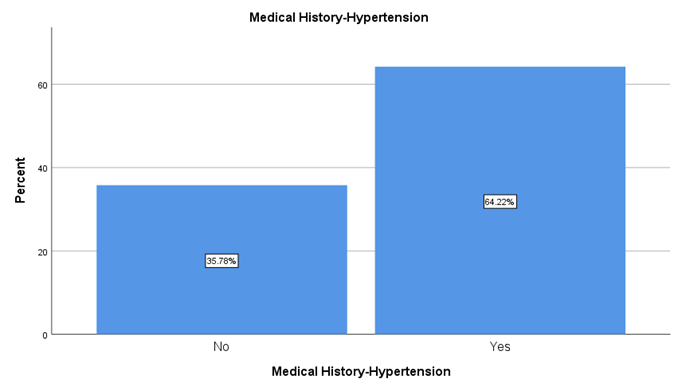
Hypertension topped the list as the most prevalent comorbidity, identified in 70 out of 109 patients (64.2%). Only 39 patients (35.8%) were normotensive. This dominance of hypertension in a cohort this young is worth underscoring it signals that blood pressure control may be a particularly pressing concern in young adults vulnerable to AMI.
Table 5.2.1: Prevalence of Hypertension Among Young AMI Patients (N=109)
|
Hypertension |
n |
% |
|
Yes |
70 |
64.2 |
|
No |
39 |
35.8 |
Diabetes mellitus was present in 49 patients (45.0%), with 60 (55.0%) free of the disease. While not a majority, having nearly half the cohort affected by diabetes at such young ages is clinically noteworthy and mirrors rising diabetes trends in the South Asian population.
Table 5.2.2: Prevalence of Diabetes Mellitus Among Young AMI Patients (N=109)
|
Diabetes Mellitus |
n |
% |
|
Yes |
49 |
45.0 |
|
No |
60 |
55.0 |
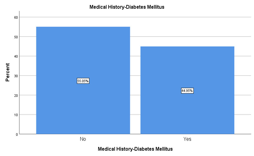
Figure 5.2.2: Medical History – Diabetes Mellitus
The prevalence of dyslipidemia was almost evenly split: 53 patients (48.6%) had abnormal lipid profiles versus 56 (51.4%) who did not. The near-equal division suggests that while dyslipidemia is common in this population, it does not dominate in isolation rather, it typically clusters with other metabolic risk factors.
Table 5.2.3: Prevalence of Dyslipidemia Among Young AMI Patients (N=109)
|
Dyslipidemia |
n |
% |
|
Yes |
53 |
48.6 |
|
No |
56 |
51.4 |
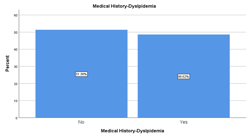
Figure 5.2.3: Medical History – Dyslipidemia
A prior history of heart disease was relatively less common, present in 30 patients (27.5%). The remaining 79 (72.5%) had no documented cardiac history, meaning the current AMI event was, for most, their first major cardiac episode.
Table 5.2.4: Prevalence of Previous Heart Disease Among Young AMI Patients (N=109)
|
Previous Heart Disease |
n |
% |
|
Yes |
30 |
27.5 |
|
No |
79 |
72. 5 |
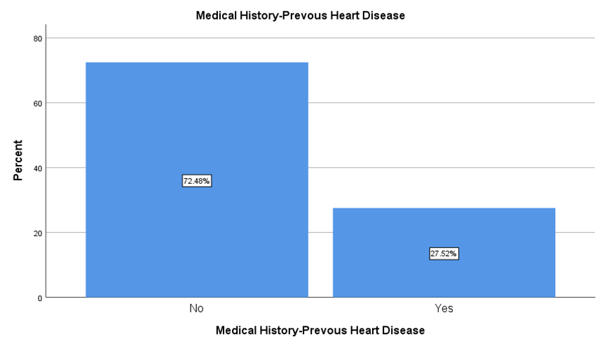
Figure 5.2.4: Medical History – Previous Heart Disease
More than half the patients 63 out of 109 (57.8%) reported a family history of premature CAD. This high figure reinforces the well-established genetic underpinning of coronary disease and its outsized role when the disease strikes early in life.
Table 5.2.5: Family History of Premature CAD Among Young AMI Patients (N=109)
|
Family History of CAD |
n |
% |
|
Yes |
63 |
57.8 |
|
No |
46 |
42.2 |
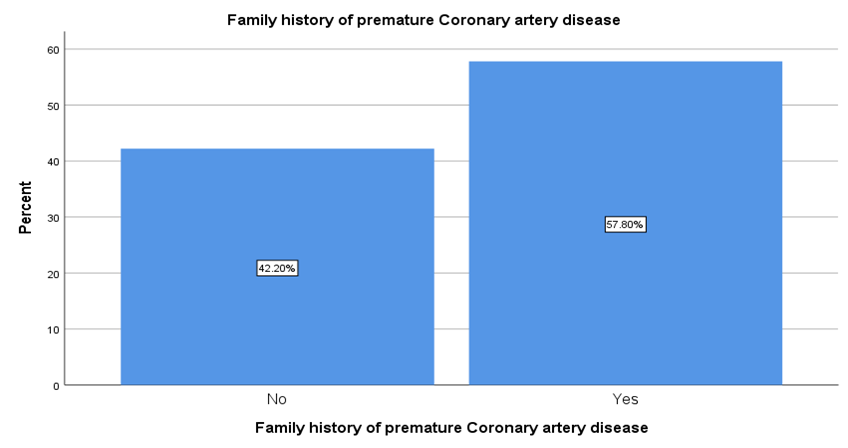
Figure 5.2.5: Family History of Premature Coronary Artery Disease
The majority of patients (64, or 58.7%) had never smoked. However, 28 (25.7%) were former smokers and 17 (15.6%) were current smokers meaning that collectively, 41.3% had some degree of tobacco exposure. This is no small fraction and warrants attention in preventive cardiology strategies targeting the young.
Table 5.2.6: Smoking Status Among Young AMI Patients (N=109)
|
Smoking Status |
n |
% |
|
Never |
64 |
58.7 |
|
Former |
28 |
25.7 |
|
Current |
17 |
15.6 |
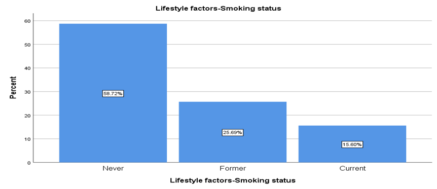
Figure 5.2.6: Lifestyle Factors – Smoking Status
Alcohol use was exceedingly rare: only 3 patients (2.8%) reported consumption, while 106 (97.2%) denied it. This near-universal abstinence likely reflects the sociocultural context of the study population, making alcohol a negligible risk factor in this particular cohort.
Table 5.2.7: Alcohol Consumption Among Young AMI Patients (N=109)
|
Alcohol Consumption |
n |
% |
|
No |
106 |
97.2 |
|
Yes |
3 |
2.8 |
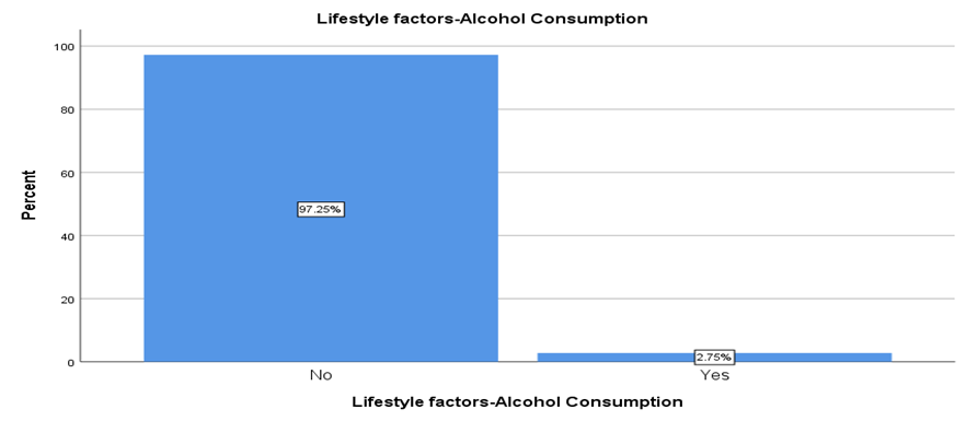
Figure 5.2.7: Lifestyle Factors – Alcohol Consumption
Physical activity levels were distributed across sedentary (46 patients, 42.2%), moderate (48, 44.0%), and active (15, 13.8%). The fact that over four in ten patients led a sedentary lifestyle is concerning, especially considering that physical inactivity is an independently modifiable risk factor for cardiovascular disease.
Table 5.2.8: Physical Activity Level Among Young AMI Patients (N=109)
|
Physical Activity |
n |
% |
|
Sedentary |
46 |
42.2 |
|
Moderate |
48 |
44.0 |
|
Active |
15 |
13.8 |
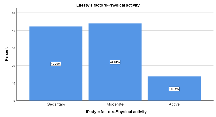
Figure 5.2.8: Lifestyle Factors – Physical Activity
Dietary habits leaned heavily toward unhealthy patterns: 67 patients (61.5%) consumed a high-fat diet. Only 28 (25.7%) maintained a balanced dietary pattern, and 13 (11.9%) reported other dietary habits. The overwhelming prevalence of high-fat intake in this young AMI cohort underscores the critical need for nutritional counselling as a frontline preventive measure.
Table 5.2.9: Dietary Pattern Among Young AMI Patients (N=109)
|
Dietary Pattern |
n |
% |
|
High Fat |
67 |
61.5 |
|
Balanced |
28 |
25.7 |
|
Other |
13 |
11.9 |
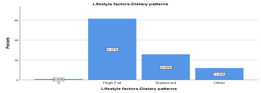
Figure5.2.9: Lifestyle Factors – Dietary Patterns
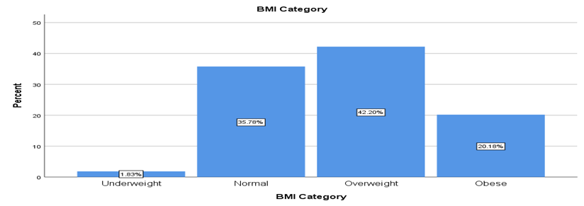
Most participants were overweight (42.20%) or of normal weight (35.78%), while 20.18% were obese and only 1.83% were underweight, indicating that over 60% of the cohort had a BMI above the normal range.
Table 5.2.10: BMI Category Distribution Among Young AMI Patients (N=109)
|
BMI Category |
n |
% |
|
Underweight |
2 |
1.8 |
|
Normal |
39 |
35.8 |
|
Overweight |
46 |
42.2 |
|
Obese |
22 |
20.2 |
When troponin I was dichotomized at the 0.04 ng/mL threshold, 69 patients (63.3%) had elevated levels indicative of myocardial necrosis, while 40 (36.7%) had values within the normal range. This roughly two-thirds positive rate is consistent with what one expects in an acute MI cohort, though the presence of normal troponin in over a third of patients may partly reflect timing of sample collection or smaller infarcts.
Table 5.2.11: Troponin I Elevation Status Among Young AMI Patients (N=109)
|
Troponin I Status |
n |
% |
|
Elevated (>0.04 ng/mL) |
69 |
63.3 |
|
Normal (≤0.04 ng/mL) |
40 |
36.7 |
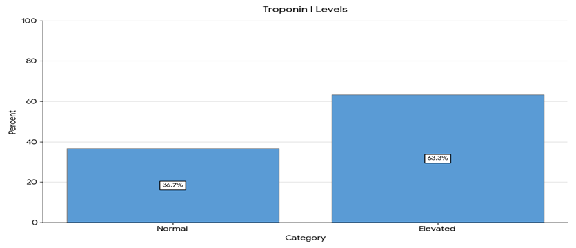
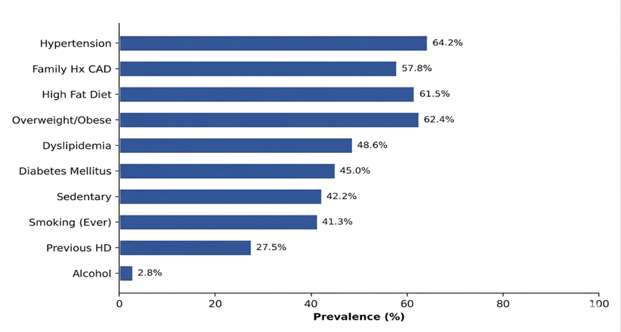
Figure 5.2.12: Prevalence of Risk Factors in Young AMI patients (N=109)
Figure 5.2.1 illustrates the prevalence of various risk factors in young AMI patients (N=109). Hypertension (64.2%) was the most common risk factor, whereas alcohol consumption (2.8%) was the least common. Other major risk factors included family history of CAD (57.8%), high fat diet (61.5%), and overweight/obesity (62.4%).
Chest pain was the dominant presenting complaint, reported by 91 patients (83.5%). Shortness of breath followed at 56.0% (n=61), sweating at 55.0% (n=60), nausea or vomiting at 49.5% (n=54), and palpitations at 38.5% (n=42). The classical triad of chest pain, diaphoresis, and dyspnea thus held true for the majority of these young patients, though palpitations a less "typical" AMI symptom were still present in more than a third.
Table 5.3.1.1: Clinical Presentation Symptom Profile (N=109)
|
Symptom |
Present n (%) |
Absent n (%) |
|
Chest Pain |
91 (83.5) |
18 (16.5) |
|
Shortness of Breath |
61 (56.0) |
48 (44.0) |
|
Sweating |
60 (55.0) |
49 (45.0) |
|
Nausea/Vomiting |
54 (49.5) |
55 (50.5) |
|
Palpitations |
42 (38.5) |
67 (61.5) |
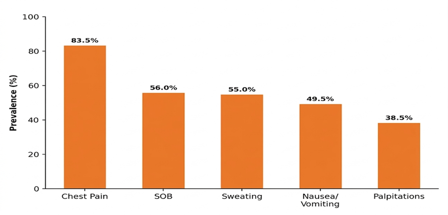
Figure 5.3: Symptom Profile of Young AMI Patients (N=109)
Table 5.3.1.2: ECG Findings Among Young AMI Patients (N=109)
|
ECG Finding |
n |
% |
|
ST Elevation |
51 |
46.8 |
|
ST Depression |
45 |
41.3 |
|
T Wave Inversion |
10 |
9.2 |
|
Q Waves |
2 |
1.8 |
|
Normal ECG |
1 |
0.9 |
ST-segment elevation was the most frequent ECG abnormality, observed in 51 patients (46.8%), followed closely by ST depression in 45 (41.3%). T-wave inversion was noted in 10 cases (9.2%), pathological Q waves in 2 (1.8%), and a normal ECG tracing in just 1 patient (0.9%). The near-equal split between ST elevation and ST depression suggests that this young cohort experienced a substantial burden of both STEMI and NSTEMI presentations.
Table 5.3.1.3: Coronary Angiography and Treatment Profile (N=109)
|
Variable |
Category |
n |
% |
|
Angiography |
Yes |
70 |
64.2 |
|
|
No |
39 |
35.8 |
|
Treatment |
Medications |
68 |
62.4 |
|
|
Thrombolysis |
21 |
19.3 |
|
|
PCI |
20 |
18.3 |
Coronary angiography was performed in 70 patients (64.2%), while the remaining 39 (35.8%) were managed without invasive evaluation. In terms of treatment, the largest proportion received conservative medical management: 68 patients (62.4%). Thrombolysis was administered to 21 (19.3%), and percutaneous coronary intervention (PCI) was carried out in 20 (18.3%). The relatively high proportion managed with medications alone may reflect resource availability, timing of presentation, or clinical judgement regarding infarct severity.
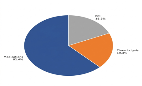
Figure 5.4: Diagnostic and treatment profile
When risk factors were compared across genders, most comorbidities showed no statistically significant difference. Hypertension was present in 67.7% of males and 59.6% of females (χ²=0.776, p=0.378). Diabetes affected 45.2% of males and 44.7% of females (χ²=0.002, p=0.960). Dyslipidemia rates were virtually identical between the sexes (48.4% vs. 48.9%; χ²=0.003, p=0.955). Previous heart disease trended higher in females (36.2%) than males (21.0%), though this did not reach statistical significance (χ²=3.097, p=0.078). Family history of CAD was comparable: 56.5% in males versus 59.6% in females (χ²=0.107, p=0.744).
Table 5.5.1: Gender-wise Distribution of Risk Factors (N=109)
|
Risk Factor |
Male |
Female |
χ² |
p-value |
Cramér's V |
|
Hypertension |
42 (67.7) |
28 (59.6) |
0.776 |
0.378 |
0.084 |
|
Diabetes Mellitus |
28 (45.2) |
21 (44.7) |
0.002 |
0.960 |
0.005 |
|
Dyslipidemia |
30 (48.4) |
23 (48.9) |
0.003 |
0.955 |
0.005 |
|
Previous Heart Disease |
13 (21.0) |
17 (36.2) |
3.097 |
0.078 |
0.169 |
|
Family History of CAD |
35 (56.5) |
28 (59.6) |
0.107 |
0.744 |
0.031 |
|
Alcohol |
3 (4.8) |
0 (0.0) |
2.339 |
0.126 |
0.146 |
|
Smoking (Never) |
23 (37.1) |
41 (87.2) |
28.881 |
<0.001 |
0.515 |
|
Smoking (Former) |
26 (41.9) |
2 (4.3) |
|
|
|
|
Smoking (Current) |
13 (21.0) |
4 (8.5) |
|
|
|
Table 5.5.2: Age Group-wise Distribution of Risk Factors (n=106)
|
Risk Factor |
≤30 yrs |
31–35 yrs |
36–40 yrs |
41–45 yrs |
χ² |
p-value |
|
Hypertension |
13 (32.5) |
12 (75.0) |
14 (77.8) |
29 (90.6) |
29.449 |
<0.001 |
|
Diabetes Mellitus |
6 (15.0) |
11 (68.8) |
8 (44.4) |
22 (68.8) |
25.541 |
<0.001 |
|
Dyslipidemia |
13 (32.5) |
4 (25.0) |
11 (61.1) |
22 (68.8) |
13.994 |
0.003 |
|
Previous HD |
5 (12.5) |
6 (37.5) |
4 (22.2) |
14 (43.8) |
9.837 |
0.020 |
|
Family Hx CAD |
27 (67.5) |
7 (43.8) |
9 (50.0) |
19 (59.4) |
3.314 |
0.346 |
The one striking gender difference emerged in smoking status. Among males, only 37.1% had never smoked, compared to 87.2% of females. Former smokers constituted 41.9% of males but merely 4.3% of females, and current smoking was reported by 21.0% of males versus 8.5% of females. This difference was highly significant (χ²=28.881, p<0.001, Cramér’s V=0.515), representing the single strongest gender-linked disparity in the dataset. Physical activity levels, dietary patterns, and BMI categories did not differ significantly between males and females.
Symptom presentation did not differ significantly between genders for any individual symptom, though some trends are worth noting. Chest pain was more common in males (88.7%) than females (76.6%), and while this difference did not reach significance (χ²=2.846, p=0.092), it echoes existing literature suggesting that women are more likely to present with atypical AMI symptoms. Nausea and vomiting trended higher in females (59.6% vs. 41.9%; χ²=3.327, p=0.068), lending further support to this observation. ECG findings, however, did differ by gender (χ²=10.004, p=0.040). Males were more likely to show ST elevation (56.5% vs. 34.0%), while females had a higher proportion of T-wave inversions (14.9% vs. 4.8%) and were the only gender to exhibit Q waves (4.3%) and normal ECGs (2.1%). This pattern suggests that males in this cohort tended toward STEMI presentations, whereas females had a more heterogeneous ECG picture.
Table 5.6: Gender-wise Clinical Presentation (N=109)
|
Symptom |
Male n (%) |
Female n (%) |
χ²(Chi-square value) |
p-value |
|
Chest Pain |
55 (88.7) |
36 (76.6) |
2.846 |
0.092 |
|
SOB |
31 (50.0) |
30 (63.8) |
2.075 |
0.150 |
|
Sweating |
37 (59.7) |
23 (48.9) |
1.246 |
0.264 |
|
Nausea/Vomiting |
26 (41.9) |
28 (59.6) |
3.327 |
0.068 |
|
Palpitations |
21 (33.9) |
21 (44.7) |
1.319 |
0.251 |
|
ECG: ST Elevation |
35 (56.5) |
16 (34.0) |
10.004 |
0.040 |
|
ECG: ST Depression |
24 (38.7) |
21 (44.7) |
|
|
|
ECG: T Wave Inv. |
3 (4.8) |
7 (14.9) |
|
|
|
ECG: Q Waves |
0 (0.0) |
2 (4.3) |
|
|
|
ECG: Normal |
0 (0.0) |
1 (2.1) |
|
|
Table 5.7: Gender-wise Angiography and Treatment (N=109)
|
Variable |
Male n (%) |
Female n (%) |
χ²(Chi-sqaure value) |
p-value |
|
Angiography: Yes |
46 (74.2) |
24 (51.1) |
6.224 |
0.013 |
|
Angiography: No |
16 (25.8) |
23 (48.9) |
|
|
|
Thrombolysis |
14 (22.6) |
7 (14.9) |
7.853 |
0.020 |
|
PCI |
16 (25.8) |
4 (8.5) |
|
|
|
Medications |
32 (51.6) |
36 (76.6) |
|
|
Coronary angiography was performed significantly more often in males (74.2%) than females (51.1%), a difference that was statistically significant (χ²=6.224, p=0.013, Cramér’s V=0.239). Treatment patterns also diverged by gender (χ²=7.853, p=0.020). Males received PCI at over three times the rate of females (25.8% vs. 8.5%) and thrombolysis more frequently (22.6% vs. 14.9%), while females were predominantly managed with medications alone (76.6% vs. 51.6%). Whether this reflects differences in disease severity, timing of presentation, or gender-based clinical decision-making merits further investigation.
Table 5.8: Age Group-wise Angiography and Treatment (n=106)
|
Variable |
≤30 yrs n (%) |
31–35 yrs n (%) |
36–40 yrs n (%) |
41–45 yrs n (%) |
χ²(Chi-square value) |
p-value |
|
Angio: Yes |
20 (50.0) |
10 (62.5) |
14 (77.8) |
23 (71.9) |
5.681 |
0.128 |
|
Angio: No |
20 (50.0) |
6 (37.5) |
4 (22.2) |
9 (28.1) |
|
|
|
Thrombolysis |
5 (12.5) |
5 (31.3) |
5 (27.8) |
6 (18.8) |
18.689 |
0.005 |
|
PCI |
3 (7.5) |
4 (25.0) |
8 (44.4) |
5 (15.6) |
|
|
|
Medications |
32 (80.0) |
7 (43.8) |
5 (27.8) |
21 (65.6) |
|
|
Angiography rates showed an upward trend with age 50.0% in the ≤30 group, 62.5% in 31–35, 77.8% in 36–40, and 71.9% in 41–45 but this gradient did not achieve statistical significance (χ²=5.681, p=0.128). Treatment modality, however, differed significantly across age groups (χ²=18.689, p=0.005). The youngest patients were overwhelmingly managed with medications (80.0%), while PCI was most common in the 36–40 age bracket (44.4%). Thrombolysis use was highest in the 31–35 group (31.3%). These patterns likely reflect the interplay between infarct type, disease burden, and therapeutic access at different ages.
Several risk factors showed meaningful associations with the likelihood of undergoing coronary angiography. Dyslipidemia was significantly associated with angiography-positive status (χ²=3.937, p=0.047; OR=2.25, 95% CI: 1.00–5.03). Hypertension showed a trend toward significance (OR=2.00, 95% CI: 0.89–4.50; p=0.092), while diabetes and family history of CAD did not demonstrate significant associations.
Table 5.9: Association of Risk Factors with Coronary Angiography (N=109)
|
Risk Factor |
Angio+ |
Angio− |
χ² |
p-value |
OR |
|
Hypertension |
49 (70.0) |
21 (53.8) |
2.844 |
0.092 |
2.00 (0.89–4.50) |
|
Diabetes Mellitus |
32 (65.3) |
38 (63.3) |
0.046 |
0.831 |
1.09 (0.50–2.40) |
|
Dyslipidemia |
39 (73.6) |
31 (55.4) |
3.937 |
0.047 |
2.25 (1.00–5.03) |
|
Family Hx CAD |
42 (66.7) |
28 (60.9) |
0.389 |
0.533 |
1.29 (0.58–2.84) |
|
Smoking: Never |
35 (54.7) |
29 (45.3) |
6.380 |
0.041 |
|
|
Smoking: Former |
21 (75.0) |
7 (25.0) |
|
|
|
|
Smoking: Current |
14 (82.4) |
3 (17.6) |
|
|
|
|
BMI: Normal |
18 (46.2) |
21 (53.8) |
10.982 |
0.012 |
|
|
BMI: Overweight |
32 (69.6) |
14 (30.4) |
|
|
|
|
BMI: Obese |
19 (86.4) |
3 (13.6) |
|
|
|
Smoking showed a dose-response relationship with angiography (χ²=6.380, p=0.041): angiography-positive rates climbed from 54.7% in non-smokers to 75.0% in former smokers to 82.4% in current smokers. BMI category demonstrated a similarly graded association (χ²=10.982, p=0.012) angiography-positive rates increased from 46.2% in normal-weight patients to 69.6% in overweight and 86.4% in obese patients.
Table 5.10: Association of Risk Factors with ECG Findings (N=109)
|
Risk Factor |
ST Elevation n (%) |
ST Depression |
T Wave Inv. n (%) |
χ² |
p-value |
|
Hypertension: Yes |
39 (55.7) |
21 (30.0) |
7 (10.0) |
11.182 |
0.025 |
|
Hypertension: No |
12 (30.8) |
24 (61.5) |
3 (7.7) |
|
|
|
DM: Yes |
29 (59.2) |
10 (20.4) |
7 (14.3) |
18.528 |
0.001 |
|
DM: No |
22 (36.7) |
35 (58.3) |
3 (5.0) |
|
|
Hypertension was significantly associated with ECG patterns (χ²=11.182, p=0.025). Among hypertensive patients, ST elevation predominated at 55.7%, whereas in non-hypertensive, ST depression was more common (61.5%). The relationship between diabetes mellitus and ECG findings was even more pronounced (χ²=18.528, p=0.001). A notable 59.2% of diabetic patients exhibited ST elevation compared to 36.7% of non-diabetics, while ST depression was far more prevalent in the non-diabetic group (58.3% vs. 20.4%). T-wave inversions were also nearly three times more common in diabetics (14.3% vs. 5.0%). These findings suggest that the presence of metabolic comorbidities may influence the electrocardiographic manifestation of acute myocardial infarction in young patients.
Table 5.11: Association of Risk Factors with Treatment Received (N=109)
|
Risk Factor |
Thrombolysis n (%) |
PCI n (%) |
Medications n (%) |
χ²(Chi-square value0 |
p-value |
|
HTN: Yes |
17 (24.3) |
18 (25.7) |
35 (50.0) |
13.154 |
0.001 |
|
HTN: No |
4 (10.3) |
2 (5.1) |
33 (84.6) |
|
|
|
Fam Hx: Yes |
9 (14.3) |
8 (12.7) |
46 (73.0) |
7.223 |
0.027 |
|
Fam Hx: No |
12 (26.1) |
12 (26.1) |
22 (47.8) |
|
|
Hypertension was significantly associated with the type of treatment administered (χ²=13.154, p=0.001). Among hypertensive patients, the distribution of treatment was relatively spread out: 24.3% received thrombolysis, 25.7% underwent PCI, and 50.0% were managed with medications. In stark contrast, non-hypertensive patients were overwhelmingly treated conservatively 84.6% received medications alone, with only 10.3% getting thrombolysis and 5.1% PCI. Family history of CAD also influenced treatment choice (χ²=7.223, p=0.027): patients without a family history received more aggressive intervention (26.1% each for thrombolysis and PCI) compared to those with a positive family history, 73.0% of whom received medications.
Table 5.12: In-Hospital Complications Among Young AMI Patients (N=109)
|
Complication Category |
n |
% |
|
None |
31 |
28.4 |
|
SOB (alone or combined) |
22 |
20.2 |
|
Chest Pain (alone or combined) |
19 |
17.4 |
|
Referred Pain (arm/jaw) |
10 |
9.2 |
|
Bradycardia |
8 |
7.3 |
|
Palpitations |
4 |
3.7 |
|
Arrhythmia (AF, VF, Reperfusion) |
3 |
2.8 |
|
Cardiogenic Shock |
2 |
1.8 |
|
Hypotension |
2 |
1.8 |
|
Nausea/Vomiting |
2 |
1.8 |
|
Other |
6 |
5.5 |
Just over a quarter of patients (31, or 28.4%) had an uncomplicated hospital stay. Among those who developed complications, shortness of breath was the most frequently recorded issue either alone or in combination affecting 22 patients (20.2%). Persistent or recurrent chest pain occurred in 19 patients (17.4%). Referred pain radiating to the arm, jaw, or both was noted in 10 cases (9.2%). Bradycardia was documented in 8 patients (7.3%), palpitations in 4 (3.7%), and various arrhythmias including atrial fibrillation, brief ventricular fibrillation, and reperfusion arrhythmia in 3 (2.8%). More severe complications were uncommon but present: cardiogenic shock in 2 patients (1.8%), hypotension in 2 (1.8%), and nausea or vomiting in 2 (1.8%). A miscellaneous group of 6 patients (5.5%) experienced other complications including heart block, heart failure, left ventricular dysfunction, mild pulmonary edema, chronic kidney disease, and minor hematoma at the catheterization site
DISCUSSION :
This study characterized the risk-factor profile and clinical presentation of 109 young patients (14–48 years) admitted with acute myocardial infarction (AMI) at Sheikh Zayed Hospital, Lahore. The findings reveal a striking burden of modifiable metabolic and lifestyle factors, a notably early age of onset, and important sex-based differences in investigation and management. Males accounted for 56.9% of the cohort, yielding a male-to-female ratio of 1.3:1, consistent with the recognized male predominance in premature coronary artery disease [11]. However, the 43.1% proportion of women exceeds figures in many Western registries and may reflect the heightened metabolic and genetic susceptibility of South Asian women [12]. The age distribution is particularly alarming: 37.7% of patients were aged 30 years or younger, and the minimum age was 14 years (mean 33.32 ± 9.57 years). While South Asian populations are known to develop coronary disease approximately a decade earlier than Western populations [13], the finding that over one-third of the cohort was below age 30 challenges conventional perceptions and underscores the need to reconsider screening thresholds in this region. Overweight or obesity (BMI ≥25 kg/m²) was present in 62.4%, with 20.2% classified as obese, reinforcing the role of excess adiposity as a driver of accelerated atherogenesis (8). Hypertension was the most prevalent comorbidity (64.2%), a figure substantially higher than the 30–50% typically reported in young AMI cohorts [11]. The age gradient was pronounced: hypertension prevalence rose from 32.5% in patients aged ≤30 years to 90.6% in those aged 41–45 years (χ²=29.449, p<0.001), illustrating cumulative vascular damage even within this young population. Diabetes mellitus affected 45.0% again exceeding Western estimates of 10–30% and climbed from 15.0% in the youngest subgroup to 68.8% in the 31–35 and 41–45 year brackets (χ²=25.541, p<0.001). These patterns are consistent with the early-onset metabolic syndrome driven by genetic predisposition, central adiposity, and dietary factors in South Asia (1,4). Dyslipidemia was documented in 48.6%, with a significant age-related increase (p=0.003). Nearly half of the patients had abnormal lipid profiles, and the absence of assessment for lipoprotein(a) and Apo lipoprotein ratios may have led to underestimation of true atherogenic risk (9). A family history of premature coronary artery disease was reported by 57.8% and was uniformly distributed across age groups (χ²=3.314, p=0.346), confirming that genetic predisposition operates independently of age [7,9].
The smoking data reveal a notable paradox. Never-smokers constituted 58.7%, diverging from the 60–80% prevalence of tobacco exposure in young AMI cohorts elsewhere [7,20]. Ever-smoking was recorded in 41.3% and displayed the strongest sex disparity in the dataset: 62.9% of males vs. 12.8% of females (χ²=28.881, p<0.001, Cramér’s V=0.515), reflecting socio-cultural norms. Alcohol consumption was negligible (2.8%), as expected in this population [12,13]. Sedentary behavior (42.2%) and high-fat dietary intake (61.5%) were common and likely fuel the high prevalence of obesity and metabolic abnormalities [14]. The symptom profile was largely classical: chest pain (83.5%), shortness of breath (56.0%), diaphoresis (55.0%), and nausea/vomiting (49.5%). Nevertheless, women less frequently reported chest pain (76.6% vs. 88.7%) and trended toward higher rates of nausea/vomiting (59.6% vs. 41.9%; p=0.068), consistent with the known phenomenon of atypical presentation in females (2,20). On electrocardiography, ST-elevation (46.8%) and ST-depression (41.3%) were nearly equally prevalent, indicating a similar burden of STEMI and NSTEMI. The sex difference in ECG patterns was significant (p=0.040): ST-elevation predominated in males (56.5% vs. 34.0%), while T-wave inversion (14.9% vs. 4.8%) and Q waves were more frequent in females, a heterogeneity that may contribute to diagnostic delays [16,17]. Notably, diabetes and hypertension were each associated with ST-elevation (p=0.001 and p=0.025, respectively), suggesting that metabolic comorbidities influence not only the occurrence but also the electrocardiographic phenotype of AMI in young patients. Coronary angiography was performed in 64.2% of patients, significantly more often in males (74.2% vs. 51.1%; p=0.013). This sex gap in invasive investigation mirrors international registry data and raises concerns about equitable access to guideline-directed care (24,33). Medical management alone was the mainstay for 62.4%, with thrombolysis and percutaneous coronary intervention (PCI) used in 19.3% and 18.3%, respectively. The reliance on conservative management may reflect single-center resource constraints and the fact that young patients with less extensive disease can often be managed medically. PCI rates were significantly higher in males (25.8% vs. 8.5%; p=0.020), and PCI utilization peaked in the 36–40-year age group (44.4%), while 80% of those aged ≤30 years received medication alone (p=0.005). Angiography-positive status, a surrogate for greater disease burden, was associated with dyslipidemia (OR=2.25, 95% CI 1.00–5.03; p=0.047), smoking (current smokers 82.4% vs. never-smokers 54.7%; p=0.041), and BMI (normal weight 46.2% vs. obese 86.4%; p=0.012), underscoring the atherogenic impact of these factors (7,22). In-hospital complications occurred in 71.6% of patients, but were predominantly mild; cardiogenic shock (1.8%) and arrhythmias (2.8%) were uncommon, consistent with the preserved cardiac reserve of younger individuals [18,19].
Limitation of the study
Several limitations must be acknowledged. The single-center design and modest sample size (n=109) may limit generalizability and statistical power for subgroup analyses. Lifestyle data relied on self-report, introducing potential recall bias. Non-traditional biomarkers (e.g., lipoprotein(a), homocysteine, high-sensitivity CRP, thrombophilia screening) were not assessed, leaving an important dimension of atherogenic and prothrombotic risk unexplored. The cross-sectional design precludes evaluation of long-term outcomes, and incomplete angiography data due to clinical or logistical reasons may introduce selection bias.
Future Recommendation
These findings have direct clinical implications. The alarmingly high prevalence of hypertension and diabetes in patients as young as 30 years supports population-level cardiovascular screening from age 20 in South Asian communities, with a focus on blood pressure, glucose, and lipid profiles. The dominant contribution of modifiable risk factors high-fat diet, physical inactivity, obesity, and smoking indicates that a substantial proportion of premature AMI is preventable through school- and workplace-based lifestyle interventions. Sex-sensitive clinical protocols are urgently needed to ensure timely diagnostic evaluation for young women, including equitable access to coronary angiography. Moreover, the uniform distribution of family history across all age groups justifies a lower threshold for cardiovascular risk assessment in young adults with a positive family history.
CONCLUSION :
Premature AMI in this Pakistani cohort is driven primarily by a cluster of modifiable metabolic and lifestyle factors, with a clear age-dependent gradient. The high proportion of patients aged ≤30 years highlights accelerated cardiovascular ageing and calls for a paradigm shift toward earlier risk stratification. Sex disparities in symptom presentation, ECG patterns, and invasive management demand systematic attention. The largely preventable nature of these events reinforces the need for aggressive, culturally adapted primordial and primary prevention strategies well before the fourth decade of life. Future multicenter prospective studies incorporating advanced biomarkers and long-term follow-up are essential to capture the full spectrum of risk and to guide region-specific interventions
REFERENCES:
1. Andersson C, Vasan RS. Epidemiology of cardiovascular disease in young individuals. Nat Rev Cardiol. 2018;15(4):230–240.
2. Angelini P. Coronary artery anomalies: a comprehensive approach. Circulation. 2007;115(10):1296–1305.
3. Arora S, Stouffer GA, Kucharska-Newton AM, Qamar A, Vaduganathan M, Pandey A, et al. Twenty year trends and sex differences in young adults hospitalized with acute myocardial infarction. Circulation. 2019;139(8):1047–1056.
4. Athyros VG, Doumas M, Imprialos KP, Stavropoulos K, Georgianou E, Katsimardou A, et al. Dyslipidemia patterns in young adults presenting with coronary artery disease. Curr Med Res Opin. 2022;38(3):431–439.
5. Aviña-Zubieta JA, Choi HK, Sadatsafavi M, Etminan M, Esdaile JM, Lacaille D. Risk of cardiovascular mortality in patients with rheumatoid arthritis: a meta-analysis of observational studies. Arthritis Rheum. 2008;59(12):1690–1697.
6. Benjamin EJ, Virani SS, Callaway CW, Chamberlain AM, Chang AR, Cheng S, et al. Heart disease and stroke statistics 2018 update: a report from the American Heart Association. Circulation. 2018;137(12):e67–e492.
7. Choo EH, Chang K, Lee KY, Lee D, Kim JG, Ahn Y, et al. Prognosis and predictors of adverse outcomes in young adults with acute coronary syndrome. PLoS One. 2019;14(3):e0213499.
8. Clarke R, Halsey J, Lewington S, Lonn E, Armitage J, Manson JE, et al. Effects of lowering homocysteine levels with B vitamins on cardiovascular disease, cancer, and cause-specific mortality. N Engl J Med. 2002;346(7):476–483.
9. Dang A, Gupta S, Sharma M. Tobacco use and early onset myocardial infarction. Int J Cardiol. 2020;319:39–44.
10. Danesh J, Wheeler JG, Hirschfield GM, Eda S, Eiriksdottir G, Rumley A, et al. C-reactive protein and other circulating markers of inflammation in the prediction of coronary heart disease. N Engl J Med. 2004;350(14):1387–1397.
11. Eckel RH, Grundy SM, Zimmet PZ. The metabolic syndrome. Lancet. 2005;365(9468):1415–1428.
12. Fox KA, Dabbous OH, Goldberg RJ, Pieper KS, Eagle KA, Van de Werf F, et al. Prediction of risk of death and myocardial infarction in the six months after presentation with acute coronary syndrome. Eur Heart J. 2002;23(15):1177–1189.
13. Gupta R, Mohan I. Epidemiology of coronary heart disease in India. Indian J Med Res. 2016;144(4):471–480.
14. Hayashi T, Kiyoshima T, Matsuura M, Ueno M, Kobayashi N, Yabushita H, et al. Plaque characteristics in young acute coronary syndrome patients assessed by optical coherence tomography. JACC Cardiovasc Imaging. 2021;14(2):377–386.
15. Hayashi T, et al. Plaque characteristics in young acute coronary syndrome patients using optical coherence tomography. JACC Cardiovasc Imaging. 2021;14(2):377–386.
16. Ismail J, Jafar TH, Jafary FH, White F, Faruqui AM, Chaturvedi N. Risk factors for non-fatal myocardial infarction in young South Asian adults. Heart. 2004;90(3):259–263.
17. Jia H, Abtahian F, Aguirre AD, Lee S, Chia S, Lowe H, et al. In vivo diagnosis of plaque erosion and calcified nodule in patients with acute coronary syndrome by intravascular optical coherence tomography. J Am Coll Cardiol. 2013;62(19):1748–1758.
18. Joshi P, Islam S, Pais P, Reddy S, Dorairaj P, Kazmi K, et al. Risk factors for early myocardial infarction in South Asians compared with individuals in other countries. JAMA. 2007;297(3):286–294.
19. Joshi P, Patel V, Islam S, Prabhakaran D, Yusuf S. Genetic determinants of premature coronary artery disease in South Asians. Circulation. 2017;135(24):1882–1895.
20. Khan S, Bhatt DL, Guedeney P, Mehran R. Non-atherosclerotic causes of acute myocardial infarction in young adults. J Am Heart Assoc. 2021;10(18):e018043.