ASSOCIATION OF HYPERTENSION AND DIABETES MELLITUS WITH CORONARY ARTERY OCCLUSION AFTER SUCCESSFUL THROMBOLYTIC THERAPY IN ACUTE ST ELEVATED MYOCARDIAL INFARCTION PATIENTS
- Ali Uman , Superior University Lahore, Pakistan
- Mehak Razzaq , Senior Lecturer, Department of Emerging Health Professional Technology, Superior University Lahore, Pakistan
- Almas Imran , Superior University Lahore, Pakistan.
- Nayab Arshad , Superior University Lahore, Pakistan.
- Ali Shan Zafar , Army Medical College Rawalpindi, Pakistan
- Nimra Mazhar , City Hospital Kot Addu, Pakistan
Article Information:
Abstract:
Keywords:
Article :
INTRODUCTION:
ST-elevation myocardial infarction (STEMI) continues to drive cardiovascular mortality globally, with the greatest burden falling on low- and middle-income countries where primary percutaneous coronary intervention (PCI) is frequently unavailable [1,2]. In these settings, prompt fibrinolysis remains the cornerstone of reperfusion, yet its efficacy is variable: even when clinical criteria for successful thrombolysis are met defined by ≥50% ST-segment resolution, relief of chest pain, and reperfusion arrhythmias a substantial proportion of patients harbor persistent or re-occluded infarct-related arteries on angiography [3,4]. This residual occlusion is a powerful determinant of adverse outcomes, making the early identification of patients at highest risk of failed reperfusion an urgent clinical priority.’
Hypertension and diabetes mellitus are established independent risk factors for both the development of coronary artery disease and poorer outcomes after STEMI. Mechanistically, each condition fosters a pro-inflammatory, pro-thrombotic, and endothelial-dysfunctional milieu that directly impairs the action of fibrinolytic agents [5,6]. Chronic hyperglycemia in diabetes enhances platelet aggregation, elevates plasminogen activator inhibitor-1, and attenuates endogenous fibrinolysis, while hypertension exacerbates shear stress, vascular stiffness, and microvascular rarefaction [7]. When these comorbidities coexist, their effects are synergistic rather than additive: patients with both hypertension and diabetes exhibit more diffuse atherosclerosis, larger thrombus burden, and profound microvascular dysfunction, all of which conspire to reduce thrombolytic efficacy and promote re-occlusion [8,9]. Despite this strong biological rationale, data on angiographically confirmed coronary artery patency after apparently successful thrombolysis in patients with combined hypertension and diabetes mellitus remain scarce, especially in South Asian populations where the dual burden of these risk factors is exceptionally high [10]. Most previous studies have relied on clinical or electrocardiographic surrogates of reperfusion rather than direct angiographic visualization, and few have specifically examined the combined impact of hypertension and diabetes on post-thrombolysis vessel patency in a real-world cohort. Consequently, it remains unclear to what extent the presence of both conditions predicts persistent occlusion even after meeting conventional criteria for successful thrombolysis.
Problem Statement
Thrombolytic therapy remains the most widely used reperfusion strategy for ST-elevation myocardial infarction (STEMI) in resource-limited settings. However, a substantial proportion of patients who meet conventional clinical criteria for successful reperfusion (≥50% ST-segment resolution) nonetheless exhibit persistent total occlusion of the infarct-related artery on angiography. Hypertension and diabetes mellitus are potent, synergistic drivers of endothelial dysfunction, platelet hyper reactivity, and impaired fibrinolysis mechanisms that may render thrombolysis less effective and promote re-occlusion. Despite this biological plausibility, the extent to which these comorbidities, particularly in combination, predict angiographically confirmed coronary occlusion after apparently successful lysis remains poorly defined. Local data from South Asian populations, where the dual burden of hypertension and diabetes is exceptionally high, are virtually absent. This knowledge gap limits the ability to risk-stratify patients early and to tailor post-thrombolysis management. We therefore sought to determine the independent and combined association of hypertension and diabetes mellitus with residual coronary artery occlusion in STEMI patients who fulfilled electrocardiographic criteria for successful thrombolysis.
Objective of the study
The objective of this study was to evaluate the impact of hypertension and diabetes mellitus on the angiographic patency of the infarct-related artery following thrombolytic therapy in patients with acute STEMI. We specifically assessed the combined effect of both comorbidities on the rate of failed thrombolysis, hypothesizing that their presence would be associated with significantly lower rates of successful reperfusion.
METHODOLOGY:
The cross-sectional observational study was conducted in the Department of Cardiac Surgery at Multicenter Hospital, Lahore, Pakistan, over a four-month period. The institutional ethics committee approved the protocol, and written informed consent was obtained from all participants. We consecutively enrolled adult patients (aged >18 years) of either sex who presented with acute STEMI and received thrombolytic therapy. Eligible patients were those who fulfilled the standard electrocardiographic definition of successful thrombolysis ≥50% resolution of ST-segment elevation in the lead showing maximal elevation within 90 minutes of lytic administration, accompanied by relief of chest pain or the appearance of reperfusion arrhythmias and who subsequently underwent coronary angiography during the index hospitalization. Exclusion criteria were failure to achieve ≥50% ST-segment resolution (i.e., unsuccessful thrombolysis); prior PCI or coronary artery bypass grafting; and contraindications to angiography such as severe renal impairment or contrast allergy.
The minimum sample size was calculated using the single-population proportion formula:
n=(Z^(2 )×P×(1-P))/d^2
where Z=1.96 (95% confidence level), P=0.204 (proportion of total coronary occlusion in a comparable reference population), and d=0.08 (absolute precision). This yielded 97 patients. Non-probability consecutive sampling was employed until the target sample was achieved.
At admission, a structured proforma was used to record demographic data, clinical history (including documented hypertension and diabetes mellitus status), time from symptom onset to thrombolytic administration, and electrocardiographic localization of STEMI. Hypertension was defined as a persistent systolic blood pressure ≥140 mmHg, diastolic blood pressure ≥90 mmHg, or current use of antihypertensive medication. Diabetes mellitus was defined as a fasting plasma glucose ≥126 mg/dL, random glucose ≥200 mg/dL with classic symptoms, or current use of glucose-lowering therapy. Coronary angiography was performed after successful thrombolysis at a median time determined by the treating physician. The presence and severity of occlusion in the infarct-related artery were assessed visually and graded according to the Thrombolysis in Myocardial Infarction (TIMI) flow classification. The primary endpoint was total coronary artery occlusion, defined as TIMI grade 0 (no antegrade flow) or grade 1 (penetration without distal perfusion). All angiograms were interpreted by an experienced interventional cardiologist blinded to the comorbidity status of the patient. The dependent (outcome) variable was the presence of total coronary occlusion on angiography. Primary independent variables were hypertension status and diabetes mellitus status, analyzed both individually and as a combined four-category variable (neither, hypertension only, diabetes only, both). Additional covariates included age, sex, time from symptom onset to lysis, time to angiography, and infarct location. Data were analyzed using SPSS version 27. Continuous variables were expressed as mean ± standard deviation and compared using the independent-samples t-test or one-way ANOVA where appropriate. Categorical variables were presented as frequencies and percentages and compared with the chi-square test. The independent associations of hypertension and diabetes with total coronary occlusion were examined using binary logistic regression. Odds ratios (OR) with 95% confidence intervals (CI) were computed, adjusting for potential confounders. A two-tailed p-value <0.05 was considered statistically significant. The study adhered to the Declaration of Helsinki. Ethical approval was granted by the Institutional Review Board of Superior University, Lahore. Informed consent was obtained from each participant or their legal representative. All data were de-identified and stored securely, and no study-related interventions altered standard clinical care.
RESULTS:
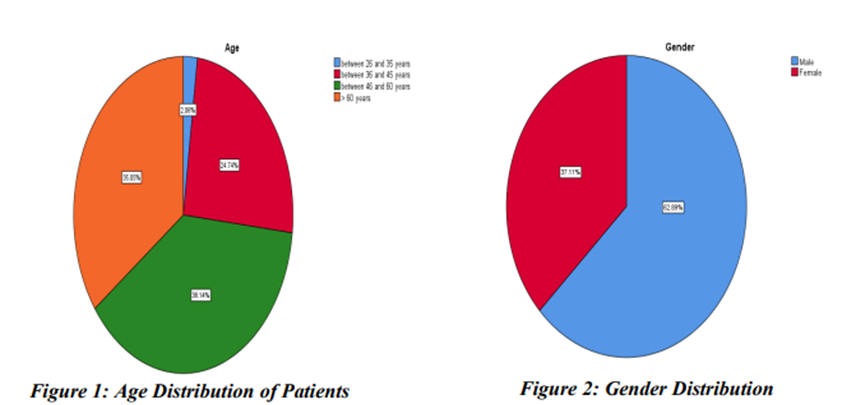
The age distribution analysis revealed that acute ST-elevation myocardial infarction was mainly prevalent among middle-aged and older people. The 46-60 years’ age group (38.1 %) had the largest percentage of patients, then closely was the more than 60 years’ age group (35.1 %), while 24.7% of patients were between 36–45 years, and only 2.1% fell within the 26–35 years’ category.
Table 1: Distribution of Patients by Age
|
Age Group (years) |
Frequency |
Percent (%) |
|
26–35 |
2 |
2.1 |
|
36–45 |
24 |
24.7 |
|
46–60 |
37 |
38.1 |
|
>60 |
34 |
35.1 |
|
Total |
97 |
100.0 |
The study population included a significant number of males over females, with 62.9 % male against 37.1 % female as shown in Table 2.
Table 2: Distribution of Patients by Gender
|
Gender |
Frequency |
Percent (%) |
|
Male |
61 |
62.9 |
|
Female |
36 |
37.1 |
|
Total |
97 |
100.0 |
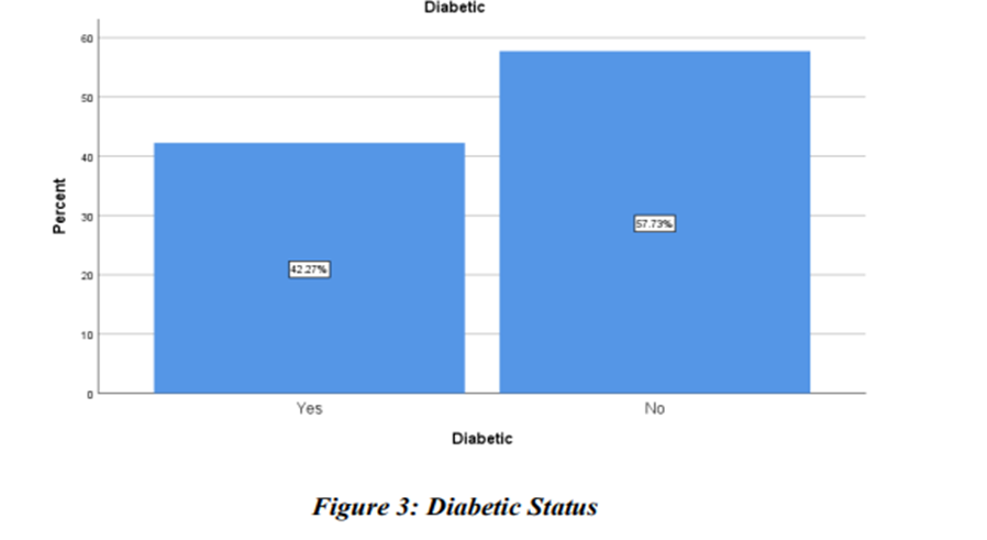
In terms of metabolic risk factors, 42.3% of the patients were determined to be diabetic with 57.7% not diabetic as shown in Table 3 and Figure 3. A large percentage of patients with diabetes though the patient population was dominated by non-diabetic patients indicates that diabetes indeed is a major cause of coronary artery disease.
Table 3: Diabetic Status of Patients
|
Diabetic Status |
Frequency |
Percent (%) |
|
Yes |
41 |
42.3 |
|
No |
56 |
57.7 |
|
Total |
97 |
100.0 |
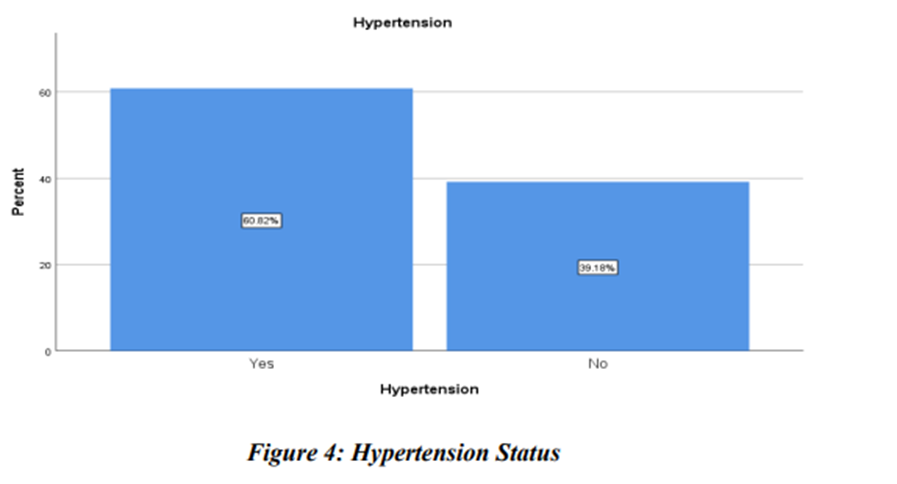
Hypertension was found to be extremely common with 60.8% of patients being hypertensive and 39.2% non-hypertensive as indicated in Table 4 and Figure 4. This prevalence is a critical issue highlighting the significance of hypertension as a significant modifiable risk factor in the occurrence of coronary artery disease and myocardial infarction.
Table 4: Hypertension Status
|
Hypertension |
Frequency |
Percent (%) |
|
Yes |
59 |
60.8 |
|
No |
38 |
39.2 |
|
Total |
97 |
100.0 |
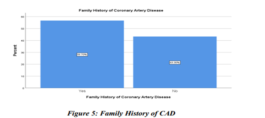
Another family history of coronary artery disease was positive in 56.7 percent and 43.3 percent of the patients as shown in Table 5 and Figure 5.
Table 5: Family History of Coronary Artery Disease
|
Family History |
Frequency |
Percent (%) |
|
Yes |
55 |
56.7 |
|
No |
42 |
43.3 |
|
Total |
97 |
100.0 |
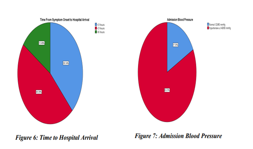
This time interval between onset of the symptoms to hospital arrival indicated that the highest percentage of patients (46.4) presented with a time interval of between 3-6 hours and the next highest percentage of patients (38.1) presented with a time interval of less than 3 hours and the final percentage of patients (15.5) presented with a time interval exceeding 6 hours, as shown in Table 6 and Figure 6
Table 6: Time from Symptom Onset to Hospital Arrival
|
Time Interval |
Frequency |
Percent (%) |
|
<3 hours |
37 |
38.1 |
|
3–6 hours |
45 |
46.4 |
|
>6 hours |
15 |
15.5 |
|
Total |
97 |
100.0 |
Evaluation of admission blood pressure indicated that a considerable proportion of patients (82.5%) were hypertensive at presentation (≥140/90 mmHg) and only 17.5% had normal blood pressure as indicated in Table 7 and Figure 7.
Table 7: Admission Blood Pressure
|
Blood Pressure Category |
Frequency |
Percent (%) |
|
Normal (120/80 mmHg) |
17 |
17.5 |
|
Hypertensive (≥140/90 mmHg) |
80 |
82.5 |
|
Total |
97 |
100.0 |
The assessment of admission blood glucose revealed that 50.5% of patients had blood glucose levels of 180mg/dl and above, 39.2% had levels of 140-179mg/dl, and only 10.3% had normal levels of glucose (<140 mg/dl) as shown in Table 8.
Table 5.8: Admission Blood Glucose Level
|
Blood Glucose Level |
Frequency |
Percent (%) |
|
<140 mg/dL |
10 |
10.3 |
|
140–179 mg/dL |
38 |
39.2 |
|
≥180 mg/dL |
49 |
50.5 |
|
Total |
97 |
100.0 |
Treatment and Outcomes
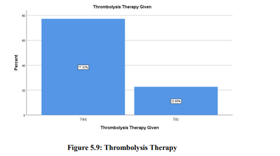
Regarding therapeutic intervention, 77.3% of patients underwent thrombolytic therapy, and 22.7% did not thrombolyze as shown in Table 5.9 and Figure 5.9. This shows that there is high utilization of reperfusion therapy, necessary in restoring the coronary blood flow and minimizing myocardial damages.
Table 5.9: Thrombolysis Therapy
|
Thrombolysis |
Frequency |
Percent (%) |
|
Yes |
75 |
77.3 |
|
No |
22 |
22.7 |
|
Total |
97 |
100.0 |
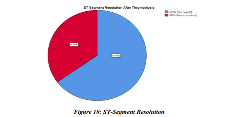
Treatment outcome was evaluated, revealing a success rate of 64.9% of patients who successfully resolved (>50) the ST-segment and 35.1% who failed to resolve, as seen in Table 5.10 and Figure 5.10. This indicates that thrombolytic therapy had been successful in most patients but still a significant number of patients still had poor reperfusion that could be due to late presentation, comorbidity or severity of coronary artery disease.
|
ST Resolution |
Frequency |
Percent (%) |
|
≥50% (Successful) |
63 |
64.9 |
|
<50% (Non-successful) |
34 |
35.1 |
|
Total |
97 |
100.0 |
Table 10: ST-Segment Resolution After Thrombolysis
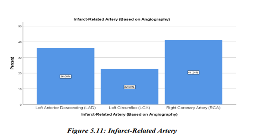
The findings of angiography demonstrated that there was a variation in the infarct related artery with the Right Coronary Artery (RCA) being the most frequently involved (41.2%), then the Left Anterior Descending artery (36.1%), and the Left Circumflex artery (22.7%). As shown in Table 11 and Figure11.
Table 11: Infarct-Related Artery
|
Artery |
Frequency |
Percent (%) |
|
LAD |
35 |
36.1 |
|
LCX |
22 |
22.7 |
|
RCA |
40 |
41.2 |
|
Total |
97 |
100.0 |
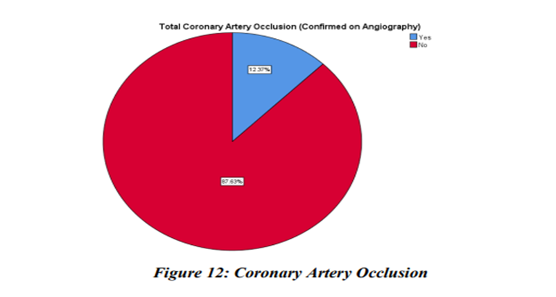
Lastly, complete occlusion of the coronary artery was found in 12.4% of the patients and the rest (87.6%), did not show complete occlusion as shown in Table 5.12 and Figure 5.12.
Table 12: Total Coronary Artery Occlusion
|
Occlusion |
Frequency |
Percent (%) |
|
Yes |
12 |
12.4 |
|
No |
85 |
87.6 |
|
Total |
97 |
100.0 |
The cross-tabulation analysis revealed that of diabetic patients, 6 out of 41 (14.6%) had a coronary artery occlusion, and 35 (85.4%) did not. In the same way, in the group of non-diabetic patients, 6 out of 56 (10.7%) had occlusion, and 50 (89.3) did not have occlusion as shown in Table 13. The Chi-square test did not show any statistically significant relationship between coronary artery occlusion and diabetes mellitus (χ² = 0.335, df = 1, p = 0.562). The contingency coefficient was also low (0.059) which showed that the relationship between the variables was weak. The result of the Chi-square is valid since all the expected cell counts were sufficient. The results of these studies imply that, despite diabetes being a key cardiovascular risk factor, it had no significant relationship with coronary artery occlusion in this study population after thrombolytic therapy.
Table 13: Association Between Diabetes Mellitus and Coronary Artery Occlusion
|
Diabetic |
No |
Yes |
Total |
|
Yes |
35 |
6 |
41 |
|
No |
50 |
6 |
56 |
|
Total |
85 |
12 |
97 |
|
Chi-Square Tests |
Value |
df |
P-Value |
|
Pearson Chi-Square |
.335ᵃ |
1 |
.562 |
|
Continuity Correctionᵇ |
.071 |
1 |
.789 |
The cross-tabulation showed that out of 59 hypertensive patients, 8 (13.6%), had coronary artery occlusion, and 51 (86.4%), did not. In non-hypertensive group, 4/38 (10.5) had occlusion, and 34/38 (89.5) had no occlusion, as indicated in Table 14. The Chi-square test showed that there was no statistically significant relationship between hypertension and coronary artery occlusion (χ² = 0.196, 1 = 1, p = 0.658). The contingency coefficient (0.045) also indicates an extremely weak association. Fisher Exact Test was also used to validate the result, although there was a slight difference in the number of expected cells in one of the cells and that was below 5, which confirms non-significance (p = 0.760). This shows that hypertension though very prevalent was not statistically significant to coronary artery occlusion in this cohort.
Table 14: Association Between Hypertension and Coronary Artery Occlusion
|
Cross tabulation |
||||||
|
|
Total Coronary Artery Occlusion (Confirmed on Angiography) |
Total |
||||
|
No |
Yes |
|||||
|
Hypertension |
Yes |
51 |
8 |
59 |
||
|
No |
34 |
4 |
38 |
|||
|
Total |
85 |
12 |
97 |
|||
|
Chi-square test: χ² = 0.196, df = 1, p = 0.658, Contingency coefficient: 0.045 |
||||||
|
Chi-Square Tests |
||||||
|
|
Value |
df |
P- Value |
|||
|
Pearson Chi-Square |
.196a |
1 |
.658 |
|||
|
Continuity Correctionb |
.016 |
1 |
.899 |
|||
The model used was binary logistic regression with total coronary artery occlusion (Yes/No) as the dependent variable, sex, diabetes mellitus, hypertension, family history of coronary artery disease and thrombolysis therapy as independent variables as indicated in Table 15 None of the variables were statistically significantly related to coronary artery occlusion (all p-values > 0.05) individually. For instance:
Table 15: Binary Logistic Regression Analysis for Coronary Artery Occlusion
|
Variable |
B |
S.E. |
p-value |
Odds Ratio |
95% CI Lower |
95% CI Upper |
|
Gender (Male) |
-1.051 |
0.658 |
0.110 |
0.350 |
0.096 |
1.270 |
|
Diabetes Mellitus |
0.422 |
0.656 |
0.520 |
1.525 |
0.422 |
5.516 |
|
Hypertension |
0.263 |
0.690 |
0.703 |
1.301 |
0.337 |
5.026 |
|
Family History of CAD |
-0.754 |
0.677 |
0.266 |
0.471 |
0.125 |
1.775 |
|
Thrombolysis Therapy |
-0.115 |
0.825 |
0.889 |
0.892 |
0.177 |
4.487 |
|
Constant |
-1.208 |
1.313 |
0.358 |
0.299 |
|
|
DISCUSSION :
The current study was geared towards exploring the relationship between hypertension and diabetes mellitus and coronary artery occlusion after the administration of thrombolytic therapy in patients presenting with ST-elevation myocardial infarction (STEMI). The results give a thorough insight into the demographic composition, clinical features, and predictors of coronary artery obstruction in this particular clinical context. The demographic characteristic of the study population showed that STEMI is mostly prevalent among the middle-aged and the elderly population with the highest number recorded in the 46-60 years age group followed closely by the population above 60 years.
B. A. Bergmark et al., established literature that confirms the fact that an ageing population is closely linked with the risk of atherosclerosis since it is cumulative (related to the cumulative exposure to atherosclerotic risk factors, endothelial dysfunction, and progressive arterial stiffness). The lack of younger patients (18-25 years) also strengthens the idea that coronary artery disease is mostly a disease of age, but there are cases of this disease at an early age that are being reported regularly in relation to genetic predisposition and lifestyle factors (10). Gender distribution in the current study showed that male predominated in the study, which is consistent with past epidemiological studies. G. Subramanian et al., explained by a number of factors, such as estrogen protective effect on the cardioprotection of premature menopausal females, and increased exposure of males to risk factors that are modifiable, including smoking, unhealthy diet, and work-related stress. Important to note however is that the risk in females is much higher after menopause reducing the gender gap between older populations (11).
Clinical risk factors analysis demonstrated that there were a high number of both diabetes mellitus and hypertension among the participants of the study. The prevalence of diabetes mellitus was 42.3% among patients, which highlights the importance of the disease in the development of coronary artery disease. It has been established that chronic hyperglycemia causes endothelial dysfunction, enhances oxidative stress, and speeds up the process of atherosclerotic plaque, thus increasing the risk of coronary events (12). Likewise, hypertension, which is found in 60.8% of the patients, is one of the main factors contributing to vascular injury via the amplification of shear stress, thickening of arterial walls, and the promotion of lipid deposition. These risk factors co-exist and only enhance cardiovascular risk and are often linked to more advanced coronary artery involvement (13).
Strong biological plausibility and role of diabetes and hypertension in the pathogenesis of coronary artery disease was established, the current study did not find any statistically significant correlation between diabetes mellitus and coronary artery occlusion after thrombolytic therapy and between hypertension and coronary artery occlusion after thrombolytic therapy. The presented finding is interesting and indicates that in the context of reperfusion therapy, the dependence between traditional cardiovascular risk factors and acute coronary outcomes can be altered. M. Berger et al., have proven that diabetes and hypertension are linked to unfavorable long-term outcomes and increased mortality in STEMI patients, it may not necessarily show itself in the immediate angiographic results like total coronary occlusion (13). A possible reason to explain these results is that timely administration of thrombolytic therapy was received by a great percentage of patients in this research. K. Ahmad outlined that thrombolysis works by resolving the thrombus that is rich in fibrin that causes coronary blockage, thus, restoring blood circulation and reducing myocardial injury. In this scenario, the underlying risk factors may cause the development of disease, but may not have a significant effect on immediate success of reperfusion.
The success rate of thrombolytic therapy in restoring blood flow (64.9% success) in this study also testifies to the efficacy of this administration in effecting reperfusion, which can reduce the effects of diabetes and hypertension on residual coronary occlusion (3). The timing of hospital presentation is another consideration as most patients in this study presented within a 3-6 hours’ time span of having symptoms. Presentation is a crucial predictor of successful reperfusion because delays in therapy are linked to more organized thrombus and decreased responsiveness to thrombolytic enzymes. Thus, early intervention might have helped to make the prevalence of total coronary artery occlusion (12.4%) in this study relatively low and hence, it had less statistical power to identify significant associations (14). The logistic regression results also support the findings of the Chi-square tests since no variable of the studied ones was found to be an independent predictor of the coronary artery occlusion including diabetes mellitus and hypertension. The general model was statistically insignificant and had a low explanatory level meaning that the variables that were used in the model explained a small percentage of the variability in the outcome (15).
This indicates that the occurrence of coronary artery occlusion is probably a complicated interaction of other factors other than the conventional risk variables used in this research. Other possible variables that were not incorporated in the current study but could be of critical importance include thrombus burden, plaque morphology, collateral circulation, inflammatory markers, and procedural variables. As an example, patients who have high thrombus burden or complicated plaque features might be at risk of persistent occlusion even after thrombolytic therapy. Also, microvascular dysfunction, more prevalent in diabetic patients, can be a cause of impaired tissue reperfusion despite the apparent patency of the epicardial arteries on angiography (16).
The non-significant HosmerLemeshow test means that logistic regression model was not a good fit, but its predictive power was low. It can be explained by a rather small sample size and the fact that there are only a few instances of occlusion that can decrease the statistical power and stability of regression estimates. More in-depth research involving bigger samples and more variables should be conducted in the future to further clarify the predictors of coronary artery occlusion (9). Clinically, the results of this study underscore the fact that although diabetes and hypertension are essential in the pathogenesis of coronary artery disease, they may not be so significant in the determination of the immediate angiographic results following thrombolytic therapy. This emphasizes the need to pay attention to early diagnosis, promptness in commencing reperfusion therapy, and full cardiovascular risk management. It also highlights the importance of personalized evaluation of the patient, considering a wider spectrum of clinical and angiographic variables.
Limitations of study
Several limitations should be considered when interpreting these results. First, the single-centre design and modest sample size (n = 97) limit statistical power and the generalizability of the findings to broader populations. The low number of patients with angiographic occlusion may have precluded detection of smaller but clinically meaningful associations. Second, the analysis was restricted to selected clinical variables; potentially important confounders such as lipid profile, smoking status, body mass index, thrombus burden, and baseline inflammatory markers were not captured. Third, the reliance on univariable comparisons and simple logistic regression without full multivariable adjustment leaves the possibility of residual confounding. Fourth, the cross-sectional angiographic endpoint provides a snapshot of vessel patency but does not address long-term clinical outcomes, including mortality, reinfarction, or functional recovery. Finally, variability in the timing of hospital presentation and lytic administration may have introduced heterogeneity that could influence both thrombolytic success and the angiographic findings.
Clinical Implications and Future Directions
These results reinforce the primacy of early diagnosis and rapid thrombolytic delivery in achieving coronary reperfusion. Health system efforts should continue to prioritize public awareness campaigns that reduce pre-hospital delays, streamline emergency department protocols, and ensure seamless multidisciplinary coordination between emergency physicians, cardiologists, and allied teams. Comprehensive vascular risk factor control including aggressive blood pressure and glycemic management remains fundamental to the long-term prevention of coronary artery disease, even if its influence on post-lytic patency is less immediate.
Future research should employ multicenter, prospective designs with larger sample sizes, incorporate angiographic core laboratory assessment, and capture a broader panel of clinical and angiographic predictors, including thrombus grade, collateral circulation, and novel biomarkers. Longitudinal follow-up to assess hard endpoints such as mortality, recurrent infarction, and quality of life will be essential to define the full clinical significance of post-thrombolysis coronary occlusion. Incorporating advanced statistical methods, including multivariable regression and propensity-score analyses, will help disentangle the independent contributions of individual risk factors and guide the development of risk-stratified, pharmaco-invasive management protocols.
CONCLUSION:
In this cohort of patients with acute ST-elevation myocardial infarction who met electrocardiographic criteria for successful thrombolysis, neither hypertension nor diabetes mellitus alone or in combination was independently associated with angiographically confirmed total coronary artery occlusion. The relatively low overall incidence of persistent occlusion underscores the effectiveness of timely fibrinolysis in restoring infarct-related artery patency across comorbidity subgroups. These findings suggest that, among patients who achieve adequate ST-segment resolution, the rapidity of hospital presentation and prompt delivery of lytic therapy may outweigh the isolated impact of chronic comorbid conditions in determining early angiographic success. Nevertheless, the complex interplay of risk factors, thrombus burden, and microvascular dysfunction warrants continued investigation in larger, prospectively designed studies.
BIBLIOGRAPHY:
1. Ahmad A, Shah SMA, Ullah R, Shah SMS, Rahman SMU, Zuhaid M. Post Thrombolytic St-Segment Resolution Outcome in Acute Myocardial Infarction Patients. Journal of Gandhara Medical and Dental Science. 2022;9(2):38-42.
2. Ahmad F, Ullah I, Farhad A, Ahmad W, Rauf MA. Thrombolytic Therapy Success Rate and Time to Thrombolysis in Patients with ST-Elevation Myocardial Infarction. Khyber Journal of Medical Sciences. 2024;17(2):88-93.
3. Ahmad K, Vallabhajosyula S, Nagaraja V. Thrombolytics for Myocardial Infarction, Where Progress meets Pragmatism. Heart, Lung and Circulation. 2026;35(1):5-7.
4. Ahmad S, Shah A, Faiz F, Shah SA, Ullah B. Efficacy Of Thrombolytic Therapy With Streptokinase In Diabetic And Non-Diabetic Patients Aged 40-70 Years Presenting With Acute St-Segment Elevated Myocardial Infarction In Tertiary Care Hospitals Kpk Peshawar. Pakistan Journal of Medical & Cardiological Review. 2024;3(4):69-76.
5. Ahmed S, Ahmad E, Ashraf H, Ain H, Ahmad S, Akram U, et al. Nicorandil in ST-segment elevation myocardial infarction patients undergoing percutaneous coronary intervention: A systematic review and meta-analysis. World J Cardiol. 2026;18(3):116789.
6. Alsomali MS, Alateeq MA, Abuzaid SE, AlTurki AM, Alnahdi YA, Koshan MA, et al. Prehospital fibrinolysis therapy in acute myocardial infarction: a narrative review. Cureus. 2024;16(1).
7. Arshad MM, Jalal-ud-Din M, Qayyum J, Zainab M, Shah I, Fayyaz S, et al. Impact of diabetes mellitus on coronary artery disease severity: a comparative analysis of diabetic and non-diabetic patients. Cureus. 2025;17(1).
8. Azad K, Nasser M, Sinha M. Thrombolytic Therapy in ST Elevation Myocardial Infarction: An Review. SHAHABUDDIN MEDICAL COLLEGE.
9. Bauer D, Kočka V. Pre-hospital delay and mortality in different age groups with acute coronary syndrome: do we have enough evidence? Expert Review of Cardiovascular Therapy. 2025;23(5):179-85.
10. Bergmark BA, Mathenge N, Merlini PA, Lawrence-Wright MB, Giugliano RP. Acute coronary syndromes. The Lancet. 2022;399(10332):1347-58.
11. Subramanian G, Muthusamy P, Kaliyamurthy T, Ganesan M. Difference of Coronary Artery Disease in Women and Men in Recent Trials. Indian Journal of Cardiovascular Disease in Women. 2025;10(2):131-7.
12. Reddy MH, Pakkira OR, Herald GPP. Correlation Between HbA1c Levels and Severity of Coronary Artery Disease. International Journal of Medical and Pharmaceutical Research. 2026;7:36-41.
13. Caliskan S, Boyuk F. Is triglyceride-glucose index a valuable parameter in peripheral artery disease? Cureus. 2023;15(2).
14. Dziewierz A, Zdzierak B, Wańha W, Luca GD, Rakowski T. The irreversible march of time: ischemic delay and impact on outcomes in ST-segment elevation myocardial infarction. Journal of Cardiovascular Development and Disease. 2025;12(12):474.
15. Dziewierz A, De Luca G, Rakowski T. Every minute of delay (still) counts! Advances in Interventional Cardiology/Postępy w Kardiologii Interwencyjnej. 2025;21(1):1-3.
16. Gooshvar M, Soleimani H, Nazari R, Senobari N, Mousavinezhad SM, Dargah AH, et al. Prehospital factors contributing to delays in the timely management of ST-elevation myocardial infarction. Jundishapur Journal of Health Sciences. 2024;16(16).