Echocardiographic Assessment of Body Position (Supine Vs Semi-Sitting) on Tr Vmax, Ivc/Rap. And Right Ventrical Indices in Chronic Obstructive Pulmonary Disease Patient
- Teba Ibrahim Assal , University of Babylon/ College of Medicine
- Ali Salih Biai , University of Babylon/ College of Medicine
Article Information:
Abstract:
Chronic obstructive pulmonary disease (COPD) is a progressive respiratory disorder characterized by persistent airflow limitation and chronic respiratory symptoms. Two-dimensional echocardiography represents a noninvasive and accessible modality for assessing right ventricular function and estimating pulmonary artery pressure in patients with COPD. Aim: To evaluate the impact of body position (supine versus semi-sitting) on tricuspid regurgitation maximum velocity (TR Vmax), inferior vena cava–derived right atrial pressure (IVC/RAP), and right ventricular echocardiographic parameters in patients with COPDPatients and Methods: This case-control study was carried out at the Echocardiographic Unit of Merjan Medical City, Babylon, Iraq, from December 2025 to April 2026. The study enrolled 40 patients older than 40 years with stable COPD and 40 healthy age- and sex-matched controls. All participants underwent detailed clinical assessment, electrocardiography, chest radiography, pulmonary function testing, and transthoracic echocardiography, including two-dimensional, M-mode, and Doppler evaluation. Echocardiographic measurements were obtained in both supine and semi-sitting positions after ensuring hemodynamic stability. The evaluated parameters included TR Vmax, inferior vena cava diameter and collapsibility for estimation of right atrial pressure, and indices of right ventricular structure and function. Results: In patients with COPD, semi-sitting position led to a statistically significant reduction in inferior vena cava (IVC) diameter (p < 0.001), right atrial pressure (p = 0.031), tricuspid regurgitation velocity (p < 0.001), pulmonary artery systolic pressure (p < 0.001), E/é ratio (p = 0.005), and right atrial area (p < 0.001). In contrast, TAPSE showed a significant improvement (p = 0.009). However, there were no significant changes in IVC collapsibility (p = 0.424), S′ wave velocity (p = 0.271), or the E/A ratio (p = 0.112). Conclusion: Body position had a significant effect on echocardiographic measurements in patients with COPD. The supine position was associated with higher right-sided cardiac pressures and increased venous return parameters compared with the semi-sitting position. In addition, COPD patients exhibited significant structural and functional abnormalities of the right ventricle compared with healthy controls. These findings highlight the importance of standardized patient positioning during echocardiographic assessment and reinforce the value of echocardiography in the early detection of pulmonary hypertension and right ventricular dysfunction in COPD patients.
Keywords:
Article :
INTRODUCTION:
Chronic obstructive pulmonary disease (COPD) is a heterogeneous respiratory disorder characterized by persistent respiratory symptoms including dyspnea, cough, sputum production, and/or recurrent exacerbations. These manifestations result from structural and functional abnormalities of the airways and/or alveoli, leading to persistent and frequently progressive airflow limitation(1). According to the World Health Organization,(COPD) was the third leading cause of disability, impaired quality of life, and mortality in 2022. It is estimated that nearly 200 million individuals worldwide are affected by COPD, with approximately 3.2 million deaths reported annually. Despite its substantial global burden, COPD is considered a common, preventable, and treatable disease(2). According to the Global Initiative for Chronic Obstructive Lung Disease (GOLD) guidelines, several factors contribute to an increased risk of developing (COPD) these include tobacco smoking, exposure to indoor and outdoor air pollutants, occupational exposures, and demographic determinants such as advancing age, sex, and low socioeconomic status(3).Subclinical dysfunction of both the left and right ventricles, involving systolic and diastolic impairment, has been observed in patients with COPD even in the absence of overt cardiovascular disease and with only mild airflow limitation, this indicates that cardiac involvement may begin early in the course of the disease and can remain clinically silent for prolonged periods(4).A key complication contributing to cardiac remodeling in COPD is pulmonary hypertension (PH). Although PH is a recognized consequence in the natural history of the disease, its true prevalence across the full spectrum of COPD remains uncertain, as most studies have focused on patients with advanced disease. Severe PH increases right ventricular (RV) afterload, thereby elevating ventricular workload and promoting RV hypertrophy, In COPD, hypoxia and inflammation-mediated pulmonary vasoconstriction further contributes to both RV dilatation and hypertrophy collectively, these changes may progress to right ventricular failure, characterized by systemic venous congestion and a diminished ability of the RV to augment cardiac output in response to increased physiological demands such as physical activity(5).Tricuspid regurgitation is not consistently present or detectable in all patients, with echocardiographic confirmation feasible in approximately 24–66% of cases. In patients with more advanced disease, pulmonary artery pressure is typically mildly to moderately elevated(6). Right heart catheterization (RHC) remains the gold standard for the diagnosis of (PH); however, its invasive nature limits its routine application and repeatability in clinical practice. As a non-invasive alternative, resting echocardiography is widely used for initial screening of PH due to its accessibility, repeatability, cost-effectiveness, and good correlation with RHC measurements, as well as its ability to detect progressive right ventricular (RV) dysfunction. Echocardiography is primarily used to estimate pulmonary arterial systolic pressure (PASP) and to assess the probability of PH(7).The inferior vena cava (IVC) is a highly distensible vessel whose size changes with breathing. These respiratory variations are greater when venous pressure is low, but become less marked when the IVC is volume overloaded or congested. Because of this behavior, the IVC is commonly used in the assessment of heart failure, where reduced respiratory variation in IVC diameter often indicates elevated central venous pressure and congestion(8).In clinical practice, right atrial pressure (RAP) is often estimated by assessing the inspiratory collapse of the inferior vena cava (IVC) and measuring its diameter throughout the respiratory cycle(9).Body position has a significant influence on cardiopulmonary mechanics. Transitioning from an upright or semi-seated posture to the supine position affects venous return, intrathoracic blood volume, diaphragmatic movement, and lung volumes. In the supine position, the increase in preload and pulmonary blood volume may lead to elevated pulmonary arterial and cardiac filling pressures(10).Positional changes have a significant effect on inferior vena cava diameter and collapsibility measurements, highlighting the influence of body position on venous return and the estimation of right atrial pressure, inferior vena cava collapsibility index (IVC-CI) measurements remain comparable in patients assessed in the semi-supine position with positional adjustments ranging from 0° to 45°. Consequently, extensive or time-consuming patient repositioning is generally unnecessary when evaluating IVC-CI(11).Although body posture is recognized to affect pulmonary function and hemodynamics, limited studies have specifically investigated its impact on echocardiographic parameters of right heart function and pulmonary pressures in patients with COPD. Understanding these positional effects is essential for ensuring accurate echocardiographic evaluation and optimizing patient positioning during assessment. this study aims to assess the effect of body position (supine versus semi-sitting) on tricuspid regurgitation maximum velocity (TR Vmax), inferior vena cava–derived right atrial pressure (IVC/RAP), and echocardiographic indices of right ventricular function in patients with chronic obstructive pulmonary disease (COPD).
Patients and Methods:
Subjects:
This study is a case-control study conducted at the Echocardiographic Unit of Merjan Medical City, Babil Health Directorate, Babylon, Iraq. Data were collected over a period of five months, from December 1st, 2025 to April 20th, 2026.The study comprised 40 over-40-year-old individuals with chronic obstructive pulmonary disease (COPD). They were selected from the respiratory outpatient clinic and inpatient wards at Marjan Teaching Hospital after obtaining verbal consent and ethical approval. All patients had a history of increased sputum production, cough, and dyspnea, and were clinically assessed as having stable chronic COPD. Review of ECG, chest X-ray (CXR), pulmonary function tests (PFTs), and transthoracic echocardiography was performed, including 2D, M-mode, and color Doppler evaluations. The study also included 40 control subjects who were comparable in terms of age, sex, weight, and height.
2.1.2 calculation of Sample size:
N=Z²*P (1- P)/d²
Where:
N: Sample size.
Z: Level of confidence interval which equals to 1.96.
P: The prevalence of chronic obstructive pulmonary disease in Iraq (15%) (12).
d.: Estimated error which equals to 5%.
The number of participants included in the study was lower than initially calculated because of the limited duration available for recruitment.
2.1.1.1. Inclusion criteria:
Participants were included if they met all of the following criteria:
• Age ≥40 years.
• Clinically stable at the time of examination.
• Able to tolerate positional changes (supine and semi-sitting at 45°).
• Confirmed diagnosis of chronic obstructive pulmonary disease (COPD) based on clinical assessment, radiological findings, and spirometry evaluation.
2.1.1.2 Exclusion criteria:
Participants were excluded if they had any of the following:
• Age <40 years.
• patients with acute exacerbation COPD.
• Hemodynamic instability or requirement for mechanical ventilation.
• Pericardial effusion or congenital heart disease.
• Significant right-sided cardiac disorders (e.g., significant valvular heart disease).
• Presence of other pulmonary diseases, including interstitial lung disease (ILD), combined pulmonary fibrosis and emphysema (CPFE), chronic pulmonary fibrosis, or obstructive sleep apnea (OSA).
• Inability to tolerate positional changes.
• Poor acoustic window on echocardiography Refusal to participate in the study.
•
2.1.1.3. Ethical Approval and Consent:
Every patient gave his/her formal approval before any data were collected, and all data was kept anonymous. Identification codes were used instead of names. Every piece of information is kept confidential on password secured laptop and data used exclusively for research purposes.
Approval was obtained by the hospital and college in accordance with an official document.
2.6 Data collection methods:
2.6.1 Questionnaire
The self-constructed questionnaire includes demographic characteristics for all patient's history (age, sex, weight, height and medical history). Body mass index (BMI) and body surface area (BSA) are calculated using the following formulas:
BMI(Kg/m²) = weight (in Kg)/ height²(in m), as recommended by control disease and prevention (CDC) (13).
BSA,m² by Mosteller's formula (14).
2.1.2. The apparatus
2.1.2.1: Echocardiography;
All individuals had their echocardiograms performed using a standardized technique on a GE Vivid 9 ultrasonography equipment with a sector transducer running at (1.5-5) MHz , as shown in Figure 2.1.
The patients were positioned in left lateral decubitus and constantly monitored with electrocardiographic leads. The imaging procedure includes parasternal long-axis images to assess ventricular and left atrial dimensions, as well as apical two chamber, four chamber and five-chamber views in-order to assess cardiac chambers and systolic and diastolic ventricular function Echocardiographic measures were performed in line of the recommendations and standards provided by the EACVI textbook of echocardiography.
For right-sided cardiac assessment, measurements were obtained in two body positions. Initial evaluation was performed in the supine position, followed by reassessment in the semi-sitting position after allowing sufficient time for hemodynamic stabilization. Right ventricular indices, tricuspid regurgitation maximum velocity (TR Vmax), and inferior vena cava parameters were measured in both positions for comparative analysis.
Figure 2.1: A GE Medical Systems Vivid 9 ultrasound machine equipped with a 1.5–5 MHz sector transducer probe
2.8.1 Echocardiographic Assessment of Right Ventricular Function and Hemodynamics
Continuous single-lead ECG monitoring was performed, followed by standard two-dimensional (2D), M-mode, and color Doppler echocardiographic evaluations. Transthoracic echocardiography was conducted sequentially in the supine and semi-sitting positions.
Assessment of right ventricular (RV) function was carried out according to the guidelines of the American Society of Echocardiography (2010)(15). Image acquisition included parasternal long- and short-axis views, subcostal views, and apical four-chamber views optimized for RV-focused assessment.
Tissue Doppler imaging (TDI) was performed using a high frame rate (approximately 180 frames/s). Peak TDI velocities were measured at the basal segment of the RV free wall and at the tricuspid annulus, as these regions provide the most reliable assessment of RV function.
Echocardiographic parameters included right atrial (RA) area, tricuspid annular plane systolic excursion (TAPSE), and tricuspid annular systolic velocity (TASV or S′). Tricuspid regurgitation flow was identified using color Doppler, and peak jet velocity was measured using continuous-wave Doppler.
Right ventricular systolic pressure (RVSP) was estimated using the modified Bernoulli equation and was considered equivalent to pulmonary artery systolic pressure (PASP) in the absence of right ventricular outflow tract obstruction.
Right atrial pressure (RAP) was estimated by assessing inferior vena cava (IVC) diameter and its inspiratory collapsibility in the subcostal view, according to the following criteria(15). :
• IVC diameter <2.1 cm with >50% collapsibility → RAP ≈ 3 mmHg
• IVC diameter <2.1 cm with <50% collapsibility → RAP ≈ 8 mmHg
• IVC diameter ≥2.1 cm with >50% collapsibility → RAP ≈ 8 mmHg
• IVC diameter ≥2.1 cm with <50% collapsibility → RAP ≈ 15 mmHg
Detailed methodologies for the echocardiographic assessment of right ventricular (RV) parameters are provided in the supplementary file.
Right ventricular dysfunction was defined based on the following echocardiographic criteria (15):
• Right ventricular index of myocardial performance (RIMP) >0.54 (tissue Doppler).
• Tricuspid annular plane systolic excursion (TAPSE) <17 mm.
• Two-dimensional fractional area change (FAC) <35%.
• Tricuspid annular systolic velocity (TASV or S′) <9.5 cm/s.
• Basal right ventricular strain > −20%.
The results were determined based on the average of three measurements.
The reference limits for right heart structure and function used in this study are summarized in Table 2.1.
Table 2.1: Summary of Reference Limits for Right Heart Structure and Function (15).
Variable Unit Abnormal Value
Chamber Dimensions
RA end-systolic area cm² >18
Systolic Function
TAPSE cm <1.7
Pulsed Doppler peak velocity at annulus cm/s <10
Diastolic Function
E/A ratio — <0.8 or >2.1
E/E′ ratio — >6
RA, right atrium; RV, right ventricle; TAPSE, tricuspid annular plane systolic excursion.
Left ventricular function (both systolic and diastolic) was also assessed in the control and COPD patients. Left ventricular systolic function was assessed by measuring left ventricular ejection fraction (LVEF). RV diastolic function was also assessed in both the control and COPD patients.
2.8.2 Assessment of Inferior Vena Cava Diameter and Collapsibility
The inferior vena cava (IVC) dimensions and respiratory variations were evaluated using the subcostal longitudinal view as in figure 2.8. The IVC is anatomically located to the right of the midline and adjacent to the aorta. For accurate assessment, measurements are recommended at a distance of 3–4 cm from the right atrium (RA) or just distal to the hepatic vein junction(16).
We begin by visualizing the heart using the subcostal approach in most supine patients, with a phased-array (cardiac). The probe is then rotated vertically, with the orientation marker
pointed cranially, and moved 1 to 2 cm to the right of the patient's midline, while maintaining visualization of the right atrium (RA)(17).
Measurements were obtained during quiet respiration. M-mode recordings were performed by aligning the cursor perpendicular to the long axis of the IVC, typically at a level approximately 5 cm below the diaphragm. Diameter measurements were taken using the leading-edge technique.
The IVC collapsibility index (IVC-CI), which reflects the inspiratory variation in IVC diameter, was calculated using the following formula:(18).
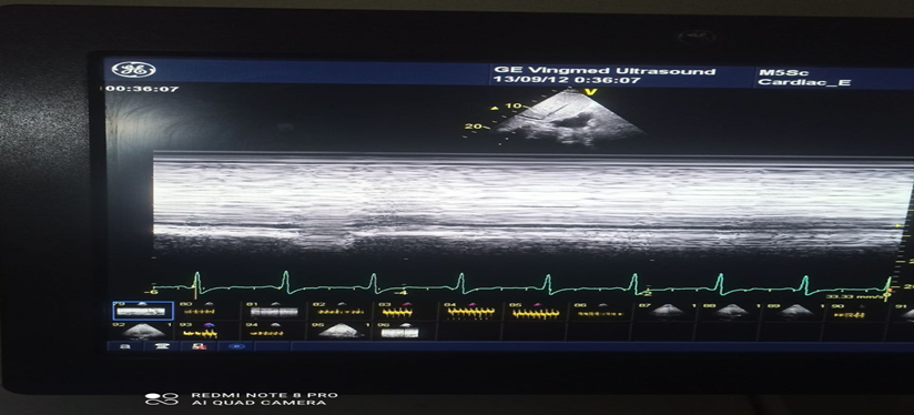
2.8: M- mode measurement of inferior vena cava(IVC) and collapsibility
2.8.3 Estimation of Right Atrial Pressure
RAP is assessed by measuring IVC size and the collapsibility during normal breathing. IVC diameter is measured at end-expiration, approximately 0.5-3.0 cm proximal to the RA ostium, from the subcostal view. Collapsibility is determined by the change in IVC diameter during sniffing and quiet respiration(9).
2.8.4 Estimation of Pulmonary Artery Systolic Pressure
The most common method for assessing systolic pulmonary artery pressure (SPAP) is to utilize the simplified Bernoulli equation to determine the RV right atrium (RA) gradient from the peak velocity of tricuspid valve regurgitation. The RV systolic pressure (RVSP) is calculated by adding this value to an estimate of the mean RA pressure.
RVSP = 4(VTR) 2 + mean RA pressure
In the absence of pulmonary stenosis or RVOT obstruction, RVSP and SPAP are evaluated equally ( normal PASP<35 mmhg )(9).
Measurement of tricuspid regurgitation velocity using continuous-wave Doppler is illustrated in Figure 2.4.
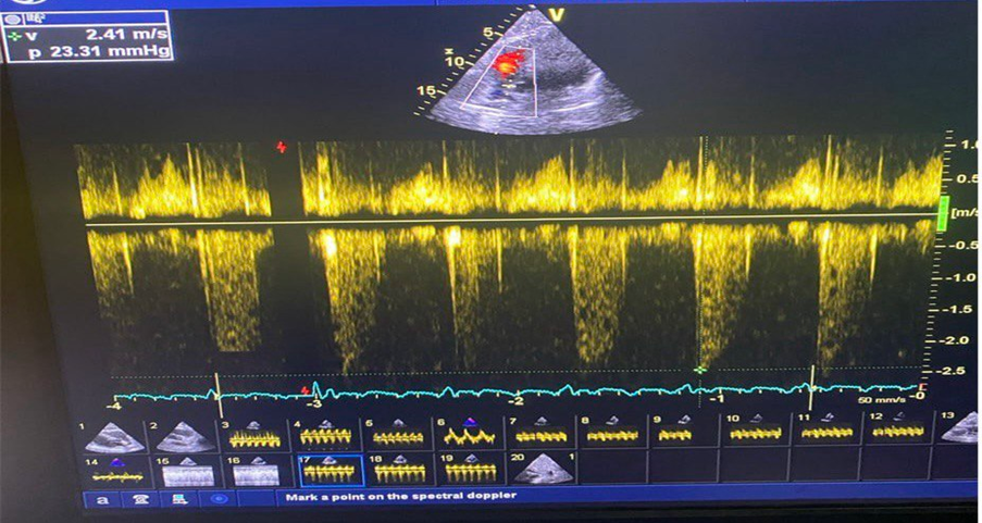
Figure 2.4: Measurement of Tricuspid Regurgitation Velocity Using Continuous-Wave Doppler
2.8.5 Tricuspid annular plane systolic excursion (TAPSE)
In the apical four-chamber view, an M-mode beam parallel to the motion of the lateral wall is oriented to cross the lateral portion of the tricuspid annulus, which is the measure of the displacement of the base of the RV towards the apex during systole(normal value of TAPSE=>17 mm)(9).
The method of TAPSE measurement is illustrated in Figure 2.3.
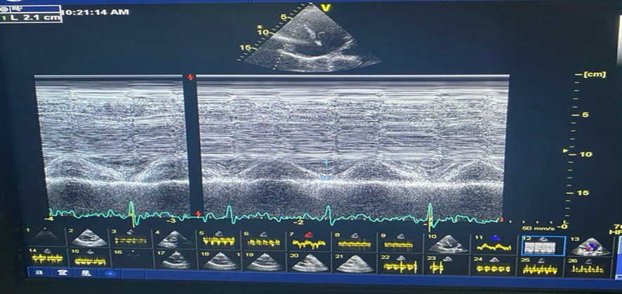
Figure 2.3: M-mode Measurement of TAPSE
Peak systolic RV annular velocity (S`): Tissue Doppler imaging (TDI) was used in the pulse-Doppler mode of the tricuspid annulus velocity at its lateral corners using the same echo unit, and systolic (S) velocity was measured (19).
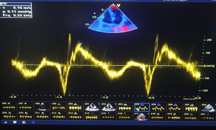
Figure (2.4): Estimation of Tissue Doppler Systolic Velocity(S`) Of Rv.
Assessment of right ventricular (RV) diastolic function.
remains less well validated because of the limited available guidelines and its complex association with various cardiac and systemic disorders. Therefore, this study evaluated specific RV diastolic function parameters using pulsed-wave (PW) Doppler of RV inflow and PW tissue Doppler imaging (PW-TDI) of the tricuspid valve (TV) annulus(20). The ratio of peak early diastolic tricuspid inflow velocity (tricuspid E) to peak late diastolic tricuspid inflow velocity (tricuspid A), expressed as the tricuspid E/A ratio, was measured using the modified apical four-chamber RV view. In addition, the early diastolic peak tricuspid annular velocity (e’) was obtained, and the tricuspid E/e’ ratio was subsequently calculated.
Doppler interrogation of tricuspid inflow:
Early (E) and late (A) trans-tricuspid inflow velocities, E/A ratio, and E deceleration time are recorded using pulsed wave Doppler at the tricuspid leaflet tips. The imaging plane should be optimized to align the beam with tricuspid inflow as in figure (2.8)
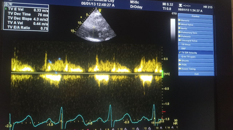
figure (2.8): Pulsed Doppler echocardiography at the tricuspid show E/A wave's ratio
Tissue Doppler at the lateral tricuspid annulus: RV E′, A′, and E′/A′ should be measured as in figure 2.9. This also permits calculation of the RV E/E` Ratio
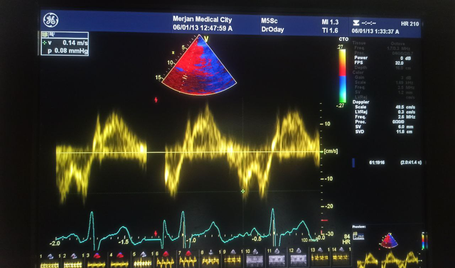
Figure (2.9) Pulsed tissue doppler imaging at the lateral annulus for measurement of E`
Right atrium area (RA): At the conclusion of systole, the right atrium area (RA) is measured from the lateral side of the tricuspid annulus to the septal aspect, following the RA endocardium and excluding the IVC, SVC, and RA appendage (19).
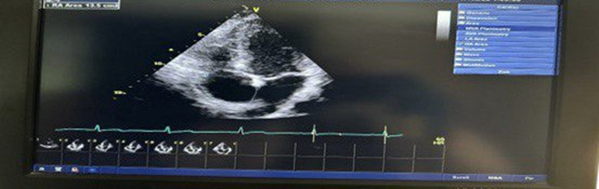
Figure (2.12) Right atrium area measured by 2DE image at end of systole.
Data analysis
Statistical analysis was carried out using SPSS version 27. Categorical variables were presented as frequencies and percentages. Continuous variables were presented as (Means ± SD). Student t-test was used to compare means between two groups . Paired t-test was used to compare means of echocardiographic parameters between supine and semi sitting positions among patients with COPD. Pearson Chi-square test was used to find the association between categorical variables. P value ≤ 0.05 was considered as significant.
Results :
Table 1: Distribution of COPD patients according to socio-demographic characteristics including (age , sex, body mass index and body surface area). Mean age of patients was (65.20 ± 9.95) years, older patient was 83.0 years and younger patient was 45.0 years. Mean BMI was (26.73 ± 2.73) Kg/m2 with maximum value was 32.27 Kg/m2 and minimum value was 21.46 Kg/m2. Only seven patients (17.5%) were obese. Mean body surface area was (1.87 ± 0.12) m2 with maximum value was 2.04 m2and minimum was 1.61 m2. Three quarters of patients were males (N=30, 75.0%).
Table 1: Distribution of COPD patients according to socio-demographic characteristics (N=40)
|
Socio-demographic characteristics |
||
|
Age (years) |
65.20 ± 9.95 |
45.0-83.0 |
|
BSA (m2) |
1.87 ± 0.12 |
1.61-2.04 |
|
Sex Male Female Total |
30 10 40 |
75.0% 25.0% 100.0% |
|
BMI (Kg/m2) Normal (18.5-24.9) Overweight (25-29.9) Obese (≥ 30) Total |
14 19 7 40 |
35.0% 47.5% 17.5% 100.0% |
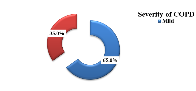
Figure 1: Distribution of COPD patients according to severity including (mild and moderate). Patients with mild COPD represent 26 patients (65.0%) and patient with moderate COPD represent 14 patients (35.0%).
Figure 1: Distribution of COPD patients according to severity (N=40)
Table 2: The comparison between COPD patients and control group according to socio-demographic characteristics including (age , body mass index, body surface area and sex). There was no significant mean differences of age, BMI and BSA between COPD patients and control group. There was no significant association between COPD and sex of patient.
Table 2: The comparison between COPD patients and control group according to socio-demographic characteristics (N=80)
|
Socio-demographic characteristics |
Study group |
P-value |
|
|
COPD (N=40) |
Control group (N=40) |
||
|
Age (years) |
65.20 ± 9.95 |
61.30 ± 8.77 |
0.067 |
|
BMI (Kg/m2) |
26.73 ± 2.73 |
26.76 ± 3.72 |
0.971 |
|
BSA (m2) |
1.87 ± 0.12 |
1.85 ± 0.10 |
0.637 |
|
Sex Male Female Total |
30 (75.0) 10 (25.0) 40 (100.0) |
28 (70.0) 12 (30.0) 40 (100.0) |
0.617 |
Table 3: The comparison between COPD patients and control group according to Forced Expiratory Volume in 1 sec and FEV1/FVC ratio . There was significant mean reduction of FEV1 and FEV1/FVC ratio among COPD patients in comparison to control group.
Table 3: The comparison between COPD patients and control group according to FEV1 and FEV1/FVC ratio (N=80)
|
Study variables |
Study group |
P-value |
|
|
COPD (N=40) |
Control group (N=40) |
||
|
FEV1 (L) |
79.28 ± 10.20 |
94.08 ± 2.60 |
<0.001* |
|
FEV1/FVC ratio |
0.64 ± 0.03 |
0.82 ± 0.02 |
<0.001* |
Table 4: The comparison between COPD patients and control group according to echocardiographic parameters measured in supine position including (Inferior Vena Cava diameter (mm), Inferior Vena Cava collapsibility (mm), Right Atrial Pressure (mmHg), Tricuspid Regurgitation Velocity (m/s), Pulmonary Artery Systolic Pressure (mmHg), Tricuspid Annular Plane Systolic Excursion (mm),
Ś (cm/s), E/A ratio, E/é and Right atrial area (cm2). There was significant mean elevation of Right Atrial Pressure (mmHg), Tricuspid Regurgitation Velocity (m/s), Pulmonary Artery Systolic Pressure (mmHg),
E/é and Right atrial area (cm2) among COPD patients in comparison to control group. There was significant mean reduction of Tricuspid Annular Plane Systolic Excursion (mm), Ś (cm/s) and E/A ratio
among COPD patients in comparison to control group.
Table 4: The comparison between COPD patients and control group according to echocardiographic parameters measured in supine position (N=80)
|
Supine Position |
Study group |
N |
Mean ± SD |
P-value |
|
Inferior Vena Cava diameter (mm) |
COPD |
40 |
16.05 ± 2.18 |
0.226 |
|
Control group |
40 |
16.60 ± 1.84 |
||
|
Inferior Vena Cava collapsibility(mm) |
COPD |
40 |
9.18 ± 1.63 |
0.076 |
|
Control group |
40 |
8.57 ± 1.34 |
||
|
Right Atrial Pressure (mmHg) |
COPD |
40 |
4.38 ± 2.26 |
<0.001* |
|
Control group |
40 |
3.00 ± 0.00 |
||
|
Tricuspid Regurgitation Velocity (m/s) |
COPD |
40 |
2.40 ± 0.41 |
<0.001* |
|
Control group |
40 |
1.56 ± 0.57 |
||
|
Pulmonary Artery Systolic Pressure (mmHg) |
COPD |
40 |
28.02 ± 7.79 |
<0.001* |
|
Control group |
40 |
16.86 ± 8.37 |
||
|
Tricuspid Annular Plane Systolic Excursion (mm) |
COPD |
40 |
22.43 ± 3.57 |
0.007* |
|
Control group |
40 |
24.38 ± 2.72 |
||
|
Ś (cm/s) |
COPD |
40 |
11.79 ± 2.36 |
<0.001* |
|
Control group |
40 |
14.00 ± 1.77 |
||
|
E/A ratio |
COPD |
40 |
0.85 ±0.27 |
0.011* |
|
Control group |
40 |
1.02 ± 0.30 |
||
|
E/é |
COPD |
40 |
5.06 ± 0.94 |
<0.001* |
|
Control group |
40 |
4.09 ± 0.35 |
||
|
Right atrial area (cm2) |
COPD |
40 |
15.21 ± 1.63 |
0.018* |
|
Control group |
40 |
14.45 ± 1.11 |
Table 5: The comparison between COPD patients and control group according to echocardiographic parameters measured in semi sitting position including (Inferior Vena Cava diameter (mm), Inferior Vena Cava collapsibility (mm), Right Atrial Pressure (mmHg), Tricuspid Regurgitation Velocity (m/s), Pulmonary Artery Systolic Pressure (mmHg), Tricuspid Annular Plane Systolic Excursion (mm),
Ś (cm/s), E/A ratio, E/é and Right atrial area (cm2). There was significant mean elevation of Inferior Vena Cava collapsibility (mm) and E/é among COPD patients in comparison to control group. There was significant mean reduction of Ś (cm/s) among COPD patients in comparison to control group.
Table 5: The comparison between COPD patients and control group according to echocardiographic parameters measured in semi sitting position (N=80)
|
Semi sitting Position |
Study group |
N |
Mean ± SD |
P-value |
|
Inferior Vena Cava diameter (mm) |
COPD |
40 |
14.73 ± 1.99 |
0.052 |
|
Control group |
40 |
15.63 ± 2.08 |
||
|
Inferior Vena Cava collapsibility(mm) |
COPD |
40 |
9.43 ± 1.80 |
<0.001* |
|
Control group |
40 |
7.78 ± 1.72 |
||
|
Right Atrial Pressure (mmHg) |
COPD |
40 |
3.38 ± 1.33 |
0.083 |
|
Control group |
40 |
3.00 ± 0.00 |
||
|
Tricuspid Regurgitation Velocity (m/s) |
COPD |
40 |
2.18 ± 0.37 |
0.102 |
|
Control group |
40 |
2.01 ± 0.50 |
||
|
Pulmonary Artery Systolic Pressure (mmHg) |
COPD |
40 |
22.70 ± 6.56 |
0.142 |
|
Control group |
40 |
20.19 ± 8.46 |
||
|
Tricuspid Annular Plane Systolic Excursion (mm) |
COPD |
40 |
23.60 ± 3.14 |
0.2 |
|
Control group |
40 |
22.68 ± 3.25 |
||
|
Ś (cm/s) |
COPD |
40 |
11.40 ± 2.20 |
<0.001* |
|
Control group |
40 |
15.13 ± 2.19 |
||
|
E/A ratio |
COPD |
40 |
0.82 ±0.20 |
0.201 |
|
Control group |
40 |
0.89 ± 0.25 |
||
|
E/é |
COPD |
40 |
4.78 ± 0.97 |
<0.001* |
|
Control group |
40 |
3.87 ± 0.36 |
||
|
Right atrial area (cm2) |
COPD |
40 |
13.73 ± 1.70 |
0.742 |
|
Control group |
40 |
13.63 ± 0.93 |
Table 6: The mean differences of echocardiographic parameters between supine and semi sitting positions among COPD patients including (Inferior Vena Cava diameter (mm), Inferior Vena Cava collapsibility (mm), Right Atrial Pressure (mmHg), Tricuspid Regurgitation Velocity (m/s), Pulmonary Artery Systolic Pressure (mmHg), Tricuspid Annular Plane Systolic Excursion (mm), Ś (cm/s), E/A ratio, E/é and Right atrial area (cm2)). There was significant mean elevation of TAPSE (mm) when measured in semi sitting position in comparison to supine position among COPD patients. There was significant mean reduction of Inferior Vena Cava diameter (mm), Right Atrial Pressure (mmHg), Tricuspid Regurgitation Velocity (m/s), Pulmonary Artery Systolic Pressure (mmHg), E/é and Right atrial area (cm2)) when measured in semi sitting position in comparison to supine position among COPD patients.
Table 6: The mean differences of echocardiographic parameters between supine and semi sitting positions among COPD patients (N=40)
|
Echocardiographic parameters |
Position |
N |
Mean ± SD |
Change(%) |
P-value |
|
Inferior Vena Cava diameter (mm) |
Supine |
40 |
16.05 ± 2.18 |
- 8.3% |
<0.001* |
|
Semi sitting |
40 |
14.73 ± 1.99 |
|||
|
Inferior Vena Cava collapsibility(mm) |
Supine |
40 |
9.18 ± 1.63 |
2.7% |
0.424 |
|
Semi sitting |
40 |
9.43 ± 1.80 |
|||
|
Right Atrial Pressure (mmHg) |
Supine |
40 |
4.38 ± 2.26 |
-22.8% |
0.031* |
|
Semi sitting |
40 |
3.38 ± 1.33 |
|||
|
Tricuspid Regurgitation Velocity (m/s) |
Supine |
40 |
2.40 ± 0.41 |
-9.2% |
<0.001* |
|
Semi sitting |
40 |
2.18 ± 0.37 |
|||
|
Pulmonary Artery Systolic Pressure (mmHg) |
Supine |
40 |
28.02 ± 7.79 |
-19.0% |
<0.001* |
|
Semi sitting |
40 |
22.70 ± 6.56 |
|||
|
Tricuspid Annular Plane Systolic Excursion (mm) |
Supine |
40 |
22.43 ± 3.57 |
5.0% |
0.009* |
|
Semi sitting |
40 |
23.60 ± 3.14 |
|||
|
Ś (cm/s) |
Supine |
40 |
11.79 ± 2.36 |
-3.3% |
0.271 |
|
Semi sitting |
40 |
11.40 ± 2.20 |
|||
|
E/A ratio |
Supine |
40 |
0.85 ±0.27 |
-3.5% |
0.112 |
|
Semi sitting |
40 |
0.82 ±0.20 |
|||
|
E/é |
Supine |
40 |
5.06 ± 0.94 |
-5.5% |
0.005* |
|
Semi sitting |
40 |
4.78 ± 0.97 |
|||
|
Right atrial area (cm2) |
Supine |
40 |
15.21 ± 1.63 |
-9.7% |
<0.001* |
|
Semi sitting |
40 |
13.73 ± 1.70 |
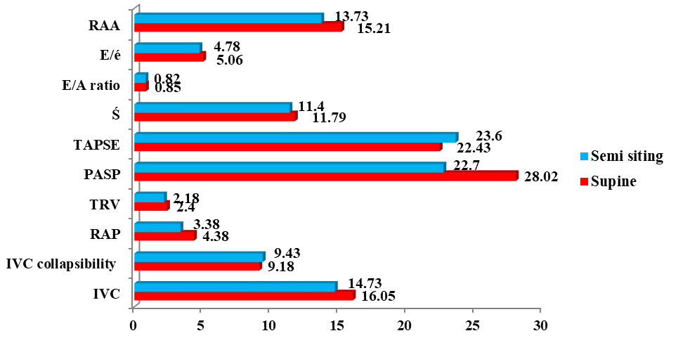
Figure 2: The mean differences of echocardiographic parameters between supine and semi sitting positions among COPD patients (N=40)
Table 7: The mean differences of echocardiographic parameters including (Inferior Vena Cava diameter (mm), Inferior Vena Cava collapsibility (mm), Right Atrial Pressure (mmHg), Tricuspid Regurgitation Velocity (m/s), Pulmonary Artery Systolic Pressure (mmHg), Tricuspid Annular Plane Systolic Excursion (mm), Ś (cm/s), E/A ratio, E/é and Right atrial area (cm2)) measure in supine position according to severity of COPD including (Mild and moderate). There was significant mean elevation of Inferior Vena Cava diameter (mm), Right Atrial Pressure (mmHg), Tricuspid Regurgitation Velocity (m/s), Pulmonary Artery Systolic Pressure (mmHg) and Right atrial area (cm2) among patients with moderate COPD in comparison to those with mild COPD.
Table 7: The mean differences of echocardiographic parameters measure in supine position according to severity of COPD (N=40)
|
Supine Position |
Severity |
N |
Mean ± SD |
P-value |
|
Inferior Vena Cava diameter (mm) |
Mild |
26 |
15.46 ± 2.14 |
0.018* |
|
Moderate |
14 |
17.14 ± 1.88 |
||
|
Inferior Vena Cava collapsibility(mm) |
Mild |
26 |
9.31 ± 1.89 |
0.412 |
|
Moderate |
14 |
8.93 ± 0.99 |
||
|
Right Atrial Pressure (mmHg) |
Mild |
26 |
3.58 ± 1.63 |
0.007* |
|
Moderate |
14 |
5.86 ± 2.57 |
||
|
Tricuspid Regurgitation Velocity (m/s) |
Mild |
26 |
2.22 ± 0.36 |
<0.001* |
|
Moderate |
14 |
2.73 ± 0.23 |
||
|
Pulmonary Artery Systolic Pressure (mmHg) |
Mild |
26 |
23.96 ± 5.80 |
<0.001* |
|
Moderate |
14 |
35.57 ± 4.78 |
||
|
Tricuspid Annular Plane Systolic Excursion (mm) |
Mild |
26 |
22.77 ± 4.26 |
0.303 |
|
Moderate |
14 |
21.79 ± 1.63 |
||
|
Ś (cm/s) |
Mild |
26 |
11.54 ± 2.26 |
0.375 |
|
Moderate |
14 |
12.25 ± 2.57 |
||
|
E/A ratio |
Mild |
26 |
0.81 ±0.24 |
0.155 |
|
Moderate |
14 |
0.93 ±0.31 |
||
|
E/é |
Mild |
26 |
5.05 ± 0.79 |
0.961 |
|
Moderate |
14 |
5.07 ± 1.21 |
||
|
Right atrial area (cm2) |
Mild |
26 |
14.64 ± 1.36 |
0.002* |
|
Moderate |
14 |
16.26 ± 1.62 |
Table 8: The mean differences of echocardiographic parameters including (Inferior Vena Cava diameter (mm), Inferior Vena Cava collapsibility (mm), Right Atrial Pressure (mmHg), Tricuspid Regurgitation Velocity (m/s), Pulmonary Artery Systolic Pressure (mmHg), Tricuspid Annular Plane Systolic Excursion (mm), Ś (cm/s), E/A ratio, E/é and Right atrial area (cm2)) measure in semi sitting position according to severity of COPD including (Mild and moderate). There was significant mean elevation of Tricuspid Regurgitation Velocity (m/s), Pulmonary Artery Systolic Pressure (mmHg) and Right atrial area (cm2) among patients with moderate COPD in comparison to those with mild COPD.
Table 8: The mean differences of echocardiographic parameters measure in semi sitting position according to severity of COPD (N=40)
|
Semi sitting Position |
Severity |
N |
Mean ± SD |
P-value |
|
Inferior Vena Cava diameter (mm) |
Mild |
26 |
14.65 ± 2.19 |
0.762 |
|
Moderate |
14 |
14.86 ± 1.61 |
||
|
Inferior Vena Cava collapsibility(mm) |
Mild |
26 |
9.62 ± 1.86 |
0.368 |
|
Moderate |
14 |
9.07 ± 1.69 |
||
|
Right Atrial Pressure (mmHg) |
Mild |
26 |
3.19 ± 0.98 |
0.331 |
|
Moderate |
14 |
3.71 ± 1.82 |
||
|
Tricuspid Regurgitation Velocity (m/s) |
Mild |
26 |
2.00 ± 0.32 |
<0.001* |
|
Moderate |
14 |
2.50 ± 0.21 |
||
|
Pulmonary Artery Systolic Pressure (mmHg) |
Mild |
26 |
19.58 ± 5.14 |
<0.001* |
|
Moderate |
14 |
28.50 ± 4.77 |
||
|
Tricuspid Annular Plane Systolic Excursion (mm) |
Mild |
26 |
23.73 ± 3.57 |
0.687 |
|
Moderate |
14 |
23.36 ± 2.24 |
||
|
Ś (cm/s) |
Mild |
26 |
11.33 ± 2.35 |
0.81 |
|
Moderate |
14 |
11.51 ± 1.97 |
||
|
E/A ratio |
Mild |
26 |
0.79 ±0.18 |
0.181 |
|
Moderate |
14 |
0.88 ± 0.22 |
||
|
E/é |
Mild |
26 |
4.77 ± 0.86 |
0.899 |
|
Moderate |
14 |
4.81 ± 1.18 |
||
|
Right atrial area (cm2) |
Mild |
26 |
13.28 ± 1.56 |
0.023* |
|
Moderate |
14 |
14.55 ± 1.69 |
DISCUSSION :
The present study investigated the effect of body position (supine versus semi-sitting) on echocardiographic parameters in patients with chronic obstructive pulmonary disease (COPD), with particular focus on right heart hemodynamics and right ventricular function. The findings revealed significant differences in pulmonary hemodynamic and right ventricular parameters between COPD patients and healthy controls. Furthermore, significant positional variations were observed between the supine and semi-sitting positions.
The present study demonstrated that the majority of COPD patients had mild disease severity (65%), while 35% were classified as having moderate COPD. This finding is consistent with Yang et al. (2022)(21), who reported that most COPD patients are commonly identified in the early stages of the disease (GOLD I and II), particularly in screening-based populations. Likewise, Bhatt et al. (2023)(22), stated that mild and moderate COPD constitute the majority of diagnosed cases and represent critical stages in disease progression. This observation may be attributed to the gradual and insidious nature of COPD progression, as many patients remain asymptomatic or only mildly symptomatic for extended periods before advancing to more severe stages.
The current study demonstrated significantly lower FEV1 and FEV1/FVC ratios in COPD patients compared with healthy controls. These findings are consistent with Higham et al. (2019)(23), demonstrated that chronic airway inflammation, small airway remodeling, mucus overproduction, and emphysematous destruction collectively drive the progressive decline in lung function seen in COPD patients. These pathological changes are mainly the result of persistent bronchial inflammation, reduced elastic recoil, narrowing of the small airways, and destruction of alveolar walls. Together, these processes lead to expiratory airflow limitation and progressive air trapping, which explain the deterioration in pulmonary function parameters over time.
Regarding echocardiographic findings in the supine position, the present study revealed significantly elevated right atrial pressure (RAP), tricuspid regurgitation velocity (TR Vmax), pulmonary artery systolic pressure (PASP), E/e′ ratio, and right atrial area in COPD patients compared with healthy controls. Conversely, tricuspid annular plane systolic excursion (TAPSE), S′ velocity, and E/A ratio were significantly reduced among COPD patients.
The finding of significantly elevated RAP among COPD patients is consistent with the study by Boerrigter et al. (2012)(24), which reported increased right-sided filling pressures and impaired right ventricular function in patients with COPD. the increase in RAP observed in COPD patients may be explained by chronic hypoxic pulmonary vasoconstriction and pulmonary vascular remodeling, both of which lead to increased pulmonary vascular resistance and elevated right ventricular afterload. Persistent right ventricular pressure overload subsequently impairs right ventricular emptying and results in elevation of right atrial pressure. In addition, lung hyperinflation and increased intrathoracic pressure may impair venous return and alter right heart filling dynamics, further contributing to elevated RAP.
The present study further revealed significantly elevated TR Vmax and PASP in COPD patients compared with healthy controls. This result aligns with Hilde et al. (2013)(25),reported pulmonary vascular remodeling and structural alterations of the right ventricle even in COPD cases without clinically established pulmonary hypertension .the rise in TR Vmax and PASP can be explained by chronic alveolar hypoxia, which promotes hypoxic pulmonary vasoconstriction and progressive vascular remodeling, ultimately increasing pulmonary vascular resistance. In addition, emphysematous destruction of the pulmonary capillary network reduces the vascular bed, further contributing to elevated pulmonary artery pressures. These pathophysiological mechanisms collectively increase right ventricular afterload, which is reflected on echocardiography as higher TR velocity and PASP values.
The present study also demonstrated significantly lower TAPSE and S′ velocity in COPD patients compared with healthy controls, suggesting impaired right ventricular systolic performance. This finding is consistent with Groussis et al. (2025)(26), who reported reduced TAPSE values among COPD patients in a systematic review and meta-analysis. The reduction in TAPSE and S′ velocity may indicate early longitudinal right ventricular systolic dysfunction resulting from chronic pulmonary hypertension and increased right ventricular afterload. Persistent pressure overload leads initially to adaptive right ventricular hypertrophy; however, prolonged overload eventually impairs myocardial contractility and systolic function. Furthermore, lung hyperinflation in COPD may alter cardiac geometry and disturb interventricular interaction, thereby reducing right ventricular longitudinal shortening and contributing to systolic dysfunction.
The current study also demonstrated a significantly larger right atrial area in COPD patients compared with healthy controls, indicating right-sided cardiac remodeling associated with chronic pulmonary disease. This finding is in agreement with Abdelalim et al. (2024)(27), who reported right atrial enlargement in COPD patients associated with elevated pulmonary artery pressure and right ventricular dysfunction. Enlargement of the right atrium may represent an adaptive mechanism in response to chronic pressure overload and increased right ventricular filling pressures secondary to sustained pulmonary vascular changes.
In the semi-sitting position, the present study showed significantly increased inferior vena cava (IVC) collapsibility and E/e′ ratio, together with reduced S′ velocity in COPD patients compared with healthy controls. The observed increase in IVC collapsibility is partly in agreement with Muller et al. (2012)(28), who highlighted that IVC behavior is strongly affected by respiratory mechanics and changes in intrathoracic pressure.
In COPD patients, this increased collapsibility may be attributed to exaggerated inspiratory effort and the generation of more negative intrathoracic pressures, which become more pronounced in the semi-upright position.
The reduction in S′ velocity observed in the semi-sitting position is consistent with Hilde et al. (2013)(25), who demonstrated impaired right ventricular systolic performance in COPD patients even during the early stages of the disease. Reduced S′ velocity may reflect increased right ventricular afterload secondary to chronic pulmonary vascular remodeling and elevated pulmonary arterial pressure. In addition, the semi-sitting position decreases preload and venous return, which may further influence tissue Doppler-derived systolic velocities because of the load-dependent nature of these parameters.
An important finding of this study was the marked effect of body position on echocardiographic measurements in patients with chronic obstructive pulmonary disease (COPD). The inferior vena cava (IVC) diameter, right atrial pressure (RAP), tricuspid regurgitation peak velocity (TR Vmax), pulmonary artery systolic pressure (PASP), E/e′ ratio, and right atrial area were all significantly reduced in the semi-sitting position compared with the supine position, whereas TAPSE was significantly increased.
These differences can be explained by the hemodynamic effects of posture on venous return, cardiac filling, and intrathoracic pressure. The semi-sitting position decreases venous return due to gravity-induced pooling of blood in the lower extremities, resulting in lower right atrial pressure and reduced IVC dimensions. Consequently, the reduction in preload may lead to lower estimated pulmonary artery pressures and smaller right atrial measurements.
The increase in TAPSE observed in the semi-sitting position may reflect improved right ventricular longitudinal function, secondary to reduced volume loading and decreased diaphragmatic compression in a more upright posture. In addition, semi-sitting positioning may enhance lung mechanics and reduce hyperinflation, which can further improve right ventricular performance.
The reduction in IVC diameter observed in the semi-sitting position is consistent with Mookadam et al. (2011)(29), who reported that IVC diameter is greater in the supine position, independent of the cardiac cycle. These positional differences may be attributed to changes in intra-abdominal pressure and potential mechanical effects, including hepatic compression of the IVC, particularly in the left lateral decubitus position. In the semi-sitting posture, gravitational pooling of blood in the lower extremities decreases venous return and reduces right atrial filling pressure. This finding reflects the normal physiological effect of body position on preload and central blood volume distribution, where upright positioning leads to reduced central venous volume, a smaller IVC diameter, and lower estimated right atrial pressure.
In patients with COPD, these posture-related hemodynamic changes may be more pronounced due to the presence of lung hyperinflation and abnormal fluctuations in intrathoracic pressure, both of which already compromise venous return and right ventricular filling. Hyperinflated lungs can mechanically compress intrathoracic vascular structures, thereby reducing cardiac preload, while exaggerated respiratory pressure swings further disrupt venous return dynamics. As a result, alterations in body position can significantly modify right-sided cardiac loading conditions and, consequently, echocardiographic measurements in this patient population.
The reduction in RAP observed in the semi-sitting position is in line with Kovacs et al. (2014)(30), who showed that right-sided filling pressures and pulmonary hemodynamics are significantly affected by preload status and body posture. In the supine position, venous return to the thorax increases due to blood redistribution, leading to higher right atrial filling pressures. In contrast, the semi-sitting position promotes peripheral venous pooling in dependent areas, which reduces venous return, resulting in lower preload and consequently decreased RAP.
Similarly, TR Vmax and PASP were significantly reduced in the semi-sitting position compared with the supine position. This is consistent with Berlier et al. (2022)(31), who demonstrated that pulmonary hemodynamic parameters are affected by body posture and preload changes.
Physiologically, the supine position increases venous return, enhancing right ventricular preload and pulmonary blood flow, which may elevate PASP and TR Vmax. In contrast, the semi-sitting position reduces central blood volume and pulmonary congestion, leading to lower values of these parameters. Additionally, improved respiratory mechanics in COPD patients in the semi-sitting position may further reduce pulmonary vascular stress and right ventricular workload.
Moreover, exercise and postural changes can alter venous return and pulmonary vascular resistance, potentially unmasking subclinical right ventricular dysfunction in COPD due to limited cardiopulmonary reserve.
Interestingly, TAPSE was significantly higher in the semi-sitting position compared with the supine position. This finding may be explained by the favorable effects of a semi-upright posture on right ventricular mechanics, including reduced intrathoracic pressure and enhanced diaphragmatic movement. These physiological adjustments can improve ventricular compliance and facilitate more efficient longitudinal contraction of the right ventricle, thereby increasing TAPSE values.
Furthermore, the semi-sitting position may reduce diaphragmatic compression on the heart, which can improve right ventricular filling and myocardial motion. The associated decrease in pulmonary artery pressure in this posture may also lower right ventricular afterload, contributing to improved systolic performance.
Since TAPSE is a load-dependent parameter of right ventricular longitudinal function, it is highly sensitive to changes in both preload and afterload. Consequently, variations in body position can substantially affect its measurement. These findings underscore the importance of standardizing patient positioning during echocardiographic assessment of right ventricular function in patients with COPD.
The present study also showed that the right atrial area was significantly reduced in the semi-sitting position compared with the supine position. This observation aligns with Khedr et al. (2023)(18), who reported that right atrial size is closely dependent on right-sided filling pressures and preload status. The reduction in venous return associated with semi-sitting posture leads to decreased right atrial filling and less chamber distension, which consequently results in lower right atrial area measurements.
The semi-sitting position promotes gravitational pooling of blood in the lower extremities, thereby reducing thoracic blood volume and right atrial preload. In patients with COPD, these positional effects may be further amplified by lung hyperinflation and abnormal intrathoracic pressure dynamics, which already impair venous return and right-sided cardiac filling. As a result, right atrial dimensions can vary considerably depending on body posture.
These findings underscore the important role of patient positioning in influencing right heart chamber measurements and hemodynamic evaluation in COPD. Accordingly, standardization of body position during echocardiographic assessment is essential to ensure accurate interpretation and reproducibility of right atrial size measurements.
Regarding disease severity, the present study demonstrated significantly higher IVC diameter, RAP, TR Vmax, PASP, and right atrial area among patients with moderate COPD compared with those with mild COPD. These findings are consistent with Abdelalim et al. (2024)(27), and Jatav et al. (2017)(32), all of whom reported progressive pulmonary hypertension and worsening right heart dysfunction with increasing COPD severity. Progressive airflow limitation and chronic hypoxemia in moderate COPD increase pulmonary vascular resistance through hypoxic pulmonary vasoconstriction and structural remodeling of the pulmonary vasculature. As a result, right ventricular afterload progressively increases, leading to elevated pulmonary artery pressure, impaired right ventricular filling, and enlargement of right-sided cardiac chambers.
In addition, persistent pressure overload may contribute to increased right atrial pressure and dilation of the inferior vena cava, reflecting progressive impairment of right heart hemodynamics. Chronic pulmonary vascular changes and reduced pulmonary vascular compliance further aggravate right ventricular workload and promote structural remodeling of the right atrium and right ventricle. These findings suggest that echocardiographic abnormalities may become more evident as COPD severity progresses, even before advanced clinical manifestations develop.
Overall, the findings of the present study emphasize that COPD is associated with early right heart hemodynamic and structural abnormalities, even during mild and moderate stages of the disease. Furthermore, body position significantly affects echocardiographic assessment of pulmonary pressures, preload-related parameters, and right ventricular function. Therefore, standardization of patient posture during echocardiographic evaluation is essential to improve the accuracy, consistency, and reproducibility of right heart measurements in COPD patients.
Limitations of Study:
1-The study included a relatively small sample size, which restricts the applicability of the findings to the wider COPD population.
2- Poor acoustic windows in COPD patients, resulting from lung hyperinflation, compromised image quality and limited the acquisition of certain echocardiographic parameters.
3- Only patients with mild and moderate COPD were enrolled, while severe and very severe stages were not assessed; therefore, the results do not reflect the entire spectrum of the disease.
4-The study design limits the ability to determine causality or evaluate the long-term progression of right ventricular changes.
5-Echocardiographic examinations were conducted at a single center, which may introduce inter-observer variability despite the use of standardized protocols.
6-Advanced echocardiographic modalities, such as strain imaging, speckle tracking, and three-dimensional echocardiography, were not utilized, which may have reduced the sensitivity for detecting subtle right ventricular dysfunction.
CONCLUSION :
transthoracic echocardiography is considered a dependable noninvasive technique for assessing right heart involvement in patients with chronic obstructive pulmonary disease (COPD). The results of this study demonstrated that patient positioning has a significant influence on echocardiographic parameters, as the supine position produced higher measurements of right-sided cardiac pressures and venous return than the semi-sitting position.
In addition, patients with COPD showed substantial changes in right ventricular structure and function when compared with healthy subjects. These abnormalities became increasingly evident with greater disease severity, likely reflecting progressive pulmonary vascular remodeling and the persistent effects of chronic hypoxia.
these findings highlight the importance of standardizing patient positioning during echocardiographic examination in COPD patients to ensure accurate assessment. They also reinforce the clinical usefulness of echocardiography in the early identification of pulmonary hypertension and right ventricular dysfunction.
Recommendations:
-Echocardiographic assessment in COPD should standardize patient body position, as it significantly affects right heart and venous return measurements.
-The semi-sitting position may provide more stable and physiologically representative evaluation of right ventricular function.
-Routine echocardiography is recommended in moderate to severe COPD for early detection of pulmonary hypertension and right ventricular dysfunction.
- Clinicians should recognize that increasing COPD severity is associated with progressive right-sided cardiac alterations; therefore, closer cardiopulmonary monitoring is required in advanced stages of the disease.
- Further large-scale studies are needed to confirm the impact of body position on echocardiographic parameters in COPD and to establish standardized assessment protocols.
BIBLIOGRAPHY:
1. (GOLD). GI for COLD. Global Initiative for Chronic Obstructive Lung Disease (GOLD). Global Strategy for the Diagnosis, Management, and Prevention of Chronic Obstructive Pulmonary Disease (2026 Report). GOLD; 2026. 2026;
2. Jarhyan P, Hutchinson A, Khaw D, Prabhakaran D, Mohan S. Prevalence of chronic obstructive pulmonary disease and chronic bronchitis in eight countries: a systematic review and meta-analysis. Bull World Health Organ. 2022;100(3):216.
3. Gudi N, Mahmood A, Roy MP, Nayak P, Verma A. Burden of COPD among population above 30 years in India: protocol for a systematic review and proposed meta-analysis. Can J Respir Ther CJRT= Rev Can la Thérapie Respir RCTR. 2021;57:14.
4. NV R, S SK, Desai N, PA M, N C, Chalasani SH, et al. Speckle-tracking echocardiographic abnormalities in chronic obstructive pulmonary disease: a systematic review and meta-analysis. J Cardiovasc Imaging. 2025;33(1):4.
5. Gupta NK, Agrawal RK, Srivastav AB, Ved ML. Echocardiographic evaluation of heart in chronic obstructive pulmonary disease patient and its co-relation with the severity of disease. Lung India. 2011;28(2):105–9.
6. Blanco I, Tura-Ceide O, Peinado VI, Barberà JA. Updated perspectives on pulmonary hypertension in COPD. Int J Chron Obstruct Pulmon Dis. 2020;1315–24.
7. Velidakis N, Khattab E, Gkougkoudi E, Kadoglou NPE. Pulmonary hypertension in left ventricular valvular diseases: A comprehensive review on pathophysiology and prognostic value. Life. 2023;13(9):1793.
8. Kouraichi C, Sekma A, Bel Haj Ali K, Chamtouri I, Sassi S, Toumia M, et al. Value of inferior vena cava collapsibility index as marker of heart failure in chronic obstructive pulmonary disease exacerbation. BMC Cardiovasc Disord. 2023;23(1):579.
9. Mukherjee M, Rudski LG, Addetia K, Afilalo J, D’Alto M, Freed BH, et al. Guidelines for the echocardiographic assessment of the right heart in adults and special considerations in pulmonary hypertension: recommendations from the American Society of Echocardiography. J Am Soc Echocardiogr. 2025;38(3):141–86.
10. Mizumi S, Goda A, Takeuchi K, Kikuchi H, Inami T, Soejima K, et al. Effects of body position during cardiopulmonary exercise testing with right heart catheterization. Physiol Rep. 2018;6(23):e13945.
11. Bondarsky E, Rothman A, Ramesh N, Love A, Kory P, Lee YI. Influence of head‐of‐bed elevation on the measurement of inferior vena cava diameter and collapsibility. J Clin Ultrasound. 2020;48(5):249–53.
12. Al Lami F, Salim Z. Prevalence and determinants of chronic obstructive pulmonary disease among a sample of adult smokers in Baghdad, Iraq, 2014. East Mediterr Heal J = La Rev sante la Mediterr Orient = al-Majallah al-sihhiyah li-sharq al-mutawassit. 2017 Mar;23(2):67–72.
13. Guevara-Tirado A. Calculation of BMI based on waist circumference, age, and height in the Peruvian population using a regression equation. Rev Fac Med la Univ Nac Colomb. 2025;73.
14. Pelletier KJ. A Quick and Reliable Mental Formula to Calculate the BSA of a Patient. J Extracorpor Technol. 2022;54(3):255–6.
15. Rudski LG, Lai WW, Afilalo J, Hua L, Handschumacher MD, Chandrasekaran K, et al. Guidelines for the echocardiographic assessment of the right heart in adults: a report from the American Society of Echocardiography: endorsed by the European Association of Echocardiography, a registered branch of the European Society of Cardiology, and . J Am Soc Echocardiogr. 2010;23(7):685–713.
16. Caplan M, Durand A, Bortolotti P, Colling D, Goutay J, Duburcq T, et al. Measurement site of inferior vena cava diameter affects the accuracy with which fluid responsiveness can be predicted in spontaneously breathing patients: a post hoc analysis of two prospective cohorts. Ann Intensive Care. 2020;10(1):168.
17. Kaptein MJ, Kaptein EM. Inferior vena cava collapsibility index: clinical validation and application for assessment of relative intravascular volume. Adv Chronic Kidney Dis. 2021;28(3):218–26.
18. Nakao S, Come PC, McKay RG, Ransil BJ. Effects of positional changes on inferior vena caval size and dynamics and correlations with right-sided cardiac pressure. Am J Cardiol. 1987;59(1):125–32.
19. Echocardiography TET of. The EACVI Textbook of Echocardiography. 2021; Available from: https://books.google.iq/books/about/The_EACVI_Textbook_of_Echocardiography.html?id=EGtuDQAAQBAJ&redir_esc=y
20. Zaidi A, Knight DS, Augustine DX, Harkness A, Oxborough D, Pearce K, et al. Echocardiographic assessment of the right heart in adults: a practical guideline from the British Society of Echocardiography. Echo Res Pract. 2020;7(1):G19–41.
21. Yang IA, Jenkins CR, Salvi SS. Chronic obstructive pulmonary disease in never-smokers: risk factors, pathogenesis, and implications for prevention and treatment. Lancet Respir Med. 2022;10(5):497–511.
22. Bhatt SP, Washko GR, Hoffman EA, Newell Jr JD, Bodduluri S, Diaz AA, et al. Imaging advances in chronic obstructive pulmonary disease. Insights from the genetic epidemiology of chronic obstructive pulmonary disease (COPDGene) study. Am J Respir Crit Care Med. 2019;199(3):286–301.
23. Higham A, Quinn AM, Cançado JED, Singh D. The pathology of small airways disease in COPD: historical aspects and future directions. Respir Res. 2019;20(1):49.
24. Boerrigter BG, Bogaard HJ, Trip P, Groepenhoff H, Rietema H, Holverda S, et al. Ventilatory and cardiocirculatory exercise profiles in COPD: the role of pulmonary hypertension. Chest. 2012;142(5):1166–74.
25. Hilde JM, Skjørten I, Grøtta OJ, Hansteen V, Melsom MN, Hisdal J, et al. Right ventricular dysfunction and remodeling in chronic obstructive pulmonary disease without pulmonary hypertension. J Am Coll Cardiol. 2013;62(12):1103–11.
26. Groussis M, Tran QK, Hoffer M, Ahari J, Pourmand A. Tricuspid annular plane systolic excursion (TAPSE) in chronic obstructive pulmonary disease patients: a systematic review and meta-analysis. Clin Exp Emerg Med. 2024;12(2):121.
27. Abdelalim MAF, Khalil MA, Sharshr RS, Abdelzaher AH. The role of transthoracic echocardiography in evaluating right ventricular parameters in chronic obstructive pulmonary disease. Egypt J Bronchol. 2024;18(1):71.
28. Muller L, Bobbia X, Toumi M, Louart G, Molinari N, Ragonnet B, et al. Respiratory variations of inferior vena cava diameter to predict fluid responsiveness in spontaneously breathing patients with acute circulatory failure: need for a cautious use. Crit Care. 2012;16(5):R188.
29. Mookadam F, Warsame TA, Yang HS, Emani UR, Appleton CP, Raslan SF. Effect of positional changes on inferior vena cava size. Eur J Echocardiogr. 2011;12(4):322–5.
30. Kovacs G, Avian A, Tscherner M, Foris V, Bachmaier G, Olschewski A, et al. Characterization of patients with borderline pulmonary arterial pressure. Chest. 2014;146(6):1486–93.
31. Berlier C, Saxer S, Lichtblau M, Schneider SR, Schwarz EI, Furian M, et al. Influence of upright versus supine position on resting and exercise hemodynamics in patients assessed for pulmonary hypertension. J Am Heart Assoc. 2022;11(4):e023839.
32. Jatav VS, Meena SR, Jelia S, Jain P, Ajmera D, Agarwal V, et al. Echocardiographic findings in chronic obstructive pulmonary disease and correlation of right ventricular dysfunction with disease severity. Int J Adv Med. 2017;4(2):476–80.