TO ASSESS ROLE OF CIRCULATING MICRORNAs IN APLASTIC ANEMIA PATIENT
- irfan Ahmed , Assistant professor Pumhs&w Nawabshah
- Muhammad Sarwar Khan , Assistant Professor,Pathology Department,LIMHS Thatta
- Farhan Ahmed Shaikh , Assistant Professor,Pathology Department, Bilawal medical College,LUMHS Jamshoro
- Aamir Ramzan , Assistant Professor, Pathology Department, LIMHS Thatta.
- kiran Memon , Asistant professor Indus medical college,Tando Muhammad Khan.
- Noor Rahman , Lab Technologist Khyber Medical University, Institute of pathology and diagnostic medicine (KMU- IPDM) Peshawar
- Fatima Zahid , University of Agriculture, Faisalabad.
Article Information:
Abstract:
Aplastic anemia is an acquired bone marrow failure categorized by marrow hyperplasia due to immune mediated effect on bone marrow. Objective: To assess role of plasma micro RNA’s (miR-150-5p) and (miR-146b-5p) in patients of aplastic anemia by (real time PCR technique) and to correlate plasma microRNA’s with severity in patients of aplastic anemia.Methods: Eight ml blood sample was collected from patients, and then distributed into two EDTA tubes. 3 ml EDTA tube was used for Complete blood count (CBC) and 5 ml EDTA sample was used for RNA isolation for performing MicroRNA testing. Bone marrow examination was performed at PATHOLOGY Department PUMHS for diagnosis of Aplastic anemia. All the data was recorded via self-made proforma and analyzed by using SPSS version 21.Results: Out of 72 cases, 36 were patients and 36 were controls, males were most common in both groups. According to severity of aplastic anemia 19 (52.8%) patients seen with very severe aplastic anemia, 10(27.8%) found with non-severe aplastic anemia and 7 (19.4%) found with severe aplastic anemia. Micro RNA 150.5p was positive among all patients, while negative in all controls. Micro RNA 146b.5p was positive among 5 patients, while it was negative in all controls p-value 0.001. Mean of micro RNA 150.5p was higher 34.89+13.99 in very severe aplastic anemia, 29.33+1.99 in severe and 28.76+2.37 in non-severe aplastic anemia. There was a negative correlation between Micro RNA 150.5p and hemoglobin, r-value 0.029, negative correlation between Micro RNA 150.5p and WBC, r-value 0.008, strong negative correlation between Micro RNA 150.5p and MCV, r-value 0.408 and weak negative correlation between Micro RNA 150.5p and platelets, r-value 0.017.Conclusion: It was concluded that plasma micro RNA’s (miR-150-5p) is a potential diagnostic marker of aplastic anemia. It was positive among all patients of aplastic anemia and showed a strong negative correlation with haemoglobin level, WBC, MCV and platelets.
Keywords:
Article :
INTRODUCTION:
Aplastic anemia (AA) is an acquired marrow deficiency marked as marrow hypoplasia, a lack of progenitor (HSPCs) and hematopoietic stem cells, and peripheral blood pancytopenia because of bone marrow immunity attack. In AA, Immune biomarkers development is a major challenging situation to monitor this medical condition.1 The general incidence is 2.3 incidents per million people in a year, it increases with age and 2 to 3 fold higher in Asia. Most commonly, AA occurs among young adults and at old age.2
AA may result from immune&genetic disorders, or drugs, chemicals, or radiation exposures. Nonetheless, the underlying reason is uncertain in around 50% of cases. Geographic variability in AA's epidemiological patterns is considered primarily because of ecological instead of genetic factors.3 Aplastic anemia patients respond to Immunosuppressive therapy (IST) which is showing that it is an “immune-mediated disease”.
Mostly, patients show improvement in condition after by giving just “Anti-Thymocyte globulins (ATG’S)” which causes transient T- cell depletion.7 The condition, AA result from immune-mediated disruption of HSPCs in most cases, causing peripheral blood pancytopenia and hypoplasia of the trilineage marrow.
The response of a substantial number of AA cases to IST is a strongest grounds for an intrinsic immune pathophysiology: following only temporary T-cell loss by ATGs, most patients display hematological change.4,5 Even though AA's immune pathophysiology is very well defined, no biomarkers are present to assist in better comprehension of a particular AA patient's immunological status, including therapy response and severity of disease.5,6
It would also be helpful for clinical trials and specific treatment decisions to have effective biomarkers that associated with severity of condition or response.5It is important to develop biomarker to evaluate the disease.1 Real time PCR- is done for diagnosis of specific MicroRNAs (miRNAs) in AA patients.4
Micro RNA are “short non- coding RNA’s that play crucial role in gene expression through complementary binding to 3’ untranslated regions of target mRNA’s, leading to translational repression”.8 Previous study in 2008 shows that miRNAs are also present in constituents of blood like in plasma, RBCs, WBCs and platelets.8 miRNAs have much importance and it serve as a novel biomarker in many diseases. Very few studies done on this regarding biomarkers, so it will approach severity of disease.
One study also show difference of some miRNAs concentration in serum and plasma. Meanwhile, serum contains higher miRNAs concentration than plasma when specific miRNAs levels were measured with an individual QPCR Taqman primers. MiR-150-5p and miR-146b-5p are Up regulated in AA. MiR-150-5p is an immuno- miRNAs which is regulator of T-cell processes.9
MiRNA-150-5p is involved in (induction of T-cell differentiation), its target is NOTCH 3, the NOTCH pathway regulation via miR-150-5p can possibly influence the development of T- cells.9 MiR-150-5p is found in nascent T-cells and afterwards highly enforced with the development of T-cell differentiation and maturation.9 C-MYB transcription factor is one more targeted factor of miR-150-5p, which contributes significantly in differentiation of T-cell.10 Within THP-1 monocytes the MiR-146b-5p reduces TNFa expression through selective suppression of TRAF and IRAK 1 in addition miR-146b-5p is engaged in response modulation of Innate immunity.1,12
A recent study reported miR-146b-5p regulatory functions in erythropoiesis. The miRNAs that Circulate in blood, mimic pathophysiological and physiological conditions and are high stable in preserved specimens of subjects, enabling them to be used as biological markers for different diseases.12 The identification of miRNA concentrations in blood serum and plasma, in general, can potentially diagnose the cancer timely and can predict the therapy response and prognosis.13,14
Recent research have also established miRNAs as biological markers for tracking the status of autoimmune disorders mediated by T-cell, like myasthenia gravis and MS.15,16 The researchers' findings indicate the role of miR-150-5p in immune-mediated malfunction by evidencing the application of miRNAs to differentiate AA from healthy (control) cases. More study is required into the related aims of current and further miRNAs. The researchers provide a little understanding of this by pathway research, which recognizes potential targets of such miRNAs linked to an immune system. For future research, these goals should be tested with classically applied molecular biology methods to evaluate miRNA biology.
Hosokawa and associates conclusively suggested a valuable new strategy for testing immunosuppression responses in AA, which is an autoimmune-mediated condition without effective biomarkers.17 The miRNAs contribution in AA pathophysiology as well as other syndromes of bone marrow dysfunction remained largely unclear.17
Furthermore, in AA's plasma or serum, miRNAs are still to be investigated. Therefore, this study was conducted to assess the contribution of plasma miRNAs as non-invasive biomarkers (MiR-150-5p)&(MiR-146b-5p) in AA and to compare miRNAs levels with severity of disease.
Material And Methods:
This study is conducted at Department of Pathology at PUMHS Shaheed Benazirabad, and Department of Medicine and Pediatrics at Civil Hospital Shaheed Benazirabad. This study took around six months post synopsis approval. It is descriptive research with a cross-sectional study. A non-probability, and Purposive Sampling Method was used during the study.
Inclusion Criteria
Those patients are included in this study who meet any of these 2 parameters:
• Hemoglobin<10.0 gm/ dl OR hematocrit<30%.
• White blood cells<1.5 ×10⁹ /L
• Platelets<50 × 109/ L
Exclusion Criteria
The sample size was considered by using open Epi software which stands to be n=36 by using proportion of 95% Confidence interval and 5% margin of error. Prevalence of Aplastic anemia is 2.34%.
After taking aseptic measures 6 ml blood sample is collected from patients, and then divided into two EDTA tubes. One tube containing 3 ml of sample is used for Complete blood count (CBC) and performed on XN 1000 by Sysmex Japan, and in other tube 3 ml of sample is used for extraction of RNA for MicroRNA testing.Bone marrow biopsy is performed at Pathology Department of LUMHS for diagnosis of Aplastic anemia. All the data was recorded via self-made proforma and analyzed by using SPSS version 21.
RESULTS:
Out of 72 cases 36 were patients and 36 were controls, mean age of patients was 18.58+13.94 years and mean age of controls was 30.13+18.31 years, p-value 0.004 (Table No 1).
Table-1: Age of the presented cases
|
Age (years) |
Study groups |
P-value |
|
|
Patients |
Control |
0.004 |
|
|
Mean + Std. Deviation |
18.58+13.94 |
30.13+18.31 |
|
Out of all study participants 21(58.3%) were males and 15(41.7%) were females in study group. 24 (66.7%) were males and 12(33.3%) were female in control group. Findings regarding gender were statistically insignificant p-value 0.465 (Table No 2).
Table-2: Gender of the presented cases
|
Gender |
Study groups |
P-value |
|
|
Patients |
Control |
0.465 |
|
|
Male |
21(58.3%) |
24(66.7%) |
|
|
Female |
15(41.7%) |
12(33.3%) |
|
|
Total |
36(100.0%) |
36(100.0%) |
|
|
Variable |
Study groups |
P-value |
|
|
Patients |
Control |
||
|
Water Intake |
|
|
|
|
Tape water |
30(83.3%) |
30(83.3%) |
0.135 |
|
Water supply |
0(0.0%) |
3(8.3%) |
|
|
Pump water |
6(16.7%) |
3(8.3%) |
|
|
Total |
36(100.0%) |
36(100.0%) |
|
Results regarding water intake, exposure to radiation and drug history were statistically insignificant according to both groups p-value were quite insignificant, results showed in table no. 5-8.
Table No 5. Patient’s Distribution According to Water Intake
n=72
Table No 6. Patient Distribution According to Exposure to Radiationn=72
|
Variable |
Study groups |
P-value |
|
|
Patients |
Control |
||
|
Exposure radiation |
|
|
|
|
Yes |
6(16.7%) |
0(0.0%) |
0.011 |
|
No |
30(83.3%) |
36(100.0%) |
|
|
Total |
36(100.0%) |
36(100.0%) |
|
Fig 4.5. Patient’s Distribution According to Exposure to Radiationn=72
Table No 7. Patient’s Distribution According to Past Medical History n=72
|
Variable |
Study groups |
P-value |
|
|
Patients |
Control |
||
|
Past Medical History |
|
|
0.011 |
|
Antimalarial |
2(5.6%) |
0(0.0%) |
|
|
HCV treatment |
4(11.1%) |
0(0.0%) |
|
|
No history |
30(83.3%) |
36(100.0%) |
|
|
Total |
36(100.0%) |
36(100.0%) |
|
Table No 8. Patient’s Distribution According to Water Intake, Exposure to Radiation, Drug Historyn=72
|
Variable |
Study Groups |
P-value |
|
|
Patients |
Control |
||
|
History of Concomitant |
|
|
|
|
Yes |
2(5.6%) |
0(0.0%) |
0.151 |
|
No |
34(94.4%) |
36(100.0%) |
|
|
Total |
36(100.0%) |
36(100.0%) |
|
Mean haemoglobin, WBC, MCV and platelets were significantly decreased among patients as compared to controls p-value 0.001 respectively (Table No 9).
Table No 9. Patient’s Distribution according to HB, WBC, MCV and Platelets n=72
|
Variables |
Study groups |
P-value |
|
|
Patients |
Control |
|
|
|
HB |
|
|
|
|
Mean +Std deviation |
6.61+2.09 |
12.10+0.69 |
0.001 |
|
WBC |
|
|
|
|
Mean +Std deviation |
2.10+0.94 |
6.57+1.01 |
0.001 |
|
MCV |
|
|
|
|
Mean +Std deviation |
74.95+20.86 |
86.30+4.29 |
0.045 |
|
Platelets |
|
|
|
|
Mean +Std deviation |
26.64+20.52 |
283.45+86.70 |
0.0001 |
According to the clinical presentation 32(52.8%) patients had epistaxis and bleeding, 14(19.4%) had bruises and 16(27.8%) had petechial (Table No 10).
Table No. 10. Cases Distribution according to Clinical Presentation n=36
|
|
Frequency (%) |
|
Clinical Presentation |
|
|
Epistaxis and bleeding |
32(52.8%) |
|
Bruises |
14(19.4%) |
|
Petechial |
16(27.8%) |
According to severity of aplastic anemia 19 (52.8%) patients seen with very severe aplastic anemia, 10(27.8%) found with non-severe aplastic anemia and 7(19.4%) found with severe aplastic anemia (Table No 11).
Table No. 11. Cases Distribution according to Severity of Aplastic Anemia n=36
|
|
Frequency (%) |
|
Severity of Aplastic Anemia |
|
|
Very severe aplastic anemia |
19 (52.8%) |
|
Severe aplastic anemia |
7(19.4%) |
|
Non-severe aplastic anemia |
10(27.8%) |
|
Total |
36(100.0%) |
Micro RNA 150.5p was positive among all patients, while negative in all controls. Micro RNA 146b.5p was positive among 5 patients, while it was negative all controls p-value 0.001 (Table No 12).
Table no 12. Patient’s Distribution According to Micro RNA
n=36
|
Micro RNA |
Study groups |
P-value |
||
|
Patients |
Control |
0.0001
0.020 |
||
|
Micro RNA |
Positive |
36(100.0%) |
0(0.0%) |
|
|
150.5p |
Negative |
0(0.0%) |
36(100.0%) |
|
|
Micro RNA |
Positive |
5(13.9%) |
0(0.0%) |
|
|
1466.5p |
Negative |
31(86.1%) |
36(100.0%) |
|
|
Total |
36(100.0%) |
36(100.0%) |
||
Very severe aplastic anemia was most common among 19 patients, severe aplastic anemia in 7 patients and non-severe aplastic anemia was in 10 patients. Mean of micro RNA 150.5p was 34.89+13.99 among patients with very severe aplastic anemia, followed by mean of micro RNA 150.5p 29.33+1.99 among severe aplastic anemia and 28.76+2.37 in non-severe aplastic anemia, these findings were statistically insignificant with a p-value of 0.252 (Table No 13).
Table no 13. Micro RNA According to Severity of Aplastic Anemia n=36
|
Severity of Aplastic Anemia |
N |
Mean+SD |
p-value |
|
Very severe aplastic anemia |
19 |
34.89+13.99 |
|
|
Severe aplastic anemia |
07 |
29.33+1.99 |
0.252 |
|
Non-severe aplastic anemia |
10 |
28.76+2.37 |
|
|
Total |
36 |
32.11+10.57 |
|
There was no significant difference in severity of aplastic anemia according age, gender, marital status and socioeconomic status p-values were quite insignificant, as showed in (Table No 14-16).
Table No. 14. Severity of Aplastic Anemia According to Gender Total 21 n=36
|
|
Gender |
Total |
p-value |
|
|
Severity of Aplastic Anemia |
Male |
Female |
||
|
Very severe aplastic anemia |
11 30.6% |
8 22.2% |
19 52.8% |
|
|
Severe aplastic anemia |
3 8.3% |
4 11.1% |
7 19.4% |
0.535
|
|
Non-severe aplastic anemia |
7 19.4% |
3 8.3% |
10 27.8% |
|
|
Total |
21 58.3% |
15 41.7% |
36 100.0% |
|
Table No. 15. Severity of Aplastic Anemia According to Age Groups n=36
|
|
Age groups |
Total |
p-value |
|||
|
Severity of Aplastic Anemia |
5-15 years |
15-30 years |
31-45 years |
45-60 years |
||
|
Very severe aplastic anemia |
8 22.2% |
9 25.0% |
0 0.0% |
2 5.6% |
19 52.8% |
|
|
Severe aplastic anemia |
4 11.1% |
1 2.8% |
2 5.6% |
0 .0% |
7 19.4% |
0.189
|
|
Non-severe aplastic anemia |
4 11.1% |
5 13.9% |
1 2.8% |
0 .0% |
10 27.8% |
|
|
Total |
16 44.4% |
15 41.7% |
3 8.3% |
2 5.6% |
36 100.0% |
|
Table No. 16. Severity of Aplastic Anemia According to Marital Status n=36
|
|
Marital status |
Total |
p-value |
|
|
Severity of Aplastic Anemia |
Un-married |
Married |
||
|
Very severe aplastic anemia |
11 30.6% |
8 22.2% |
19 52.8% |
|
|
Severe aplastic anemia |
3 8.3% |
4 11.1% |
7 19.4% |
0.718
|
|
Non-severe aplastic anemia |
7 19.4% |
3 8.3% |
10 27.8% |
|
|
Total |
21 58.3% |
15 41.7% |
36 100.0% |
|
There was a negative correlation between Micro RNA 150.5p and haemoglobin, r-value 0.029 (Figure No 1).
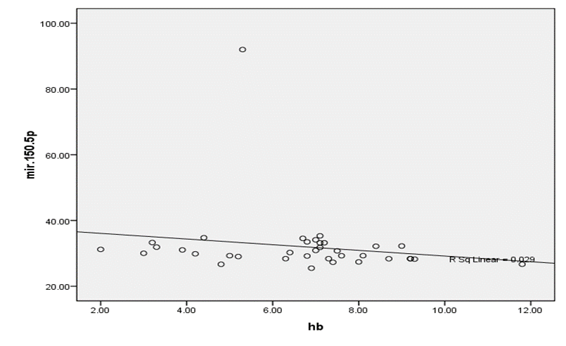
Fig 1. Negative Correlation between Micro RNA 150.5p and Hemoglobin r-value 0.029
We found weak negative correlation between Micro RNA 150.5p and WBC, r-value 0.008 (Figure No 2).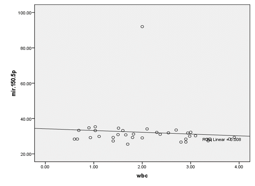
Fig 2. Weak Negative Correlation between Micro RNA 150.5p and WBC r-value 0.008
There was a strong negative correlation between Micro RNA 150.5p and MCV, r-value 0.408 (Figure No 3).
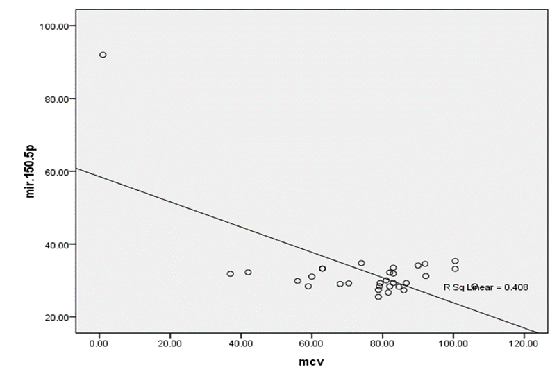
Fig 3. Strong Negative Correlation between Micro RNA 150.5p and MCV r-value 0.408
In this study a weak negative correlation between Micro RNA 150.5p and platelets, r-value 0.017 (Figure No 4).
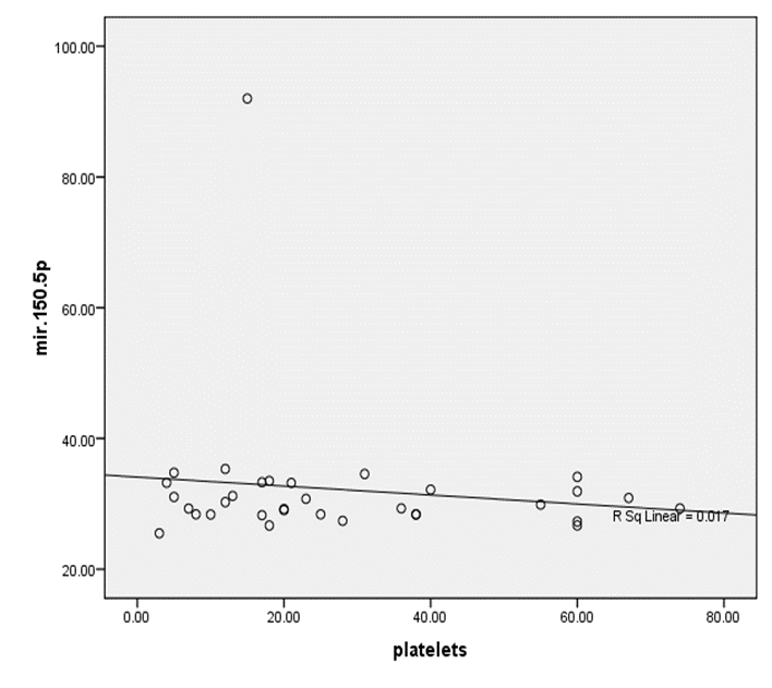
Fig 4. Weak Negative Correlation between Micro RNA 150.5p and Platelets r-value 0.017
DISCUSSION :
Aplastic Anemia (AA) has been classified as a rare disease, in which bone marrow has been observed to produce lower number of blood cells in contrast to the requirement of the body. It has been suggested that it majorly affects the Asians; two to three folds more than the people living in other regions of the world. There are specific microRNAs that have been reported as a reliable bio-makers of various solid and hematologic malignancies. In this particular study, microRNA 150-5p has been observed as a useful bio-maker for the AA, as it resulted positive among all the cases, whereas, it was negative in all controls. Likewise, in various other studies, circulating microRNAs have been suggested as a non-invasive bio-maker for various diseases that includes the Aplastic Anemia. 18-20 However, in the study conducted by Hosokawa K et al21 reported that microRNA 150-5p; the induction of T-Cell’s differentiation), and microRNA 146b-5p were elevated in the plasma of AA, the said plasmas in AA can help in diagnosis, whereas, microRNA 150-5p for monitoring of the disease.
MicroRNAs (miR-150-5p and miR-146b-5p) as well as miR-1 were observed to be significantly elevated, and decreased in the AA patient’s plasma, respectively. 22 While conducting a pathway analysis, it was revealed that above mentioned three microRNAs were targeting key immune-specific
functions among the patients. It was revealed that among others, one of the targets of miR-150-5p is NOTCH3; T-Cell development was heavily impacted during the regulation of Notch pathway through miR-150-5p. another target was identified is the C-MYB: a transcription factor, which plays a vital role in differentiation of T-Cells. MiR-150-5p, a immune-miRNA, is considered as a key regulator during the processes of T-Cells. 23 Meanwhile, it has been stated that after the identification of nineteen (19) dysregulated miRNA in a discovery set of 179 miRNAs, the researchers went on the validate their findings in a total of 108 cases, and identified the mentioned three miRNAs dysregulated with at least a 1.5 fold changes.
The miRNAs (miR-150-5p and miR-146b-5p) were upregulated in the group of AA, have previously had roles in development of T-Cells, and regulation of innate immunity, whereas, the role of miR-1 downregulated in the group of AA may play a vital role in auto-immunity. 24,25
In this study mean age of patients was 18.58+13.94 years and 21(58.3%) were males and 15(41.7%) were females. Similarly in the study of Ehsan et al26 reported that there were 49 (59.8%) males and 33 (40.2%) females and mean age of the patients was 27.93±18.7 years. This mean age was higher as compared to this study and this may because difference in sample size and selection of age range. Hanif et al27 reported male preponderance with a male to female ratio 3.4:1 and most of their patients were also found in second decade of life. A higher number of male is considered to be a significant finding, there may be a factor of social biasness, as male child in majority of rural areas are still considered to be a precious child. And are preferentially brought for proper treatments.
In this study mean haemoglobin, WBC, MCV and platelets were significantly decreased among patients as compared to controls p-value 0.001 respectively. However in the study of Biswajit H et al28 reported that the total count of RBC, WBC and platelets were lower than the normal value in the present study.
In this series according to the clinical presentation 32(52.8%) patients had epistaxis and bleeding, 14(19.4%) had bruises and 16(27.8%) had petechial. In the study of Ehsan A Et al29 reported that bleeding from gums was a predominant symptom seen in 43 patients followed by petechiae/ bruises in 38, epistaxis in 14, haematuria in 6 cases. Recently Ahmed P et al30 also found comparable results. Mechanisms of Immune are involved in its pathogenesis, which are resulting in immune destruction of hematopoietic stem cells. Majority of the patients may die due to infections and internal bleeding, if not treated. 44 patients (30.6%) presented with the issues of bleeding, from which 17 from gums, 11 had epistaxis, 08 purpura, 02 presented with per rectal bleeding, and 03 from other sides, among them 09 were facing life threatening bleeding (Adil S et al).31 Moreover, 30 patients (20.8%) had symptomatic anemia i.e. increasing pallor, tiredness, and shortness in breath.
In this study according to severity of aplastic anemia 19 (52.8%) patients seen with very severe aplastic anemia, 10(27.8%) found with non-severe aplastic anemia and 7(19.4%) found with severe aplastic anemia. In the study of Mex RH et al32 reported that Eleven, 28 and 10 patients had non-severe, severe, and very severe AA, respectively. Gutiérrez-Serdán R et al32 reported that eleven patients (22%) had non-severe AA, 28 (57%) severe AA, and 10 cases (20%) were categorized as very severe AA. Tan-Lim CS et al33 stated that thirteen patients had non-severe disease, 15 had severe, and 11 had very severe aplastic anemia
In this study, it was observed that no significant difference in the severity of AA appeared according to age, gender, marital status and socioeconomic status p-values were quite insignificant. Furthermore, it was revealed that there is a negative correlation between miR-150-5p and haemoglobin, r-value 0.029. We found also found strong negative correlation of Micro RNA 150.5p with MCV, and platelets. This correlation not found in published studies in the literature.
CONCLUSION:
It was concluded that plasma micro RNA’s (miR-150-5p) is a potential diagnostic marker of aplastic anemia. It was positive among all patients of aplastic anemia and showed a strong negative correlation with haemoglobin level, WBC, MCV and platelets.
BIBLIOGRAPHY:
1. Hosokawa K, Kajigaya S, Feng X, Desierto MJ, Ibanez MD, Rios O, Weinstein B, Scheinberg P, Townsley DM, Young NS. A plasma microRNA signature as a biomarker for acquired aplastic anemia. Haematologica. 2017 Jan 1;102(1):69-78.
2. Montane E, Ibanez L, Vidal X, Ballarin E, Puig R, Garcia N, Laporte JR. Epidemiology of aplastic anemia, a prospective multicenter study . Haematologica April 2008 93: 518-523; Doi: 10.3324/ haematol. 12020.
3. Adil SN, Burney IA, Kakepoto GN, Khurshid M. Epidemiologic features of aplastic anemia in Pakistan. J Pak Med Assoc 2001;51:443–5
4. Young NS, Calado RT and Scheinberg P. Current concepts in pathophysiology and treatment of aplastic anemia. Blood 2006 108:2509-2519.
5. Scheinberg P, Nunez O, Weinstein B, Biancotto A, Wu CO, Young NS. Horse versus rabbit antithymocyte globulin in acquired aplastic anemia. N Engl J Med. 2011;365(5):430–438.
6. Hosokawa K, Kajigaya S, Feng X, Desierto MJ, Ibanez MD, Rios O, Weinstein B, Scheinberg P, Townsley DM, Young NS. A plasma microRNA signature as a biomarker for acquired aplastic anemia. Haematologica. 2017 Jan 1;102(1):69-78.
7. Hosokawa K, Muranski P, Feng X, et al. Memory stem T cells in autoimmune disease: high frequency of circulating CD8+ memory stem cells in acquired aplastic anemia. J Immunol. 2016;196(4):1568–1578.
8. Tijsen AJ, Pinto YM, Creemers EE. Circulating microRNA’s as diagnostic biomarkers for cardiovascular diseases. American Journal of Physiology - Heart and Circulatory Physiology Published 1 November 2012 Vol. 303 no. 9, H1085-H1095.
9. Kroesen BJ, teteloshvli N, SmigielskaCzapielK, et al. Immuno-miR’s: critical regulators of T- cell development, function and aging. Immunology.2015:144(1):1- 10.
10. Ghisi M, Corradin A, Basso k, et al. Modulation of microRNA expression in human T- cell development: targeting of NOTCH 3 by miR- 150. Blood 2011 117:7053-7062.
11. lieu YK, kumar A, Pajerowski AG, Rogers TJ, Reddy EP. Requirement Of c- myb in T- cell development and in mature T-cell function. ProcNatlAcaDSci USA 2004; 101 (41): 14853- 14858.
12. Grasedieck S, Sorrentino A, Langer C, et al. Circulating microRNAs in hematological diseases: principles, challenges, and perspectives. Blood. 2013;121(25):4977–4984.
13. Schwarzenbach H, Nishida N, Calin GA, Pantel K. Clinical relevance of circulating cell-free microRNAs in cancer. Nat Rev Clin Oncol. 2014;11(3):145–156.
14. Zuo Z, Calin GA, de Paula HM, et al. Circulating microRNAs let-7a and miR-16 predict progression-free survival and overall survival in patients with myelodysplastic syndrome. Blood. 2011;118(2):413–415.
15. Gandhi R, Healy B, Gholipour T, et al. Circulating microRNAs as biomarkers for disease staging in multiple sclerosis. Ann Neurol. 2013;73(6):729–740.
16. Punga T, Le Panse R, Andersson M, Truffault F, Berrih-Aknin S, Punga AR. Circulating miRNAs in myasthenia gravis: miR-150-5p as a new potential biomarker. Ann Clin Transl Neurol. 2014;1(1):49–58.
17. Bell JB, Abedin S, Platanias LC. Circulating microRNAs: promising biomarkers in aplastic anemia. haematologica. 2017 Jan;102(1):1.
18. Grasedieck S, Sorrentino A, Langer C, et al. Circulating microRNAs in hematological diseases: principles, challenges, and perspectives. Blood. 2013;121(25):4977–4984.
19. Schwarzenbach H, Nishida N, Calin GA, Pantel K. Clinical relevance of circulating cell-free microRNAs in cancer. Nat Rev Clin Oncol. 2014;11(3):145–156.]
20. Punga T, Le Panse R, Andersson M, Truffault F, Berrih-Aknin S, Punga AR. Circulating miRNAs in myasthenia gravis: miR-150-5p as a new potential biomarker. Ann Clin Transl Neurol. 2014;1(1):49–58.
21. Hosokawa K, Kajigaya S, Feng X, Desierto MJ, Ibanez MD, Rios O, Weinstein B, Scheinberg P, Townsley DM, Young NS. A plasma microRNA signature as a biomarker for acquired aplastic anemia. Haematologica. 2017 Jan 1;102(1):69-78.
22. Hosokawa K, Kajigaya S, Feng X, Desierto MJ, Ibanez MD, Rios O, Weinstein B, Scheinberg P, Townsley DM, Young NS. A plasma microRNA signature as a biomarker for acquired aplastic anemia. Haematologica. 2017 Jan 1;102(1):69-78.
23. Kroesen BJ, Teteloshvili N, Smigielska-Czepiel K, et al. Immuno-miRs: critical regulators of T-cell development, function and ageing. Immunology. 2015;144(1):1–10.
24. Bell JB, Abedin S, Platanias LC. Circulating microRNAs: promising biomarkers in aplastic anemia. haematologica. 2017 Jan;102(1):1.
25. Ehsan A, Shah SA, Ibrahim T. Epidemiology of acquired aplastic anaemia in Pakistan. Journal of Ayub Medical College Abbottabad. 2011 Mar 1;23(1):102-5.
26. Hanif S, Naz F, Siddiqui E, Raza J. Acquired Aplastic anemia: Treatment in a developing country. Pak J Med Sci 2007;23:37–4.
27. Biswajit H, Pratim PP, Kumar ST, Shilpi S, Krishna GB, Aditi A. Aplastic anemia: a common hematological abnormality among peripheral pancytopenia. North American journal of medical sciences. 2012 Sep;4(9):384.
28. Ehsan A, Shah SA, Ibrahim T. Epidemiology of acquired aplastic anaemia in Pakistan. Journal of Ayub Medical College Abbottabad. 2011 Mar 1;23(1):102-5.
29. Ahmed P, Chaudhry QU, Satti TM, Mahmood SK, Ghafoor T, Shahbaz N, Khan MA, Satti HS, Akram Z, Iftikhar R. Epidemiology of aplastic anemia: a study of 1324 cases. Hematology. 2020 Jan 1;25(1):48-54.
30. Adil S, Kakepoto GN, Khurshid M, Burney IA. Epidemiological features of aplastic anaemia in Pakistan. Journal of Pakistan Medical Association. 2001;51:443.
31. Mex RH. Acquired aplastic anemia: a demographic, clinical, and therapeutic survey of a single institution in Mexico City. HEMATOLOGÍA Revista de. 2016 Apr;17(2):81.
32. Gutiérrez-Serdán R, Lopez-Karpovitch X. Anemia aplástica: estudio demográfico, clínico y terapéutico de una institución en la Ciudad de México. Revista de Hematología. 2016 Aug 15;17(2):81-9.
33. Tan-Lim CS, Melendres EM. Clinical and Demographic Profile of Pediatric Patients with Aplastic Anemia Seen in the Philippine National Tertiary Hospital from 2006 to 2013. Asp J Pediatrics Child Health. 2019 Dec 22;1(1):40-45.