Prevalence, Molecular Diagnosis and Phylogenetic Analysis of Fasciola hepatica Isolated from Ruminants in Erbil City, Kurdistan Region, Iraq
- Hamad Mustafa Saleh , Medical Laboratory Technology Department, Erbil Health and Medical Technical College, Erbil Polytechnic University, Erbil/ Iraq.
- Karwan S. N. AL-Marjan , Medical Laboratory Technology Department, Erbil Health and Medical Technical College, Erbil Polytechnic University, Erbil/ Iraq.
- Mustafa Ibrahim Mustafa , Medical Laboratory Technology Department, Erbil Health and Medical Technical College, Erbil Polytechnic University, Erbil/ Iraq.
- Sara Yousf Mustafa , Medical Laboratory Technology Department, Erbil Health and Medical Technical College, Erbil Polytechnic University, Erbil/ Iraq.
- Chnwr Farhan MuhammadAmin , Medical Laboratory Technology Department, Erbil Health and Medical Technical College, Erbil Polytechnic University, Erbil/ Iraq.
- Dilan Huner Omer , Medical Laboratory Technology Department, Erbil Health and Medical Technical College, Erbil Polytechnic University, Erbil/ Iraq.
- Muhammed Abdulrazzaq Amin , Medical Laboratory Technology Department, Erbil Health and Medical Technical College, Erbil Polytechnic University, Erbil/ Iraq.
Article Information:
Abstract:
Among liver flukes, Fasciola hepatica is the most important parasite because their impact on the economy through damaging ruminant productivity and a zoonotic risk. Accurate identification of species is still challenging, so, integration of molecular tools is essential. This study aimed to assess the molecular characteristics of liver flukes isolated from ruminants in Erbil City. A total of 85 slaughtered ruminants (sheep, cattle, and goats) were examined in the period between January and March 2026. Genomic DNA was extracted, followed by COI gene amplification using Polymerase Chain Reaction (PCR), DNA sequencing and bioinformatic analysis including BLASTN alignment and phylogenetic reconstruction. Results confirm the overall prevalence 7.1% of the infection which was statistically significant (P = 0.034) with highest occurrence in sheep (16.7%), then cattle (3.3%), while no infections were identified in goats. Five isolates were sequenced and were submitted to the GenBank database of NCBI and recorded under these accession numbers, PZ327390, PZ327391, PZ327392, PZ327393, and PZ327394. BLASTN analysis showed a high similarity (100.00% identity) between isolated sequences and reference F. hepatica sequences, confirmed the identity of species. Phylogenetic analysis supported that all isolates clustered within the clade of F. hepatica, and showed close association with global reference strains with a significant bootstrap values. In conclusion, this study provides a molecular insight into liver fluke isolates from ruminants in Erbil City, gives another important part for limited genetic data available from Kurdistan Region of Iraq.
Keywords:
Article :
INTRODUCTION:
Congenital melanocytic nevi (CMN) are pigmented Liver flukes are major parasitic pathogens affecting ruminant livestock worldwide, exerting serious effects on animal health and overall productivity (Hayward et al., 2021). Significant economic losses are caused by liver fluke infections through reduced growth and productivity, increased illness and mortality rate, growth retardation, sterility, inefficient nutrient utilization, low quality of products, condemnation of infected livers and rising expenditures associated with control measures (Regasa & Seboka, 2021). Liver flukes are members of the trematodes, with Fasciola hepatica and F. gigantica being the predominant species reported in Iraq. Infection by liver flukes has effects on the liver, gallbladder and biliary tree, and also represents an important zoonotic disease (Ali et al., 2021). Over the last three decades, liver fluke infection, especially human fascioliasis, has emerged as a significant global concern for public health. The disease is often underestimated in humans due to frequent misdiagnosis, resulting in delayed therapeutic intervention. In some cases, ectopic migration of parasites occurs, and flukes may localize in atypical sites such as skin, brain, eyes, and neck (Lan et al., 2024).
Identification of liver fluke isolates is essential due to variations in intermediate and definitive hosts, pathogenicity, response to anti-parasitic drugs, and epidemiological patterns (Ashrafi et al., 2006). Liver fluke species cannot be reliably differentiated using clinical, pathological, or immunological approaches. In addition, serological techniques lack species specificity, and traditional morphological identification methods are constrained by substantial limitations (Othman et al., 2023).
Consequently, molecular diagnostic approaches have become increasingly essential, and several studies have employed various molecular markers to accurate identification (Bozorgomid et al., 2020). Commonly applied markers include ITS-1, ITS-2, 28S rRNA, COI, and nad1, although most existing studies focus on individual species (Hasanpour et al., 2020). Polymerase chain reaction (PCR)-restriction fragment length polymorphism (RFLP) is widely used because it is rapid, cost-effective, and uncomplicated (Galavani et al., 2024). However it does not provide insight into genetic diversity and haplotype structure, necessitating the use of mitochondrial DNA sequencing (Bozorgomid et al., 2020).
Accordingly, this study aimed to investigate liver fluke infections among ruminants in Erbil City, Kurdistan Region-Iraq. The study focused on molecular identification and characterization of liver fluke isolates using PCR-based techniques and COI molecular marker, assessment of genetic relationships among isolates using phylogenetic analysis, determining prevalence of liver fluke infection and potential zoonotic risk evaluation.
Materials and Methods :
Erbil City, in Kurdish known as Hawler and historically as Arbela, is the capital and most populous city in the Kurdistan Region in northern Iraq. It lies at latitude 36.206293 and longitude 44.008870 (36°12′22.6548″ N, 44°00′31.932″ E).
Between January 2026 and March 2026, a total of 28 adult liver flukes were collected from the infected livers from 85 examined slaughtered ruminants (sheep, n = 30; cattle, n = 30; goats, n = 25).
The general liver condition was initially evaluated visually, followed by incision and compression of the hepatic parenchyma toward the bile ducts to facilitate the release of flukes. Following collection, adult liver flukes were stored at −20 °C until further processing and molecular analysis.
Liver flukes were examined macroscopically, as well as microscopically under a light microscope to confirm their morphological features, and photographs were captured at 4x and 10x magnification (Figure 2). Some liver flukes were stored at −20 °C for molecular analysis, while the remaining parasites were fixed in 10% buffered formalin for morphological reference and educational purposes.
Morpholoical Identification and Sample Preservation:
|
4
|
|
3 |
|
1 |
|
2 |
|
4
|
|
3 |
|
1 |
|
2 |
Figure 1: Satellite map of Erbil City indicating the locations where sampling was conducted. 1- Bnaslawa, 2- Chwarchira, 3- Zheyan and 4- Erbil Modern Slaughterhouse.
Morphological Identification and Sample Preservation:
Liver flukes were examined macroscopically, as well as microscopically under a light microscope to confirm their morphological features, and photographs were captured at 4x and 10x magnification (Figure 2). Some liver flukes were stored at −20 °C for molecular analysis, while the remaining parasites were fixed in 10% buffered formalin for morphological reference and educational purposes.
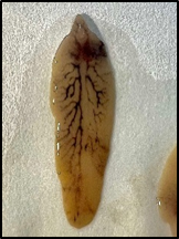
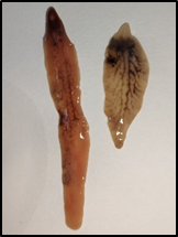
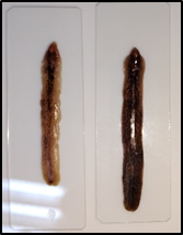
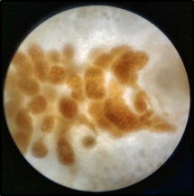
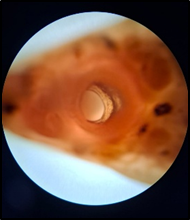
Figure 2: Morphological characteristics of adult liver flukes recovered from infected ruminants. Gross appearance showing dorsoventrally flattened body and internal branching structures (A, B, and C), and microscopic views demonstrating internal organs and eggs within the uterus (D and E).
DNA Extraction:
Genomic DNA was extracted from adult liver fluke tissues using the AddPrep Genomic DNA Extraction Kit (AddBioMeditek, Daejeon, South Korea), following the manufacturer’s instructions with minor adjustments.
DNA Quantification and Qualification:
DNA concentration and purity were measured using a NanoDrop spectrophotometer (ND-1000, USA). Samples with concentrations above 0.5 µg/µl and absorbance ratios greater than 1.7 were selected for molecular applications.
Polymerase Chain Reaction (PCR) Amplification:
Polymerase chain reaction targeting the cytochrome c oxidase subunit I (COI) gene was conducted. Reactions were prepared in 50 μl volumes containing 2x Taq DNA Polymerase Master Mix (AMPLIQON A/S, Stenhuggervej 22), 20 picomole (pmol) primers, DNase-free water, and template DNA (Table 1 and Table 2). Amplification was conducted using Bioresearch PTC-200 Gradient thermocycler.
Table 1: Reagents of Polymerase Chain Reaction (PCR) amplification.
|
No. |
PCR components |
Concentration |
Volume (µl) |
|
1 |
Master Mix |
2x |
25 |
|
2 |
Forward Primer |
20 pmol |
2 |
|
3 |
Reverse Primer |
20 pmol |
2 |
|
4 |
DNase free Water |
50 µl |
16 |
|
5 |
Template DNA |
50 ng/µl |
5 |
|
Total |
50 |
||
Table 2: Pair of primers used in COI gene amplification.
|
Primer |
Sequence 5’-3’ |
Amplicon size (bp) |
PCR Condition |
|
COI-F |
5’- ACGTTGGATCATAAGCGTGT -3’ |
490 bp |
95°-5 min; 95°-38 sec, 59°-38 sec, 72°-1 min; 72°-10 min; 4° ∞ |
|
COI-R |
5’- CCTCATCCAACATAACCTCT -3’ |
Separation of DNA fragments based on their molecular weight is done using Agarose gel electrophoresis, which is a procedure that is essential part of almost any standard experiments performed in molecular biology. The three main steps include agarose gel preparation, and electrophoresis of DNA fragments, followed by visualization of them.
DNA Sequencing:
ABI Prism Terminator Sequencing Kit (Applied Biosystem) was used for sequencing the PCR product samples of COI partial gene at Macrogen Molecular Company in South Korea. Then, for base calls confirmation and editing the chromatogram of genes, the Finch TV tool software is used.
Bioinformatics
To get accession numbers, the isolated sequences had been submitted to National Center for Biotechnology Information (NCBI). Sequence alignment was done at Basic Local Alignment Search Tool (BLAST) using (https://blast.ncbi.nlm.nih.gov/Blast.cgi), to comparing and alignment laboratory or query sequence with the same sequencing fragment marker (COI) to find out more similarity with liver flukes. The phylogenetic location of isolated sequences was determined by repeated alignment among a number of related liver fluke sequences, and these sequences obtained from NCBI, and then aligned with each other (adding Fasciolopsis buskii, with accession number of KX449331.1 as an out group) using Muscle model within MEGA software v.11 (Program Files\MEGA11 \MEGA_64.exe.).
Results:
A total of 85 ruminants (sheep, n = 30; cattle, n = 30; goats, n = 25) were examined, of which 6 (7.1%) were positive for liver fluke infection. The highest prevalence was observed in sheep, with 5 out of 30 animals (16.7%) infected, followed by cattle with 1 out of 30 (3.3%), while no infections were detected among goats.
Table 3: Occurrence and prevalence of liver fluke infection among slaughtered ruminants examined in Erbil City, Kurdistan Region, Iraq.
|
Hosts |
Positive |
Negative |
Prevalence % |
P value |
|
Sheep |
5 |
25 |
16.7% |
0.034 |
|
Cattle |
1 |
29 |
3.3% |
|
|
Goat |
0 |
25 |
0.0% |
|
|
Total |
6 |
79 |
7.1% |
PCR Amplification and Sequencing Results:
The mitochondrial Cytochrome C Oxidase subunit I (COI) gene was amplified from liver fluke isolates. The expected PCR product size was approximately 500 base pairs (bp), and after sequence editing and trimming, a final fragment length of 470 bp was obtained (Figure 3). The quality of the obtained sequences was confirmed by clear chromatogram peaks (Figure 4).
|
100bp |
|
3000bp |
|
1500bp |
Figure 3: Agarose gel electrophoresis showing sequence amplification of the COI gene sequence, with size of 470bp, isolated from liver flukes.
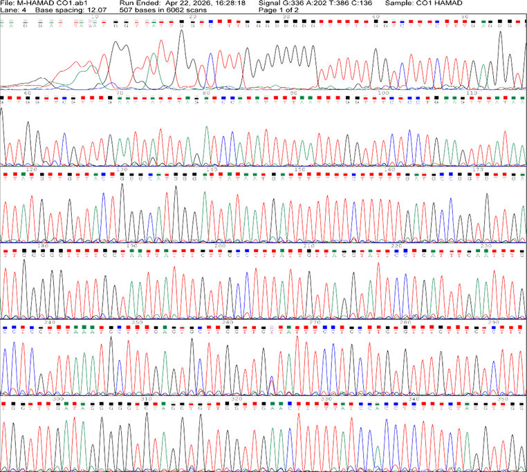
Figure 4: Representative graph shows summary profile of isolated COI gene sequences.
Sequence Data and GenBank Submission:
A total of five liver fluke isolates were sequenced. The obtained nucleotide sequences were edited and saved in FASTA format, then submitted to the GenBank database of the National Center for Biotechnology Information (NCBI) and recorded under the following accession numbers, PZ327390, PZ327391, PZ327392, PZ327393, and PZ327394.
BLASTN analysis revealed that the obtained sequences showed high similarity to Fasciola hepatica sequences available in GenBank. The query sequences demonstrated 100.00% identity with reference sequences, with a query coverage of 100% and E-value of 0.0, indicating a highly significant match (Table 4).
Table 4: BLASTN alignment results of liver fluke isolates based on COI gene sequences compared with reference sequences in GenBank.
|
Strain Description |
Accession (Reference sequences) |
||||
|
100% |
0.0 |
100.00% |
|||
|
F. hepatica isolate CO48 cytochrome oxidase subunit 1 gene, partial cds; mitochondrial |
100% |
0.0 |
100.00% |
||
|
F. hepatica haplotype FhC2 cytochrome oxidase subunit I (COI) gene, partial cds; mitochondrial |
100% |
0.0 |
100.00% |
||
|
F. hepatica isolate FhCHL-04 cytochrome c oxidase subunit I (COX1) gene, partial cds; mitochondrial |
100% |
0.0 |
100.00% |
||
|
100% |
0.0 |
100.00% |
|||
|
F. hepatica isolate CO12 cytochrome oxidase subunit 1 gene, partial cds; mitochondrial |
100% |
0.0 |
100.00% |
||
|
F. hepatica isolate CO14 cytochrome oxidase subunit 1 gene, partial cds; mitochondrial |
100% |
0.0 |
100.00% |
||
|
F. hepatica isolate CO21 cytochrome oxidase subunit 1 gene, partial cds; mitochondrial |
100% |
0.0 |
100.00% |
||
|
F. hepatica isolate CO7 cytochrome oxidase subunit 1 gene, partial cds; mitochondrial |
100% |
0.0 |
100.00% |
MK447944.1 |
Sequence Alignment:
Multiple sequence alignment of the obtained isolates with reference sequences demonstrated a high degree of nucleotide similarity, with only minor variations observed at specific positions. Overall, the alignment confirmed the close genetic relationship between the studied isolates and known F. hepatica sequences (Figure 5).
Phylogenetic analysis was performed using the Neighbor-Joining method based on COI gene sequences. The constructed phylogenetic tree showed that all five isolates clustered within the Fasciola hepatica clade, demonstrating close genetic relatedness to reference strains retrieved from GenBank. The isolates included in this study are grouped with previously reported F. hepatica sequences, indicating minimal genetic divergence among the local isolates and global reference strains (Figure 6)
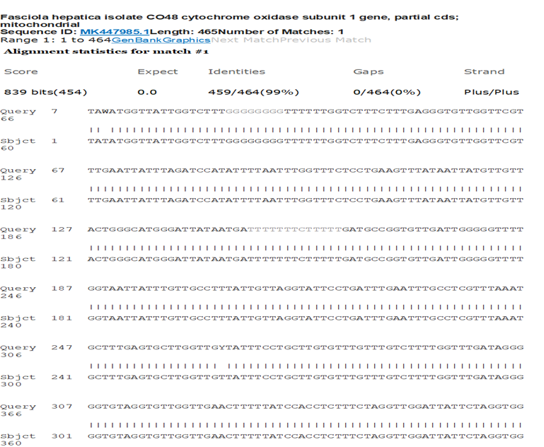
Figure 5: Pairwise alignment of the COI gene sequence from the studied isolate (Query) with a reference Fasciola hepatica sequence from GenBank (Subject).
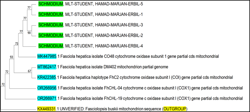
Figure 6: Phylogenetic tree shows the relationship between the isolated liver fluke (green) and reference Fasciola hepatica sequences (blue) based on COI gene sequences.
DISCUSSION :
liver fluke isolates collected from ruminants in Erbil City, Kurdistan Region, Iraq. We found that the overall prevalence of liver fluke infection among examined ruminants was 7.1%, which was statistically significant (p = 0.034). Previously, Muhammad & Hassan, 2021 reported a 7.93% prevalence rate of F. hepatica in sheep, cattle and goats, in Erbil, and which was close to our finding.
The limitations of conventional diagnostic methods increase the need for molecular identification. For molecular methods, the well-known mitochondrial Cytochrome C Oxidase subunit I (COI) gene was used as a reliable molecular marker for species identification and phylogenetic analysis, as Garcia-Corredor et al., 2023 stated, among number of different molecular markers used, COI is one of the two markers that is informative and can be used to evaluate genetic variations through phylogenetic analysis. The variable sites of the COI gene are higher than other markers by three to four times (Semyenova et al., 2006), offering enough genetic variation for identifying closely related species and strains of liver flukes. Moreover, mitochondrial genes are maternally inherited and do not undergo recombination, and have high mutation rates, which increases their stability and usefulness in phylogenetic studies (Shafiei et al., 2014; Xing et al., 2025). In our study, amplification and sequencing of the COI gene enabled the identification of liver fluke isolates. BLAST analysis showed a high similarity (100.00%) between the generated sequences and reference sequences of F. hepatica from GenBank, verifying the identification.
Different strains of F. hepatica show pathological differences, such as ability for adaptation to a wide range of hosts and environments, affected by frequent F. hepatica gene non-synonymous polymorphisms, and these variations influence excretory-secretory molecules, for example cathepsin L proteases, that responsible for severity of tissue degradation during liver parenchyma migration (Galtier et al., 2009; Lalor et al., 2021; Herrera-Torres et al., 2024). Some strain that show resistance to treatments like Triclabendazole (TCBZ), has specific molecular mutations, and pose a higher risk because of prolonged infection compared to susceptible strains (Raina et al., 2015; Flores-Velázquez et al., 2023). Also, it was suggested that primoinfection and reinfections, may stimulate varying levels of Foxp3+ T cells, and parasite’s ability to induce activation of M2 macrophage is a key survival mechanism which facilitates chronic infection (Herrera-Torres et al., 2024). For these reasons, it is important to identify different strains of F. hepatica.
Additional insight into genetic relationships within the isolates studied was provided by phylogenetic analysis based on COI gene sequences. The generated phylogenetic tree showed that all five isolates clustered within the clade of F. hepatica and demonstrated a strong relationship with reference sequences from GenBank. The isolates in our study were grouped with previously reported strains, showing minimal genetic variation and a significant level of similarity between local and global strains, as reported by Husch et al., 2020 and X. Wang et al., 2022. This observation indicates that the circulating liver fluke strains in the study area are genetically conserved and may share a common evolutionary origin, additionally, Alvi et al., 2023 reported similar findings.
CONCLUSION :
This study provides a molecular insight into liver fluke isolates from ruminants in Erbil City, gives another important part for limited genetic data available from Kurdistan Region of Iraq. Besides 7.1% overall prevalence of the infection. The findings indicate that COI gene sequencing was an accurate and reliable marker for identifying species, overcoming conventional method limitations. This study highlights the importance of adding molecular methods into routine parasitological investigations to improve the accuracy of diagnosis and epidemiological surveillance support.
BIBLIOGRAPHY:
1. Ali, K. N., Marif, H. F., Kakarash, N. A., & Mohammed, H. O. (2021). Liverfluke Coprological Cross-Sectional Survey in Cattle, Sheep and Goats in Sharazur District Kurdistan-Iraq. Al-Anbar Journal of Veterinary Sciences, 14(1).
2. Alvi, M. A., Khalid, A., Ali, R. M. A., Saqib, M., Qamar, W., Li, L., Ahmad, B., Fu, B.-Q., Yan, H.-B., & Jia, W.-Z. (2023). Genetic variation and population structure of Fasciola hepatica: An in silico analysis. Parasitology Research, 122(9), 2155–2173.
3. Ashrafi, K., Valero, M. A., Panova, M., Periago, M. V., Massoud, J., & Mas-Coma, S. (2006). Phenotypic analysis of adults of Fasciola hepatica, Fasciola gigantica and intermediate forms from the endemic region of Gilan, Iran. Parasitol Int, 55(4), 249–260. https://doi.org/10.1016/j.parint.2006.06.003
4. Bozorgomid, A., Rouhani, S., Harandi, M. F., Ichikawa-Seki, M., & Raeghi, S. (2020). Genetic diversity and distribution of Fasciola hepatica haplotypes in Iran: Molecular and phylogenetic studies. Veterinary Parasitology: Regional Studies and Reports, 19, 100359.
5. Flores-Velázquez, L. M., Ruiz-Campillo, M. T., Herrera-Torres, G., Martínez-Moreno, Á., Martínez-Moreno, F. J., Zafra, R., Buffoni, L., Rufino-Moya, P. J., Molina-Hernández, V., & Pérez, J. (2023). Fasciolosis: Pathogenesis, host-parasite interactions, and implication in vaccine development. Frontiers in Veterinary Science, 10, 1270064.
6. Galavani, H., Raeghi, S., Karamian, M., Torabi, N., & Haniloo, A. (2024). Morphometric and Molecular Characterization of Fasciola spp. In Livestock From Northwestern Provinces of Iran. Veterinary Medicine and Science, 10(6), e70097.
7. Galtier, N., Nabholz, B., Glémin, S., & Hurst, G. (2009). Mitochondrial DNA as a marker of molecular diversity: A reappraisal. Molecular Ecology, 18(22), 4541–4550.
8. Garcia-Corredor, D., Alvarado, M., Pulido-Medellín, M., Muñoz, M., Cruz-Saavedra, L., Hernández, C., Giraldo, J. C., Vásquez-Arteaga, L. R., Morillo Coronado, A. C., & Ramírez, J. D. (2023). Molecular characterization of Fasciola hepatica in endemic regions of Colombia. Frontiers in Veterinary Science, 10, 1171147.
9. Hasanpour, H., Falak, R., Naddaf, S. R., Mas-Coma, S., Rokni, M. B., Badirzadeh, A., Mokhtarian, K., Mohebali, M., Azami, S. J., & Fadavi, A. (2020). Molecular characterization of Fasciola spp. From some parts of Iran. Iranian Journal of Public Health, 49(1), 157.
10. Hayward, A. D., Skuce, P. J., & McNeilly, T. N. (2021). The influence of liver fluke infection on production in sheep and cattle: A meta-analysis. International Journal for Parasitology, 51(11), 913–924.
11. Herrera-Torres, G., Ruiz-Campillo, M. T., Bautista, M. J., Martínez-Moreno, F. J., Zafra, R., Buffoni, L., Rufino-Moya, P. J., Martínez-Moreno, Á., Molina-Hernández, V., & Pérez, J. (2024). Liver histopathological and immunohistochemical evaluation from Fasciola hepatica experimentally infected and reinfected sheep. Animals, 14(12), 1833.
12. Husch, C., Sattmann, H., Haefeli, I., Prosl, H., & Walochnik, J. (2020). Genetic diversity of Fasciola hepatica in Austria. Parasitology Research, 119(5), 1697–1701.
13. Lalor, R., Cwiklinski, K., Calvani, N. E. D., Dorey, A., Hamon, S., Corrales, J. L., Dalton, J. P., & De Marco Verissimo, C. (2021). Pathogenicity and virulence of the liver flukes Fasciola hepatica and Fasciola gigantica that cause the zoonosis Fasciolosis. Virulence, 12(1), 2839–2867.
14. Lan, Z., Zhang, X. H., Xing, J. L., Zhang, A. H., Wang, H. R., Zhang, X. C., Gao, J. F., & Wang, C. R. (2024). Global prevalence of liver disease in human and domestic animals caused by Fasciola: A systematic review and meta-analysis. J Glob Health, 14, 04223. https://doi.org/10.7189/jogh.14.04223
15. Muhammad, M. J., & Hassan, Z. I. (2021). Molecular Diagnosis of Fasciola hepatica in livestock using cox1 gene in Erbil Provence-Kurdistan Region/Iraq. Zanco Journal of Pure and Applied Sciences, 33(4), 36–42.
16. Othman, V. S., Hama, A. A., Zorab, R. H., & Dalimi, A. (2023). Molecular Characterization of Liver Fluke Isolated from Sheep, Goat and Cattle in Sulaymaniyah, Iraq. Iran J Parasitol, 18(4), 554–562. https://doi.org/10.18502/ijpa.v18i4.14264
17. Raina, O., Jacob, S. S., Sankar, M., Bhattacharya, D., Bandyopadyay, S., Varghese, A., Chamuah, J. K., & Lalrinkima, H. (2015). Genetic characterization of Fasciola gigantica from different geographical regions of India by ribosomal DNA markers. Journal of Parasitic Diseases, 39(1), 27–32.
18. Regasa, A., & Seboka, M. (2021). Review on Fasciolosis, its effect on meat quality/hazards and economical importances. Entomol Ornithol Herpetol, 10, 245.
19. Semyenova, S. K., Morozova, E. V., Chrisanfova, G. G., Gorokhov, V. V., Arkhipov, I. A., Moskvin, A. S., Movsessyan, S. O., & Ryskov, A. P. (2006). Genetic differentiation in eastern European and western Asian populations of the liver fluke, Fasciola hepatica, as revealed by mitochondrial nad1 and cox1 genes. The Journal of Parasitology, 92(3), 525–530.
20. Shafiei, R., Sarkari, B., Sadjjadi, S. M., Mowlavi, G. R., & Moshfe, A. (2014). Molecular and morphological characterization of Fasciola spp. Isolated from different host species in a newly emerging focus of human fascioliasis in Iran. Veterinary Medicine International, 2014(1), 405740.
21. Wang, X., Zhang, K., Guowu, Z., Li, Z., Yunxia, S., Chengcheng, N., Chunhui, J., Qiao, J., Qingling, M., & Xuepeng, C. (2022). Molecular characteristics and genetic diversity of Fasciola hepatica from sheep in Xinjiang, China. Journal of Veterinary Research, 66(2), 199–207.
22. Xing, B., Lin, L., & Wu, Q. (2025). Application of mitochondrial genomes to species identification and evolution. Electronic Journal of Biotechnology, 76, 39–48.